
Why Is My CA 19-9 High Despite Normal Imaging?
It feels like a contradiction: your CT scan or MRI came back clean, your doctor said the pancreas, liver, and bile ducts all "look normal," and yet your CA 19-9 blood test is sitting well above the reference range. That mismatch is unsettling, but it is also far more common than most patients are told, and it usually has nothing to do with a tumor hiding just out of view. CA 19-9 is a carbohydrate marker released by the cells lining your bile ducts, pancreas, and gut lining, and dozens of ordinary, non-cancerous conditions can push it upward — a blocked bile duct from a gallstone, a flare of pancreatitis, fatty liver disease, even uncontrolled diabetes. Imaging and blood work are simply answering two different questions: a scan asks "is there a visible mass," while CA 19-9 asks "how much of this particular sugar-protein is circulating in your blood right now," and a normal answer to the first question does not automatically produce a normal answer to the second. This article walks through exactly why that gap exists, which benign explanations are the most statistically likely, and what a careful next step actually looks like.
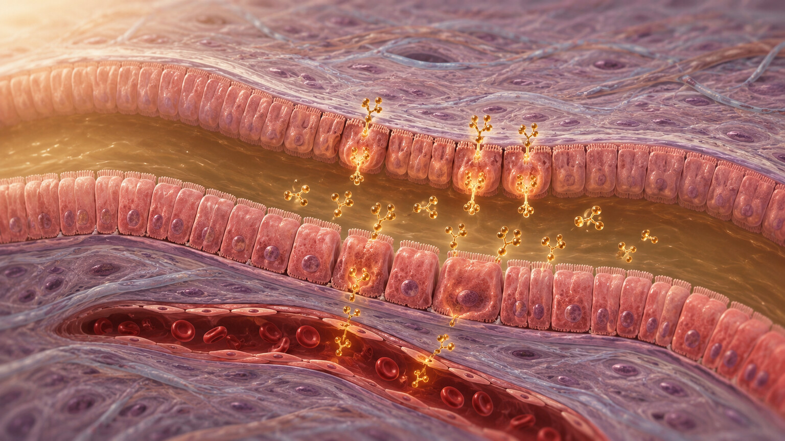
Figure 1. Epithelial cells lining the bile duct release CA 19-9 glycoprotein into surrounding bile and the bloodstream as part of normal cell turnover.
What CA 19-9 Actually Is — and Why a Clear Scan Doesn't Automatically Clear It
CA 19-9 stands for "carbohydrate antigen 19-9," and despite the word "antigen" sounding alarming, it is simply a sugar molecule attached to a protein — a glycoprotein — that gets manufactured and shed by the epithelial cells that line your bile ducts, pancreatic ducts, gallbladder, stomach lining, and parts of your intestines. These cells make small amounts of it as a routine part of normal cellular turnover, the way skin cells shed dead layers constantly. A tiny, steady trickle of CA 19-9 always ends up in your bloodstream, which is exactly why the test has a "normal" range in the first place, usually under about 37 units per milliliter, rather than a threshold of zero.
The test became clinically useful because pancreatic cancer cells, in particular, tend to overproduce CA 19-9 dramatically — sometimes to levels in the hundreds or thousands. That association is real and well studied, which is why doctors order the test when pancreatic cancer is already suspected or being monitored. But "cells that make CA 19-9 can become cancerous and overproduce it" is a very different statement from "anything that raises CA 19-9 must be cancer." The same ductal cells get inflamed, irritated, blocked, or stretched by a long list of benign problems, and inflamed or obstructed cells frequently make more CA 19-9 too, cancer or no cancer. Imaging, meanwhile, is built to detect a physical abnormality — a mass, a dilated duct, a cyst — above a certain size, typically several millimeters at minimum. A number on a lab report and a shadow on a scan are measuring fundamentally different physical realities, and each has blind spots the other doesn't share.
Imaging and Blood Work Are Answering Two Different Questions
It helps to think of a CT or MRI scan as a structural inspection, similar to a home inspector walking through a house checking for visible cracks, water stains, or a sagging roofline. If nothing looks structurally wrong, the inspector reports the house as sound. CA 19-9, by contrast, is more like testing the water that comes out of the tap — it tells you something is or isn't dissolved in the system, but it doesn't tell you where in the plumbing the problem is, or whether there's a problem in the pipes at all versus something upstream in the water source itself. A structurally normal house can still have unusual mineral content in its water for reasons that have nothing to do with a cracked pipe: hard water from the municipal supply, an old water heater, nearby soil composition. In the body, "unusual mineral content in the water" is the elevated CA 19-9, and there are a lot of upstream explanations that never show up as a visible structural defect on a scan.
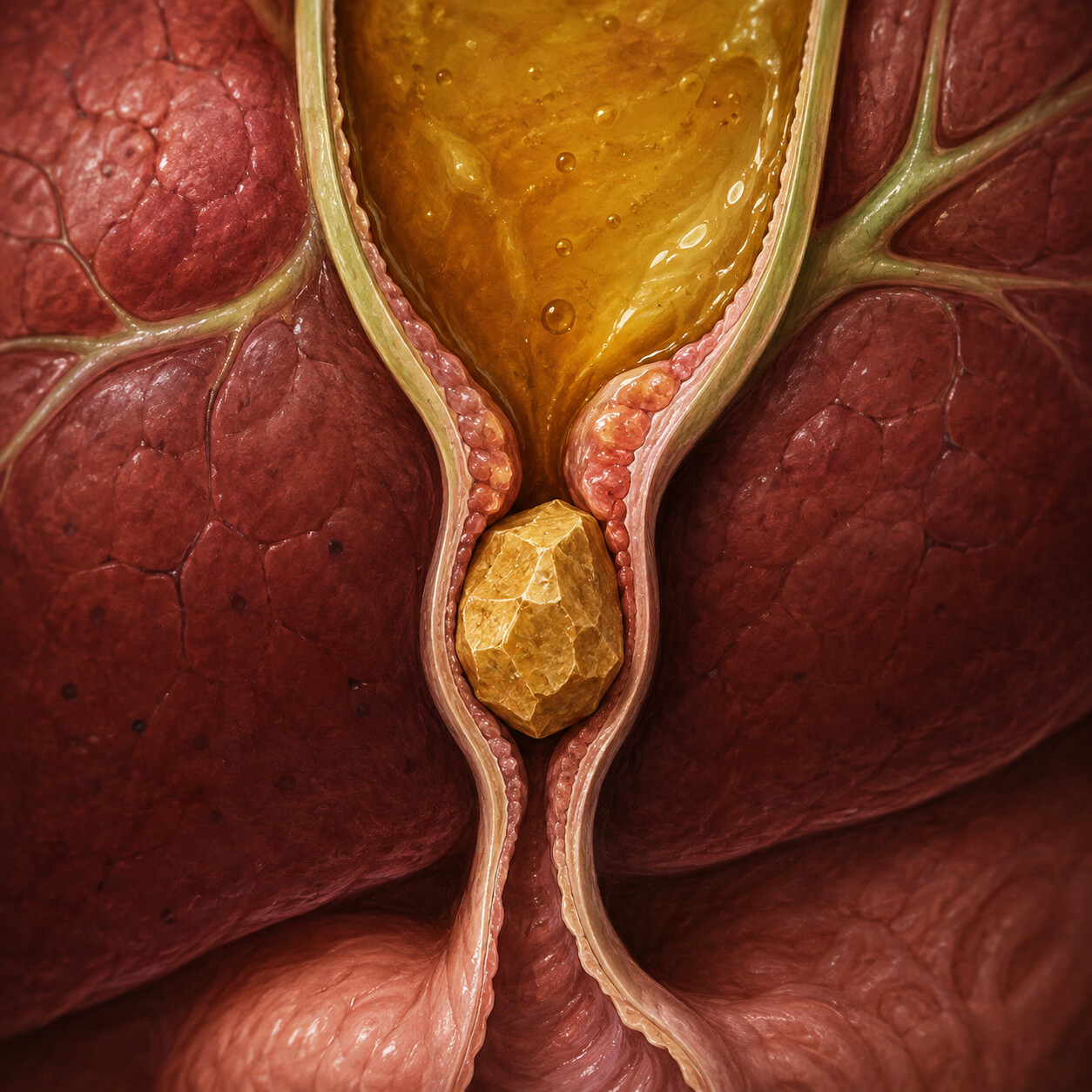
Figure 2. A gallstone lodged in the common bile duct causes bile to back up, distending and irritating the duct lining above the obstruction.
One of the most common upstream explanations is also one of the smallest: a gallstone that has slipped into the common bile duct, the tube that carries bile from the liver and gallbladder into the small intestine. A stone can be tiny — a few millimeters — and still partially or fully block that tube. Below the blockage, bile literally backs up, and the ductal cells sitting in that backed-up bile become distended and irritated, ramping up CA 19-9 production as a direct physical response to pressure and inflammation, not to malignancy. Many gallstones this small pass on their own or resolve without ever appearing on a standard abdominal CT, especially if the scan happens to be taken between episodes, after the stone has already moved or dissolved. Liver enzyme tests (like ALT, AST, and especially alkaline phosphatase and bilirubin) are often the more telling clue here, since they reflect the actual backup of bile far more directly and sensitively than a snapshot image does.
This is part of why doctors will frequently order — or re-order — liver function tests alongside a repeat CA 19-9 rather than jumping straight to another round of imaging: the liver panel can catch evidence of a passing obstruction that a scan taken at the wrong moment might miss entirely.
Curious what your liver panel, CA 19-9, and the rest of your labs actually mean side by side? Upload your results and get a complete, plain-language breakdown in under 15 minutes.
Analyze My ResultsThe Most Common Benign Reasons CA 19-9 Rises With a Clean Scan
Beyond a passing gallstone, several other everyday, non-cancerous conditions are well documented to elevate CA 19-9, sometimes substantially, without ever producing a visible mass:
Pancreatitis — inflammation of the pancreas, most often from gallstones, alcohol use, high triglycerides, or certain medications, directly irritates the same duct-lining cells that produce CA 19-9. Levels can rise during an active flare and then settle back down over the following weeks as the inflammation resolves, all while imaging shows nothing more than mild, nonspecific swelling or nothing abnormal at all once the acute episode has passed.
Cholangitis and biliary tract infections — inflammation or infection anywhere along the bile duct system produces the same irritation-driven overproduction, often with a fever or right-upper-abdomen pain that resolves with antibiotics long before a follow-up scan is even scheduled.
Chronic liver disease — cirrhosis, fatty liver disease, and viral hepatitis are all associated with elevated CA 19-9, likely because chronically inflamed or scarred liver tissue and the small bile ductules running through it behave similarly to actively obstructed ducts, oozing out extra marker over a long period rather than a sudden spike.
Poorly controlled diabetes — this one surprises most people, but several studies have found a meaningful association between elevated blood sugar and elevated CA 19-9, independent of any pancreatic structural disease. The mechanism isn't fully settled, but it may relate to how hyperglycemia affects mucin and glycoprotein production throughout the digestive tract's lining.

Figure 3. A patient reviewing an elevated CA 19-9 result on a lab portal shortly after a normal imaging study, the exact mismatch this article addresses.
Recent endoscopic procedures — an ERCP (a procedure that passes a scope and dye through the bile duct system) is well known to cause a temporary, sometimes dramatic spike in CA 19-9 for days to weeks afterward simply from the mechanical irritation of the ducts, unrelated to whatever the procedure was originally investigating.
Ovarian and other benign cysts — in women, benign ovarian cysts, endometriosis, and even pregnancy can raise CA 19-9, since the marker isn't produced exclusively by pancreatic or biliary tissue.
It's genuinely common for a patient to sit in the exact scenario this article is about — a scan read as normal, a CA 19-9 modestly elevated in the 40s, 60s, or even low hundreds — and for the ultimate explanation to be one of the conditions above, something already resolving or already being managed, rather than a cancer that imaging simply failed to catch.
Could This Still Be an Early Cancer That the Scan Simply Missed?
This is the question underneath the question, and it deserves a direct, honest answer: it's possible, but it is the less likely explanation, and there are specific reasons a scan can miss something small. Standard imaging has real resolution limits — a CT scan typically needs a lesion to be roughly 1 to 2 centimeters before it becomes reliably visible, and even MRI, which is more sensitive for soft tissue, can miss very small or very subtly infiltrating tumors, particularly in the pancreas, an organ that sits deep in the abdomen surrounded by bowel gas and other structures that can obscure small changes. Some early pancreatic cancers grow in a diffuse, infiltrative pattern rather than forming a single discrete mass, which makes them notoriously harder to see even in retrospect.
This is precisely why a persistently or progressively rising CA 19-9 with a normal initial scan doesn't end the investigation — it usually triggers either a repeat scan after a defined interval (often 4 to 12 weeks, timed to let any benign, inflammation-driven elevation settle on its own) or a more sensitive test called endoscopic ultrasound, which threads a small ultrasound probe through the stomach and duodenum to image the pancreas from just millimeters away, catching small lesions that a CT scan taken from outside the body cannot resolve. The distinguishing feature that worries a clinician far more than a single elevated number is a trend: a value that keeps climbing on repeat testing, especially alongside new symptoms like unexplained weight loss, jaundice, or persistent abdominal or back pain, is treated very differently from a value that rises once and then plateaus or falls.
Genetics — Some People Simply Run Higher, and Some Never Produce Any CA 19-9 at All
One of the least widely known facts about this test is that your baseline CA 19-9 level is partly determined by genetics, specifically by a set of genes that control the Lewis blood group antigen system — a completely separate classification from the ABO blood type most people are familiar with. The enzyme responsible for attaching the CA 19-9 sugar structure onto proteins requires a functional copy of a gene called FUT3. Roughly 5 to 10 percent of people, depending on ancestry, inherit two non-functional copies of this gene and are classified as "Lewis-negative." These individuals are physically incapable of producing CA 19-9 in any meaningful amount — their level will read near zero even in the presence of an aggressive, advanced pancreatic cancer, which is a critical and separate clinical pitfall from the one this article focuses on.
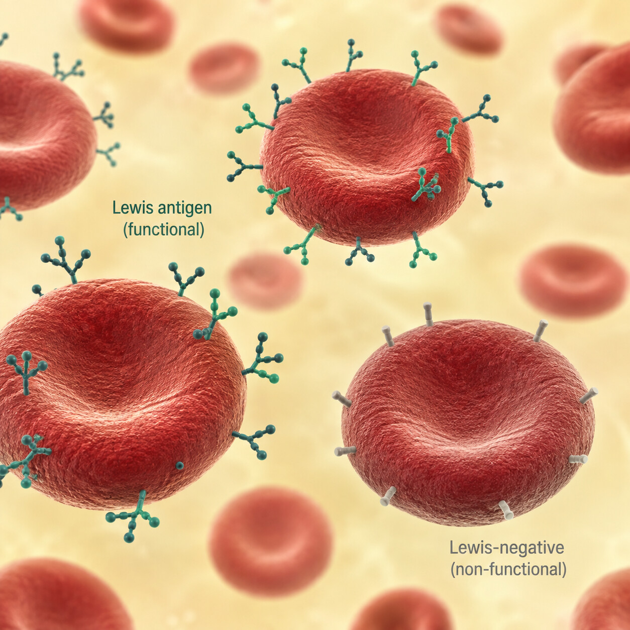
Figure 4. Lewis blood group antigens on red blood cell surfaces reflect FUT3 gene activity, the same gene that determines how much CA 19-9 a person's ductal cells can produce.
The flip side matters for exactly the situation this article addresses: the roughly 90 to 95 percent of people who are Lewis-positive naturally vary in how much baseline CA 19-9 their ductal cells produce, the same way people naturally vary in resting heart rate or baseline cholesterol without any of them being unhealthy. Some Lewis-positive individuals simply sit higher in the normal physiological range than others as a matter of inherited biology, with no disease process involved at all. When that higher personal baseline gets compared only against a generic population reference range rather than against that individual's own prior results, it can look like an "abnormal" finding on paper when it's actually just where that person's body normally runs. This is one of the strongest arguments for tracking CA 19-9 as a trend over multiple tests rather than reacting to any single isolated number, since a person's own historical baseline is a far more meaningful comparison point than a population-wide cutoff.
What Usually Happens Next — More Information, Not Immediate Alarm
When a doctor is faced with an elevated CA 19-9 and a normal scan, the typical next step is rarely a dramatic escalation. It usually looks like a short, structured process of ruling things out in order of likelihood: reviewing recent history for anything that could explain a temporary spike (a recent procedure, a gallbladder flare, a stretch of poorly controlled blood sugar), checking liver function tests and a complete blood count for corroborating signs of inflammation or obstruction, and then repeating the CA 19-9 itself after several weeks to see whether the number is falling, holding steady, or continuing to climb.
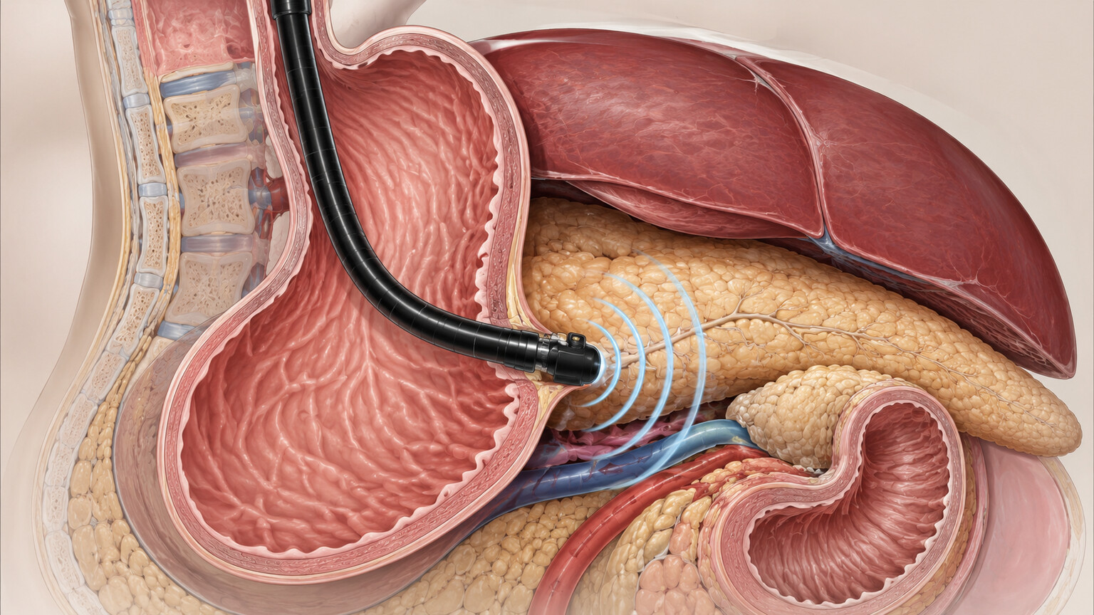
Figure 5. Endoscopic ultrasound positions the imaging transducer directly against the stomach wall adjacent to the pancreas, resolving small lesions a standard CT scan can miss.
If the number trends downward or stabilizes and no new symptoms appear, most clinicians are comfortable attributing the elevation to a resolved or resolving benign cause and simply monitoring from there. If the number continues rising, or if new symptoms emerge — unintended weight loss, new-onset diabetes in someone without risk factors for it, jaundice, or persistent pain radiating to the back — the next step is typically the endoscopic ultrasound described earlier, sometimes paired with an MRI using a contrast protocol specifically optimized for the pancreas (an MRCP), rather than a repeat of the same CT that already came back clean. Each of these steps exists specifically because the first-line scan and a single blood draw, taken together, still leave a gap that only a more targeted look can close.
Frequently Asked Questions
How high does CA 19-9 need to be before it's genuinely concerning?
There's no single cutoff that applies to everyone, but as a general pattern, mild elevations in the 40s to low 100s are far more often explained by benign causes like a recent gallbladder issue, inflammation, or an individual's naturally higher baseline. Levels in the hundreds or thousands, especially alongside symptoms or a rising trend on repeat testing, carry substantially more clinical weight and warrant closer follow-up.
Should I get another scan right away if my CA 19-9 is high but my CT was normal?
Not necessarily immediately. Because many causes of a temporary elevation resolve on their own within weeks, many doctors will first repeat the CA 19-9 and basic liver tests after a defined interval to see the trend before ordering another scan. An endoscopic ultrasound, which is more sensitive than a standard CT for small pancreatic changes, is often prioritized over simply repeating the same type of imaging.
Can CA 19-9 be high just because of my blood type or genetics?
Yes, indirectly. The Lewis blood group system, controlled by the FUT3 gene, determines whether your body can produce CA 19-9 at all and influences how much a person tends to produce as their personal baseline. This is separate from your ABO blood type and is one reason your own prior CA 19-9 results are a more useful comparison than a generic population range.
Does a normal CA 19-9 mean I definitely don't have pancreatic cancer?
No. CA 19-9 is not sensitive or specific enough to be used as a standalone screening test, and roughly 5 to 10 percent of people are genetically unable to produce it at all (Lewis-negative), meaning their level can read normal even with an aggressive cancer present. It's used to support monitoring and diagnosis alongside imaging and symptoms, never in isolation.
Conclusion
A high CA 19-9 sitting next to a normal scan is a genuinely common, and genuinely explainable, combination — not a sign that something sinister is lurking just beyond what the imaging could pick up. The marker responds to inflammation, obstruction, liver stress, blood sugar, recent procedures, and even inherited genetics, and every one of those pathways can push the number up without ever producing a mass large enough for a CT or MRI to detect. The right response isn't to assume the worst or to dismiss the number outright, but to let your doctor use the tools built for exactly this situation — liver panels, a repeat CA 19-9 to establish a trend, and, if needed, a more targeted look like an endoscopic ultrasound — to close the gap between what the scan saw and what the blood test is telling you.
Still Not Sure What Your Results Mean?
Upload your labs and get a complete, visual, plain-language interpretation of every biomarker — delivered to your inbox in under 15 minutes.
Get My ReportThis article is for educational purposes only and does not constitute medical advice. Always consult your healthcare provider regarding your specific lab results.