
The Two Biological Pathways Behind a Non-Cancer CEA Elevation
Hand a dozen different benign causes of elevated CEA to a cell biologist, and they won't see a scattered list — they'll see two mechanisms wearing different costumes. Every one of them, from an inflamed gut to a cigarette to a sluggish liver, ultimately does one of exactly two things at the cellular level: it makes a tissue produce more CEA than usual, or it slows down how quickly the body clears CEA back out of the bloodstream. This article is built around that production-versus-clearance framework specifically, walking through the molecular evidence behind each pathway rather than simply cataloguing which conditions belong on the list — that broader catalogue, with the statistics behind how common each cause actually is, lives in our companion piece on why an elevated CEA doesn't always mean cancer.
The distinction matters because "inflammation can raise this" is a satisfying-sounding explanation that often collapses the moment you ask a follow-up question — inflammation how, exactly, and through what actual switch inside the cell? This article tries to answer that follow-up question directly, tracing each condition back to the specific signaling molecules, receptors, or genes involved wherever the published research allows it, rather than leaving "inflammation" as an unexamined black box.
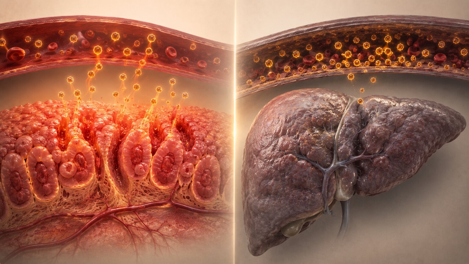
The Two Biological Pathways Behind Every Benign Elevation
The first pathway is production: tissue that's inflamed, irritated, or under mechanical stress tends to ramp up its own output of CEA and its related family of surface proteins, simply because that's part of how these particular cells behave when they're disturbed. The second pathway is clearance: CEA doesn't just sit in the bloodstream forever, it's actively removed, mainly by the liver and to a lesser degree the kidneys, and when either of those organs isn't operating at full capacity, CEA that would normally be filtered out instead lingers and accumulates. Some conditions covered in this article lean almost entirely on one pathway; others, like liver disease, genuinely involve both at once. Keeping these two mechanisms in mind while reading through the specific causes below is what turns this from a list to memorize into a system you can actually reason through.
It helps to picture the production pathway and the clearance pathway as two ends of the same pipe. Production is the tap at one end, controlling how much CEA enters the bloodstream in the first place; clearance is the drain at the other end, controlling how quickly it leaves again. A mild, temporary elevation usually means the tap has been turned up slightly, somewhere in the body, while the drain keeps working normally and eventually catches up once the underlying irritation settles down. A more stubborn, persistent elevation is more likely to involve the drain itself being at least partly blocked, which is exactly why liver disease in particular tends to produce elevations that don't resolve quickly on their own the way a passing bout of gastritis often does.
Inflammatory Bowel Disease: The Best-Mapped Mechanism of All

Of every condition in this article, Crohn's disease and ulcerative colitis have the most precisely documented mechanism behind their effect on CEA, and it's worth walking through in detail because it's genuinely elegant biology. Laboratory research has found that a cocktail of inflammatory signaling molecules — specifically TNF-alpha, interferon-gamma, and interleukin-1 beta, all of which surge during an IBD flare — rapidly switches on production of several CEA-family proteins in the cells lining the intestine, within a matter of hours. That's the production pathway in its most direct, observable form: inflammation itself, acting through specific known chemical messengers, directly commanding these cells to make more of the very protein this article is about.
There's a second, genuinely striking layer to this mechanism involving bacteria. Researchers have found that adherent-invasive E. coli, a specific strain of bacteria closely associated with Crohn's disease, actually uses one of these CEA-family proteins, CEACAM6, as a physical docking point on the intestinal cell surface, using a bacterial protein called FimH to latch onto it directly. That creates a self-reinforcing loop worth sitting with for a moment: inflammation raises CEACAM6 production, more CEACAM6 gives this particular bacteria more places to attach and colonize, and that bacterial colonization then drives further inflammation, which raises CEACAM6 again. Even ordinary diet appears to play a role in this same system — the common food emulsifier polysorbate-80 has been shown to trigger a similar rise in these proteins, while short-chain fatty acids like butyrate, produced by healthy gut bacteria fermenting fiber, have been shown to push CEACAM production back down, suppressing the same pathway other triggers switch on.
What this mechanism looks like in real patients is genuinely striking. One study following 57 people with ulcerative colitis over an average of 18 months found elevated CEA in just 12% of people with proctitis, the mildest and most limited form of the disease, but that figure climbed to 47% in left-sided colitis and 60% in more extensive, transverse or universal colitis — a number that tracks almost perfectly with how much intestinal surface area is actually inflamed. Severity mattered just as much as extent: only 24% of people in a mild flare had elevated CEA, compared with 86% during a severe flare, and a striking 92% among people with both extensive disease and a severe flare happening at the same time. The researchers' own conclusion was direct: a transiently elevated CEA in this setting reflects genuinely active mucosal inflammation, sometimes even before a patient feels dramatically different symptomatically.
Curious what your own lab results are really telling you? Upload them and get a complete, plain-language breakdown in under 15 minutes.
Analyze My ResultsPeptic Ulcer Disease, Diverticulitis, and Pancreatitis: Ordinary Digestive Irritation
Peptic ulcer disease, diverticulitis, and pancreatitis all belong to this same production-pathway family, even though the research documenting their exact molecular mechanism isn't as detailed as the IBD studies above. All three involve inflammation of tissue that sits along the same general digestive lineage responsible for CEA's normal, healthy background production — a stomach or duodenal ulcer irritating the stomach lining, diverticulitis inflaming small pouches along the colon wall, and pancreatitis inflaming the pancreas itself. The underlying logic is the same one demonstrated so clearly in IBD: irritated glandular and mucosal tissue tends to shed more CEA into the bloodstream than that same tissue would at rest, without requiring any tumor to be involved anywhere. None of these three conditions typically pushes CEA to a dramatic level on its own, but each is a well-documented, legitimate, benign explanation for a mild-to-moderate elevation, particularly during an acute flare-up of symptoms.
Peptic ulcer disease specifically has a well-known bacterial trigger worth naming directly: Helicobacter pylori, a bacterium that colonizes the stomach lining and is responsible for a large share of recurrent ulcers. Research has found higher CEA levels in people with chronic H. pylori infection, and the mechanism researchers have traced looks structurally similar to the bacterial story already covered in IBD — H. pylori invades the stomach's epithelial lining and interacts with local immune cells called macrophages, prompting them to release inflammatory signaling molecules that further damage the gastric mucosa. That ongoing cycle of bacterial irritation and immune response is a specific, named example of the same broad production pathway showing up in a completely different organ, reinforcing just how consistently this particular mechanism repeats itself across the digestive tract.
Diabetes: A Metabolic Route to the Same Molecule
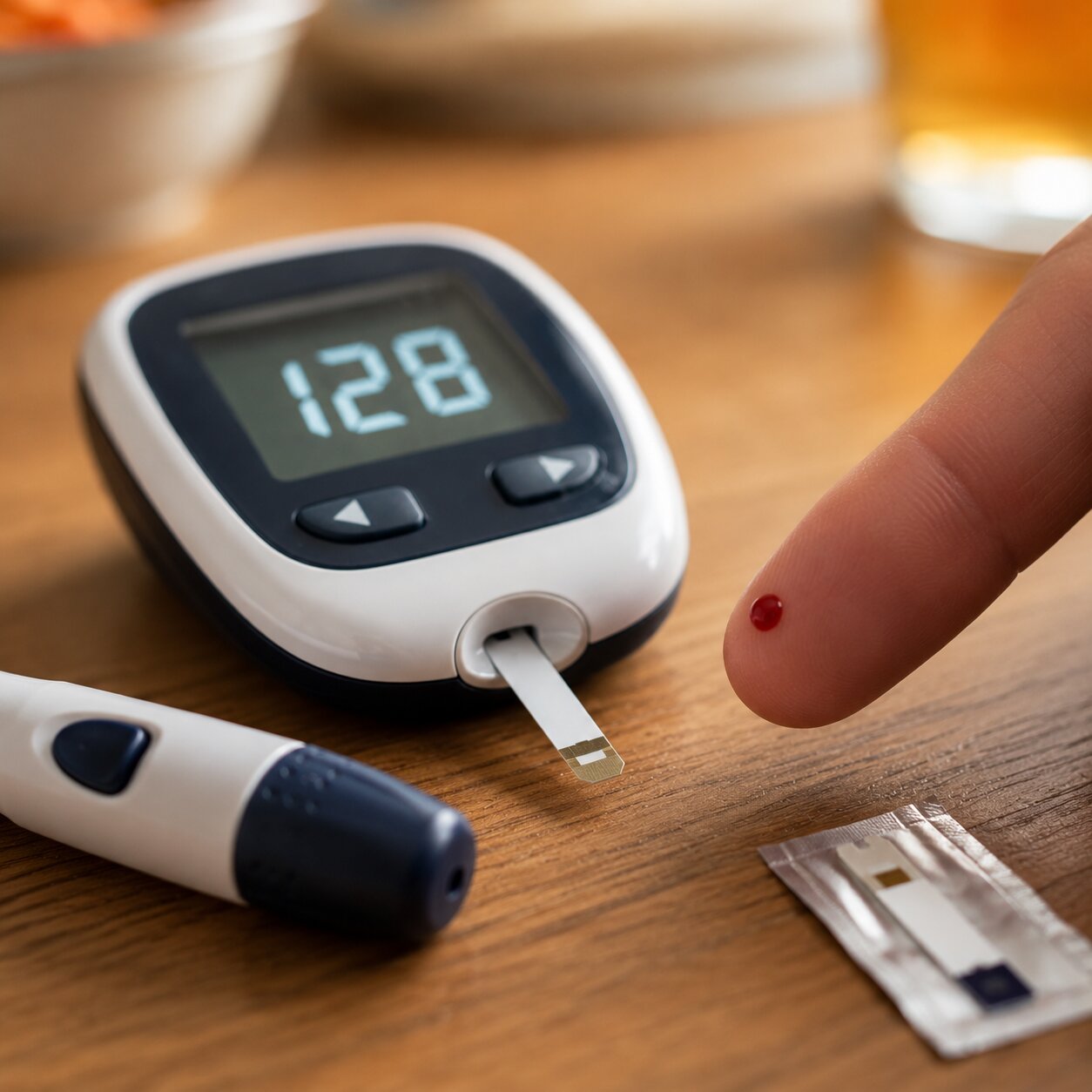
Diabetes reaches CEA through a genuinely different door than inflammation does, and the research behind it is fairly recent and specific. Studies have found serum CEA rises alongside both fasting blood glucose and HbA1c — the standard lab measurement reflecting average blood sugar control over roughly the prior three months — with a positive correlation strong enough that it held up even after researchers statistically adjusted for age, gender, and body weight. The same research found CEA moving in the opposite direction from HDL cholesterol, the "good" cholesterol type, hinting at a broader metabolic pattern rather than a single isolated effect. Encouragingly, when blood sugar control actually improved in these patients, CEA came back down again, tracking the same downward trend seen in several other inflammatory and metabolic markers measured in the same study, including CA 19-9 and CRP, a well-known general marker of inflammation.
The proposed underlying mechanism ties back to a process called glycation, in which excess circulating glucose chemically attaches itself to proteins throughout the body, including likely CEA itself, altering the molecule and how it's processed. This isn't a process unique to CEA — glycation is the same basic chemistry behind HbA1c itself, which is really just hemoglobin, the oxygen-carrying protein in red blood cells, after glucose has attached to it over time. Sustained high blood sugar means more glucose molecules are available to react with circulating proteins generally, and the byproducts of that reaction, called advanced glycation end-products, are known to interfere with a protein's normal shape and function once formed, alongside triggering their own additional inflammatory signaling once they build up. It's a genuinely different chemical pathway from the cytokine-driven story described in IBD, arriving at a similar destination — more CEA circulating in the blood — through an entirely different door.
Because this pattern has been documented clearly enough, some researchers have specifically proposed using a higher CEA cutoff for people with poorly controlled diabetes, rather than applying the same reference range used for everyone else — a genuinely practical suggestion that mirrors the smoker-adjusted reference ranges already in common use, discussed in our companion article on how smoking raises CEA levels.
Autoimmune and Collagen Vascular Disease: General Inflammatory Activity

Conditions like rheumatoid arthritis and lupus have long been recognized as legitimate, if less common, causes of a mildly elevated CEA, and it's worth being honest that the precise molecular mechanism here is less thoroughly mapped than it is for IBD specifically. What's well established is that these are conditions of chronic, body-wide inflammatory activity, driven by the immune system's own signaling molecules attacking the body's own tissue — some of the very same broad categories of inflammatory messengers, like TNF-alpha, that were shown to directly trigger CEACAM production in the intestinal studies discussed earlier. The most reasonable working explanation is that this same general production pathway is being activated more diffusely throughout the body, rather than concentrated in one specific organ, producing a milder, more generalized elevation rather than the more dramatic, localized spikes possible in a severe IBD flare or an acute pancreatitis episode.
This is also a useful place to be honest about a genuine gap in the research: unlike IBD, where researchers have measured the exact percentage of patients affected at different disease stages, no comparably detailed study was found mapping precisely how CEA tracks with rheumatoid arthritis or lupus disease activity specifically. What exists instead is a longer-standing clinical recognition that these conditions belong on the list, paired with the general, well-established principle that widespread inflammatory disease of almost any kind tends to nudge inflammation-sensitive markers upward to some degree. That's a real, legitimate benign explanation worth knowing about, even without the same level of mechanistic detail this article was able to provide for inflammatory bowel disease.
Liver Disease and Smoking: The Two Causes With Their Own Deep Dives
Two of the most well-documented non-cancer causes of elevated CEA deserve only a brief mention here, since each has already been covered in the depth it warrants elsewhere on this site. Liver disease is the clearest real-world example of the clearance pathway described at the top of this article — the liver is responsible for pulling roughly 70% of circulating CEA out of the blood within about an hour under normal conditions, and when that clearance machinery is impaired by cirrhosis, hepatitis, fatty liver disease, or a blocked bile duct, CEA backs up in the bloodstream even though production hasn't necessarily changed at all. Our companion article on how liver disease affects CEA covers that mechanism, and the specific magnitude research behind it, in full. Smoking works through the production pathway instead, directly switching on CEA gene activity in the cells lining the airways, a mechanism our companion article on smoking and CEA walks through in detail, including exactly how much of an effect it produces and how quickly it reverses after quitting.
It's worth noting that these two causes are genuinely instructive precedents for everything else in this article, precisely because they've been studied in enough depth to confirm the two-pathway framework directly rather than by inference. Liver disease didn't just correlate with elevated CEA in a general statistical sense — researchers were able to trace the exact receptor system on liver cells responsible for pulling CEA out of circulation and show how disease disrupts it. Smoking's effect was confirmed down to the level of measuring CEA gene activity rising in lung tissue exposed to smoke extract in a laboratory setting. Those two conditions are effectively the proof of concept for the entire framework this article is built around, which is part of why they're referenced repeatedly throughout rather than treated as unrelated, standalone items on a list.
Kidney Disease: A Weaker Clearance Link Than You'd Expect

Kidney disease is often listed alongside liver disease as a clearance-pathway cause of elevated CEA, and dialysis does mechanically remove CEA from the blood the same way it removes other waste products, through diffusion and ultrafiltration across the dialysis membrane. But the actual research relating kidney function specifically to CEA levels is more mixed than the confident, well-established liver story. One study comparing healthy people, dialysis patients, and people with kidney failure found only a loose tendency between a measure of kidney function called creatinine clearance and CEA levels, without reaching statistical significance — a notably weaker relationship than the same study found for two other tumor markers, AFP and CA 15-3, which did correlate significantly with kidney function. That's a genuinely useful, honest nuance: kidney impairment can plausibly contribute a mild elevation to CEA specifically, but it isn't nearly as reliable or predictable a contributor as liver impairment is, and it shouldn't be assumed to explain an elevated CEA as confidently as reduced liver function would.
Part of why kidney disease behaves so differently from liver disease in this specific context likely comes back to where CEA actually gets removed from the blood in the first place. The liver, as covered in our companion article on liver disease and CEA, handles the overwhelming majority of CEA clearance through a dedicated receptor system on hepatocyte cells built specifically to recognize and remove this molecule. The kidneys were never CEA's primary exit route the way they are for smaller waste molecules like creatinine, which is a reasonable explanation for why impaired kidney function shows up as, at most, a minor contributor rather than a dominant one. Anyone with both kidney and liver impairment together should expect the liver side of that equation to carry far more explanatory weight for an elevated CEA than the kidney side does on its own.
Why the Same Handful of Mechanisms Explain Nearly Every Cause
Stepping back across everything covered in this article, the pattern holds up remarkably well: IBD, peptic ulcer disease, diverticulitis, pancreatitis, and smoking all funnel through the production pathway, each one irritating or inflaming a specific type of tissue that responds by making more CEA. Liver disease sits mostly on the clearance side, with kidney disease contributing a much weaker version of that same clearance-based story. Diabetes and autoimmune disease occupy a middle ground — both are fundamentally inflammatory or metabolic processes that appear to nudge the production pathway upward in a more diffuse, whole-body way rather than through one clearly localized tissue. Once you see the underlying pathway rather than memorizing each condition individually, a genuinely long and initially overwhelming list becomes something closer to a short list of two ideas, applied repeatedly across different organs and diseases.
It's also worth noticing what these mechanisms have in common at a cellular level, even across such different organs. In nearly every production-pathway example in this article, the trigger is some form of ongoing irritation meeting tissue that already had the biological machinery to make CEA in small amounts under normal, healthy conditions — the inflammatory signal doesn't invent a new capability out of nowhere, it simply turns up the volume on something that tissue was already quietly doing in the background. That's a genuinely different story from what happens in cancer, where the tissue itself has changed at a deeper level, and it's part of why benign production-pathway elevations tend to stay comparatively modest and tend to fall back down once the underlying irritation resolves, rather than climbing indefinitely the way an actively growing tumor's output can.
When More Than One Cause Applies at Once
Real patients rarely fit neatly into just one category from this article, and it's worth addressing that directly rather than pretending every elevated CEA has a single, tidy explanation. Someone with poorly controlled diabetes who also smokes is being pushed by two separate production-pathway mechanisms simultaneously, and their combined effect on CEA isn't necessarily just the two individual effects added together in some predictable way — biology rarely works that cleanly. Similarly, someone with inflammatory bowel disease who also happens to have early liver impairment is dealing with a production-pathway cause and a clearance-pathway cause stacking on top of each other at the same time, which can produce an elevation that looks disproportionately high compared to what either condition alone would typically explain. This is exactly why doctors take a full history rather than reflexively attributing an elevated result to whichever single benign condition happens to be listed first in a patient's chart — and it's a genuinely good reason to mention every relevant condition you have to your doctor, even ones that seem unrelated to each other or to the digestive tract specifically.
What This Means for Interpreting Your Own Result
If your CEA came back mildly elevated, the single most useful thing you can do with the information in this article is match your own health history against the two pathways above. Ask yourself honestly whether you have a known digestive condition, diabetes that isn't well controlled, an autoimmune diagnosis, a smoking history, or any known liver or kidney impairment — and bring that specific list to your doctor rather than the general question of "why is this high." A result that lines up cleanly with one of these known, benign explanations, especially one that's mild and consistent with an active flare of a condition you already know you have, carries a very different weight than the same number with no plausible explanation in sight at all.
It's also worth asking your doctor whether any of your known conditions are currently active or in remission, since the production pathway responds specifically to active inflammation rather than to a diagnosis sitting quietly in your medical history. A well-controlled autoimmune condition or a diabetes diagnosis with consistently good glucose readings is a much weaker candidate to explain a new elevation than the same condition during an active flare or a stretch of poor control — which is exactly why your doctor will likely want to know not just what conditions you have, but how they've been behaving lately, and whether any recent lab work, symptom changes, or medication adjustments line up with the timing of your CEA result.
Frequently Asked Questions
What are the two main ways non-cancer conditions raise CEA?
Either more CEA is being produced by inflamed or irritated tissue, or less CEA is being cleared out of the bloodstream due to impaired liver or kidney function. Most benign causes work through one of these two pathways, and some, like liver disease, involve both.
How does inflammatory bowel disease actually raise CEA?
Inflammatory signaling molecules like TNF-alpha directly switch on CEA-family protein production in intestinal cells within hours. Certain bacteria associated with Crohn's disease also use one of these proteins, CEACAM6, as a docking point, creating a self-reinforcing inflammatory cycle.
Can diabetes really raise CEA on its own?
Yes. Research has found CEA correlates positively with blood glucose and HbA1c levels, and this relationship held even after adjusting for age, gender, and weight. CEA has been shown to decrease again when blood sugar control improves.
Does kidney disease raise CEA the same way liver disease does?
Not as reliably. While dialysis does mechanically remove CEA from the blood, research has found only a weak, non-significant tendency between kidney function and CEA levels, notably weaker than the well-documented relationship between liver function and CEA.
Can autoimmune diseases like rheumatoid arthritis or lupus raise CEA?
Yes, though the specific mechanism is less precisely mapped than in inflammatory bowel disease. These conditions involve chronic, body-wide inflammatory activity that likely activates the same general CEA-production pathway seen more clearly in localized digestive inflammation.
Does diet affect CEA levels through this same mechanism?
There's early evidence it might, at least in the context of gut inflammation. Research has found the food emulsifier polysorbate-80 can trigger the same CEA-family proteins that inflammation does, while short-chain fatty acids from fiber fermentation have been shown to suppress that same pathway.
Can H. pylori infection raise CEA even without a diagnosed ulcer?
Yes. Research has found higher CEA levels in people with chronic H. pylori infection, likely through the same kind of bacterial-inflammatory mechanism seen with Crohn's-associated bacteria in the intestine, where local immune cells respond to the infection by releasing inflammatory signals into the tissue.
Can having two of these conditions at once raise CEA higher than either alone?
It's possible. Someone with two active production-pathway causes, or a production-pathway and a clearance-pathway cause together, may see a combined effect that looks disproportionately high compared to either condition individually, which is why a full medical history matters when interpreting an elevated result.
Conclusion
Every non-cancer condition covered in this article, and in its companion piece on why an elevated CEA doesn't always mean cancer, ultimately traces back to one of two things: irritated tissue making more of this protein, or an impaired liver or kidney failing to clear it out fast enough. That's genuinely the whole story, mechanistically speaking, even when the full list of individual conditions capable of producing it looks long, scattered, and disconnected on the surface at first glance. From cytokines switching on genes in inflamed intestinal cells, to bacteria using CEA's own surface proteins as a foothold, to glucose molecules quietly altering circulating proteins in poorly controlled diabetes, to a struggling liver simply falling behind on its usual clearance workload, every mechanism this article traced eventually funnels back into one of those same two doors.
Understanding which pathway, or which specific condition, plausibly applies to your own situation is what turns an unexplained number into one you can actually make sense of — and it's exactly the kind of detail worth bringing directly into your next conversation with your doctor, alongside an honest account of which of your own conditions are currently active rather than quietly under control. A number stops being frightening in the abstract the moment you can trace it back to something concrete, physical, and genuinely explainable actually happening somewhere specific inside your own body.
Still Not Sure What Your Results Mean?
Upload your labs and get a complete, visual, plain-language interpretation of every biomarker — delivered to your inbox in under 15 minutes.
Get My ReportThis article is for educational purposes only and does not constitute medical advice. Always consult your healthcare provider regarding your specific lab results.