
Why Do Inflammatory Bowel Conditions Raise CEA Levels?
If you have Crohn's disease or ulcerative colitis and your doctor ran a CEA test — the same blood marker used to monitor colon cancer — and it came back a little high, the first thing to know is this: an inflamed intestine can raise CEA on its own, with no cancer anywhere in the picture. CEA, short for carcinoembryonic antigen, is made by the cells that line your digestive tract, and it rises whenever those cells are irritated, damaged, or turning over faster than normal — which is exactly what happens during a flare of inflammatory bowel disease (IBD). The inflammation doesn't just injure the surface of the intestine; it also loosens the tight seals between cells and increases blood flow to the area, both of which make it easier for CEA to leak out of the gut lining and into your bloodstream. Doctors who care for IBD patients already know this, which is why a mildly elevated CEA during an active flare is treated very differently than the same number would be treated in someone with no bowel disease at all.
What CEA Actually Is, and Why the Gut Makes It in the First Place
Carcinoembryonic antigen is a glycoprotein — a protein with sugar molecules attached to it — that's normally produced in small amounts by the cells lining your gastrointestinal tract, starting before you're even born. In a developing fetus, CEA plays a role in cell adhesion, helping cells stick together properly as tissues form. After birth, production drops off sharply, but it never disappears completely: healthy adults continue to shed small amounts of CEA from the epithelial cells (the thin layer of cells that lines the inside surface of your intestines) into the bloodstream as part of normal tissue turnover. This is why a "normal" CEA isn't zero — it's typically under 3 to 5 nanograms per milliliter (ng/mL) in a non-smoker, a small baseline hum of activity from a gut that's constantly renewing its lining. CEA became useful in oncology because certain cancers, particularly colorectal cancer, cause epithelial cells to overproduce and shed far more of it than healthy tissue does. But that's the key nuance that gets lost in translation when a patient sees "cancer marker" on a lab requisition: CEA isn't a cancer-specific molecule, it's a normal-tissue molecule that cancer happens to make in excess. Anything else that disrupts, damages, or accelerates the turnover of that same intestinal lining can push the number up too — and inflammatory bowel disease does exactly that, chronically and sometimes dramatically.
It helps to think of the intestinal lining as a floor that's constantly being replaced, tile by tile, even when nothing is wrong. A healthy gut sheds and regenerates its entire epithelial surface roughly every four to seven days — one of the fastest renewal rates of any tissue in the body, faster than skin, faster than the lining of the stomach. Each of those routine replacement cycles releases a trickle of CEA into the blood as old cells are shed and new ones take their place, which is why the baseline reference range isn't zero but a small, steady number reflecting that constant background renovation. Inflammatory bowel disease doesn't introduce a new process here — it accelerates and intensifies a process that was already happening. When the immune system attacks the lining during a flare, cells die faster than normal, get replaced faster than normal, and the entire floor-replacement analogy speeds up dramatically in the affected stretch of intestine. More tiles being torn out and reinstalled per day simply means more CEA released per day, which is the simplest possible way to understand why a flare and an elevated CEA so often show up together.
Already have a CEA number from a recent test, but not sure if it's actually meaningful for you? Just plug it in and see instantly.
🧮 Try the Free CalculatorCEA's Molecular Family: Why It's Built Into the Gut Lining in the First Place
CEA belongs to a larger family of related proteins called the carcinoembryonic antigen-related cell adhesion molecules, or CEACAMs — a group of roughly a dozen related glycoproteins that sit on the surface of epithelial cells throughout the digestive tract, among other tissues. As the family name suggests, their normal job is adhesion: helping neighboring cells recognize each other and stick together in an organized way, similar to how matching puzzle pieces know which other pieces they're supposed to connect to. CEA itself (formally CEACAM5) is anchored to the outer surface of the cell membrane, facing into the space between cells rather than staying buried inside the cell. This surface position is precisely what makes it so easy for CEA to end up in the bloodstream when that cell layer is disturbed — it isn't a molecule that has to be actively secreted or manufactured for export; it's already sitting right at the cell's outer edge, and inflammation-driven barrier damage simply lets it slough off into circulation along with dying and shedding cells. Cancer cells exploit this same molecule for a different purpose, using excess CEACAM5 production to help tumor cells cluster together and resist normal cell-death signals, which is part of why cancerous tissue produces so much more of it than healthy tissue does. But the underlying protein, and the reason it ends up detectable in a blood draw at all, traces back to this same basic adhesion role it plays in every intact intestinal lining, healthy or not.
The Mechanism: How an Inflamed Intestine Leaks CEA Into the Blood
To understand why IBD raises CEA, it helps to picture the intestinal lining the way a plumber would picture a pipe. In a healthy gut, the epithelial cells sit shoulder to shoulder, sealed together by structures called tight junctions — molecular "gaskets" that control exactly what's allowed to pass between cells and what isn't. This barrier keeps most of what's inside the gut (bacteria, digestive enzymes, and yes, CEA produced by the cells themselves) from randomly crossing into the bloodstream. During a Crohn's or ulcerative colitis flare, chronic inflammation damages this barrier directly. Immune cells flood the tissue and release inflammatory signaling molecules called cytokines — tumor necrosis factor-alpha (TNF-α) and interleukin-6 (IL-6) among the most influential — which physically loosen those tight junctions and increase what's called "gut permeability." At the same time, the inflamed tissue is more vascular than normal, meaning more small blood vessels are actively feeding the area, which shortens the distance CEA has to travel to reach circulation. Layer on top of that the sheer mechanical damage from ulceration — active IBD literally erodes and sometimes ulcerates the surface of the intestinal lining — and you have three separate physical reasons, all happening simultaneously, why more CEA than usual ends up in a blood draw: a leakier barrier, a higher blood supply, and more raw, damaged tissue actively shedding cells.
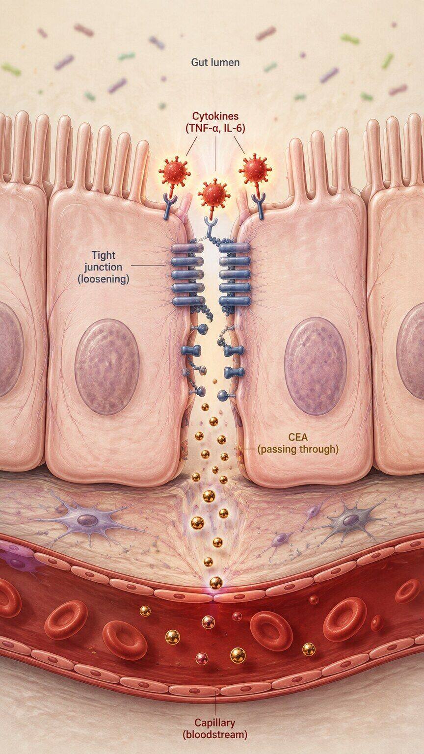
Figure 1. TNF-α and IL-6 released during an IBD flare loosen the tight junctions that normally seal intestinal epithelial cells together.
Research measuring CEA in IBD patients bears this out consistently. Studies comparing CEA levels during active flares versus periods of remission in the same patients have found meaningful, reproducible increases during active disease — not in every patient, and rarely to the dramatic levels seen in advanced colorectal cancer, but often enough to push a result from comfortably normal to mildly or moderately elevated. The severity of the elevation tends to track loosely with the extent and severity of inflammation: someone with extensive, deeply ulcerated colitis affecting a large portion of the colon is more likely to show a bump in CEA than someone with a small, localized patch of mild inflammation. This is the same general principle seen with other inflammation-sensitive lab markers, like C-reactive protein (CRP) or fecal calprotectin — the body's various "damage signals" tend to rise together when a tissue is actively inflamed, and CEA, despite its oncology reputation, behaves as one of them in this context.
It's worth being precise about scale here, because the word "elevated" can sound scarier than what's actually being described. In colorectal cancer, especially once it has spread beyond the original tumor, CEA can climb into the tens, hundreds, or even thousands of ng/mL — numbers that are unmistakably abnormal by any standard. Inflammation-driven elevations from IBD almost never approach that range. Most published series describing CEA during active IBD flares report values in the single digits to low double digits — often somewhere between 3 and 10 ng/mL, occasionally a bit higher in severe, extensive disease — which is a meaningfully different order of magnitude than what cancer-related elevations typically produce. This doesn't mean small differences don't matter clinically, but it does mean that the sheer size of an elevation is itself a piece of information: a CEA of 5.5 ng/mL in someone with a moderate ulcerative colitis flare is behaving exactly the way inflammation-driven CEA is expected to behave, while a CEA of 80 ng/mL in that same patient would immediately shift the conversation toward looking for something else entirely, flare or no flare.
Crohn's Disease vs. Ulcerative Colitis — Does It Matter Which One You Have?
Both major forms of inflammatory bowel disease can elevate CEA, but the pattern differs somewhat because the two diseases behave differently in the gut. Ulcerative colitis is confined to the colon and rectum and affects the innermost lining in a continuous, unbroken stretch — when it's active, that entire stretch of tissue is inflamed and permeable at once, which can produce a fairly proportional relationship between how much colon is involved and how high CEA climbs. Crohn's disease, in contrast, can affect any part of the digestive tract from mouth to anus, often in patchy "skip lesions" separated by normal-looking tissue, and it penetrates deeper into the intestinal wall rather than staying confined to the surface layer. This deeper, transmural inflammation in Crohn's — especially when it involves fistulas (abnormal tunnels connecting the gut to other structures) or abscesses — has in some studies been associated with even more pronounced CEA elevations than colitis limited to the surface lining, likely because the barrier disruption is more severe and involves more layers of tissue. Neither pattern is a hard rule that applies to every patient, but it's part of why a gastroenterologist interpreting a CEA result in someone with IBD will always ask which disease, how much of the gut is involved, and how active it currently is — the number alone tells only part of the story.
Before moving on from mechanism to management, it's worth naming the emotional weight this test carries, because it's real and it's common. Very few lab tests come pre-loaded with as much anxiety as one labeled a "tumor marker" or "cancer marker" on a requisition form, and for someone already living with a chronic bowel disease, seeing that phrase attached to a number outside the normal range can trigger a wave of fear that's completely understandable, even when the biology says the fear is very likely misplaced. It helps to remember that CEA was never a cancer molecule that occasionally shows up in other conditions — it's a normal-tissue molecule that cancer happens to make in excess, and IBD, as a disease that directly damages and accelerates turnover of the very tissue that produces it, is one of the most well-documented non-cancer reasons this specific marker rises. Knowing that a mildly elevated CEA during a flare is, in a very real sense, expected — a predictable consequence of the same inflammation causing your symptoms — can take some of the sting out of seeing a number flagged as "high" on a lab report.
Location within the digestive tract also matters in a way that's easy to overlook. Crohn's disease affecting the small intestine alone, without any colonic involvement, tends to produce smaller CEA elevations than Crohn's colitis or ulcerative colitis, likely because the small intestine's epithelial cells don't produce quite as much baseline CEA as the colon's do, and because small-bowel Crohn's disease is often diagnosed and monitored somewhat differently, with imaging playing a larger role than colonoscopy alone. This is one more reason CEA behaves like a blunt instrument rather than a precision tool in IBD: it's sensitive to overall inflammatory burden and to which tissue is involved, but it can't tell a clinician which specific segment of bowel is inflamed, how deep the inflammation runs, or whether it's actively worsening versus already improving. Those questions still require direct visualization, imaging, or more IBD-specific biomarkers to answer — CEA simply adds one more data point to a picture that's built from many sources at once.
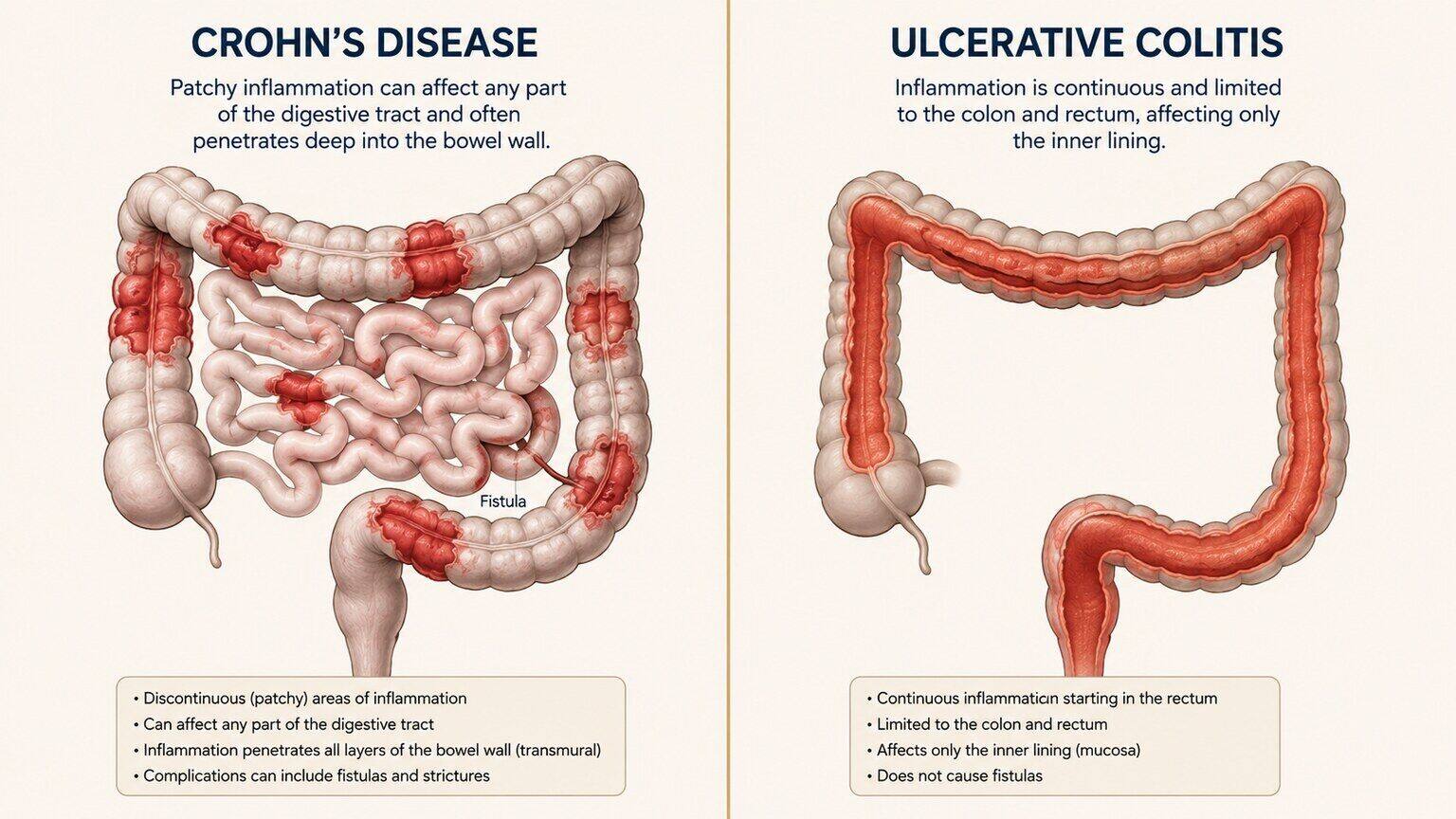
Figure 2. Crohn's disease often involves patchy, deep transmural inflammation, while ulcerative colitis produces continuous surface-level inflammation confined to the colon and rectum.
Flare vs. Remission — Why the Same Person's CEA Can Change Over Time
One of the most clinically useful things about CEA in IBD is that it isn't a fixed number for a given patient — it moves with disease activity. Someone with well-controlled Crohn's disease in deep remission may have a completely normal CEA for years, then see it creep upward during a flare, and then watch it settle back down once treatment brings the inflammation under control again. This makes CEA, in some IBD patients, a rough secondary indicator of disease activity alongside more specific tools like fecal calprotectin, CRP, and direct visualization via colonoscopy — though it's never used as a stand-alone measure of flare severity, since it's far less sensitive and specific for gut inflammation than those purpose-built markers. If your gastroenterologist has ordered a CEA test alongside other bloodwork during a flare, they're very likely looking at the overall trend and context, not treating that single number as diagnostic of anything on its own. A CEA drawn during an acute flare that resolves back to normal once the flare is treated is one of the most reassuring patterns possible — it strongly suggests inflammation, not a separate underlying process, was driving the number the whole time.
What Happens to CEA Once Biologic Treatment Takes Effect
Modern IBD management has shifted heavily toward biologic medications — drugs like anti-TNF agents (infliximab, adalimumab), anti-integrin therapies (vedolizumab), and interleukin-blocking agents (ustekinumab, risankizumab) — that work by directly interrupting the inflammatory cascade described earlier rather than just suppressing symptoms. Because these drugs act on the same cytokines (TNF-α, IL-6, and related signaling molecules) responsible for loosening the intestinal barrier and driving CEA leakage, achieving what gastroenterologists call "deep remission" — healing visible at the tissue level, not just symptom improvement — has been associated in several studies with a corresponding normalization of inflammation-sensitive lab markers, CEA included. This is actually one of the more elegant confirmations of the whole mechanism: if CEA were rising because of an emerging cancer, starting a biologic for bowel inflammation wouldn't be expected to bring it down. When CEA does fall in step with calprotectin, CRP, and endoscopic healing after a biologic takes effect, that trajectory is itself strong evidence that inflammation was the driver all along. Clinicians increasingly use this kind of multi-marker trend — several inflammatory indicators moving together in the same direction — as a more reliable read on what's happening than any single value checked in isolation.
The timeline for this normalization varies by person and by which biologic is used, but it typically unfolds over the same weeks-to-months window in which clinical symptoms and endoscopic inflammation also improve — there's no separate, faster or slower clock specifically for CEA. A patient starting infliximab for a moderate-to-severe ulcerative colitis flare, for example, might see clinical symptoms improve within days to a couple of weeks, while the deeper tissue healing that fully resolves the CEA leak can take several months to become apparent on repeat colonoscopy. This is exactly why a single CEA recheck two weeks after starting treatment isn't always the most informative test; a value checked a few months later, once deeper healing has had time to occur, tells a much more complete story about whether the elevation truly was inflammation-driven.
Why This Matters So Much for IBD Patients Specifically: Colorectal Cancer Screening
Here's the part that makes this topic more than academic curiosity: long-standing inflammatory bowel disease, particularly ulcerative colitis and Crohn's colitis involving a large portion of the colon, is itself an established risk factor for colorectal cancer, especially after eight to ten years of disease duration. This means IBD patients are a population where CEA gets checked more often and watched more closely than in the general public — and it's also a population where the marker's inflammation-driven false elevations collide most directly with its cancer-monitoring purpose. A gastroenterologist managing a patient with 15 years of ulcerative colitis has to hold two things in mind simultaneously: this patient's baseline inflammatory disease can push CEA up on its own, and this same patient is at meaningfully higher risk for the exact cancer CEA is best known for detecting. The way this gets resolved in practice is through pattern recognition over time — comparing a new CEA value against that specific patient's own prior results, correlating it with current flare status, and using colonoscopy (the actual gold-standard surveillance tool for colorectal cancer in IBD) rather than any blood marker as the primary screening method. CEA in this context functions as one supporting data point among many, never the deciding factor on its own.
This is also why current gastroenterology surveillance guidelines for long-standing IBD are built around scheduled colonoscopy intervals — typically starting around the eight-to-ten-year mark after diagnosis for extensive colitis, and repeated on a schedule tightened or loosened based on individual risk factors like disease extent, family history, and prior findings — rather than around blood-marker thresholds. A colonoscopy can directly sample any suspicious area and confirm at the cellular level whether dysplasia (precancerous cell changes) or early cancer is present; a blood test, no matter how it trends, can only ever raise or lower suspicion from a distance. Patients sometimes ask whether a normal CEA means a surveillance colonoscopy can be skipped or delayed, and the honest answer is no — CEA's poor sensitivity for early, localized colorectal cancer means a reassuringly normal value provides very little protection against missing something that direct visualization would catch. The two tools aren't interchangeable, and IBD surveillance protocols are deliberately built around the one with real diagnostic teeth.
None of this means CEA is worthless in the IBD context — it just means its role is narrower and more supportive than its "cancer marker" label suggests to most people first encountering it. Where it earns its place is in longitudinal tracking for patients already identified as higher risk: someone with fifteen years of extensive ulcerative colitis, for instance, might have CEA checked periodically as one thread in a much larger surveillance tapestry that also includes calprotectin trends, symptom review, and scheduled colonoscopy with random and targeted biopsies. A steady, unremarkable CEA across several years of checks, sitting comfortably within the range explained by whatever background inflammation is present, adds a small amount of reassurance to that broader picture. A value that starts climbing across consecutive checks, independent of flare activity, adds a small amount of additional concern — not enough to act on by itself, but enough to make the next scheduled colonoscopy feel a little more urgent to move up rather than push back. Used this way, as one thread among many rather than a stand-alone verdict, CEA does the modest job it's actually suited for.
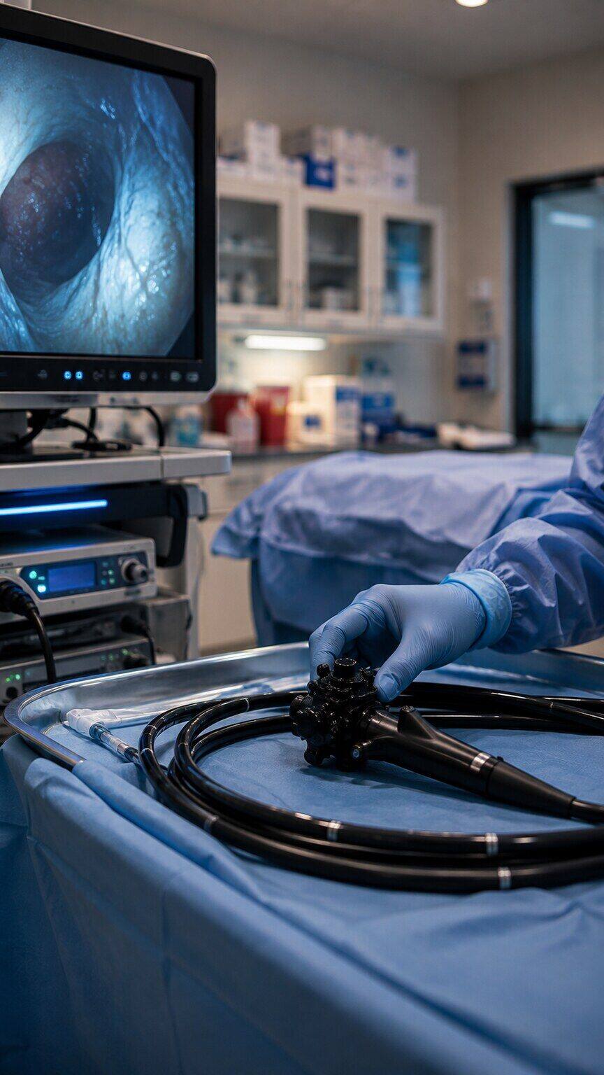
Figure 3. Colonoscopy remains the primary surveillance tool for colorectal cancer risk in long-standing IBD, with CEA serving only as supporting context.
This distinction — screening tool versus context marker — is worth sitting with, because it explains why a gastroenterologist won't panic over a CEA of 6 ng/mL in a patient with active colitis the same way they might in a healthy 50-year-old with no bowel disease and no other explanation. Context changes what a number means. A moderately elevated CEA in the middle of a documented flare, with a colonoscopy showing active but non-cancerous inflammation, is doing exactly what the biology predicts it should do. The same number appearing in someone whose IBD is currently quiet, whose colonoscopy shows minimal inflammation, and who has no other explanation is a very different conversation — one that appropriately shifts toward ruling out something else, colorectal cancer included. Smoking status matters here too, independent of IBD: cigarette smoking is well documented to raise baseline CEA on its own by several tenths to a full point or more, so a clinician reviewing an elevated result in an IBD patient who also smokes has to account for both factors before drawing any conclusion.
There's also an important asymmetry worth understanding: Crohn's disease itself, particularly small-bowel and colonic Crohn's, carries an elevated colorectal and small-bowel cancer risk profile that is separate from — but can coexist with — the direct inflammation-driven CEA elevations discussed throughout this article. Long-standing strictures (narrowed segments of bowel from chronic scarring) can occasionally harbor cancer that develops silently within the narrowed segment, sometimes making colonoscopic surveillance technically more difficult in exactly the patients who need it most. This is one more reason CEA trends are followed longitudinally in higher-risk IBD patients rather than checked once and dismissed — not because CEA is a good cancer-detection tool on its own, but because a persistent, unexplained upward trend over multiple checks, especially in a patient with strictures or long-standing extensive disease, is one of several signals that can prompt a gastroenterologist to intensify surveillance imaging or endoscopy sooner than the routine schedule would otherwise dictate.
Extraintestinal Complications of IBD That Can Independently Affect CEA
Inflammatory bowel disease is often described as a systemic condition rather than one confined strictly to the gut, and several of its extraintestinal manifestations — complications that occur outside the intestine itself — carry their own independent influence on CEA, stacking on top of whatever the bowel inflammation itself is contributing. Primary sclerosing cholangitis (PSC), a chronic disease of the bile ducts strongly associated with ulcerative colitis, is one of the more significant examples: PSC progressively narrows and scars the bile ducts, and because the liver plays a meaningful role in clearing CEA from circulation, impaired bile flow and liver function can slow that clearance and allow CEA to accumulate independent of gut activity. Fatty liver changes, which are common in IBD patients partly due to corticosteroid use and partly due to the metabolic effects of chronic inflammation itself, can have a similar, milder effect on hepatic clearance. Joint inflammation (arthritis), skin manifestations like erythema nodosum or pyoderma gangrenosum, and eye inflammation (uveitis) — all recognized extraintestinal complications of IBD — don't directly elevate CEA themselves, but they're markers of how systemically active a patient's overall inflammatory disease is at a given moment, which loosely correlates with how much gut-driven CEA leakage is likely occurring at the same time.
Everyday Factors That Can Nudge CEA Slightly, Even Outside a Flare
Beyond the disease-specific mechanisms already covered, a handful of everyday factors can add a small amount of additional variability to CEA results in anyone, IBD or not, and it helps to know about them so a borderline number doesn't get over-interpreted. Recent respiratory infections, urinary tract infections, and other unrelated sources of acute inflammation can transiently bump CEA up by a small margin while the body is actively fighting them off, simply because CEA is one of many proteins whose production is sensitive to the general inflammatory signaling happening throughout the body during any infection, not just gut-specific ones. Age plays a modest role as well — CEA tends to drift very slightly upward across the general population as people get older, independent of any disease process, which is part of why reference ranges are sometimes interpreted a little more loosely in older adults. Laboratory-to-laboratory variation in the specific assay used to measure CEA can also introduce small differences between two results drawn at different facilities, which is one more reason clinicians prefer to track values over time using the same lab whenever possible, rather than comparing numbers generated by different testing methods as if they were perfectly interchangeable. None of these factors produce dramatic swings on their own, but in someone already running a mildly elevated CEA from IBD-related inflammation, they can be enough to tip a borderline result one direction or the other on a given day — which is exactly why a single value is treated as a data point to track, not a verdict to act on immediately.
Other Non-Cancer Causes That Can Stack on Top of IBD
Because IBD patients often have overlapping conditions, it's worth knowing that inflammation isn't the only non-cancer driver of CEA that shows up in this population. Liver involvement — which can occur in IBD through conditions like primary sclerosing cholangitis, a bile duct disease strongly linked to ulcerative colitis — can independently raise CEA, since the liver plays a role in clearing this protein from the bloodstream and impaired clearance lets levels build up regardless of gut inflammation. Diverticulitis, gallbladder disease, and pancreatitis have all been documented to cause modest CEA elevations as well, and IBD patients aren't protected from developing any of these conditions in addition to their underlying bowel disease. This is part of why an elevated CEA in someone with IBD often prompts a broader look rather than an assumption that the bowel disease alone explains everything — a thorough clinician will typically ask about liver function tests, smoking history, recent infections, and other inflammatory conditions before settling on "this is just the colitis" as the final explanation.
What Should Happen Next If Your CEA Comes Back High
If you have Crohn's disease or ulcerative colitis and just got a CEA result that's higher than the reference range, the most useful next step isn't panic — it's context-gathering, and your doctor has almost certainly already started that process. They'll typically want to know how active your disease currently is (recent symptoms, recent colonoscopy findings, current calprotectin or CRP if available), whether you smoke, and what your CEA looked like in the past if you've had it checked before. A single elevated value, especially during a flare, usually just gets rechecked once the flare is under better control — if it comes back down alongside your other inflammatory markers, that's a strong signal the flare itself was the cause. If it stays elevated despite quiet disease, or if it's climbing steadily over serial tests without an inflammatory explanation, that's when additional evaluation — imaging, a closer look at colonoscopy findings, sometimes referral for further workup — becomes appropriate. The goal at every step is matching the intensity of the workup to the actual level of concern the pattern suggests, not reacting to a single number in isolation.
It's also worth knowing what questions are reasonable to ask at that appointment, since patients often leave a visit wishing they'd asked more. Useful questions include: how does this CEA compare to any prior values I've had; does my current disease activity (based on symptoms, recent calprotectin, or recent colonoscopy) reasonably explain this level; do I smoke or have any liver, gallbladder, or pancreatic issues that could also be contributing; and what would make you want to look further, versus simply rechecking this in a few months. A clinician who can answer these clearly, with specific reference to your own history rather than generic reassurance, is giving you the kind of contextual reading that a CEA value actually requires in IBD. If those answers aren't forthcoming, or if the elevation keeps recurring across multiple checks without a clear inflammatory explanation, asking directly about next steps for further evaluation is entirely reasonable and not something to feel hesitant about raising.
How CEA Fits Alongside the Other Tools Used to Monitor IBD
It's worth stepping back and placing CEA within the full toolkit gastroenterologists actually use to track inflammatory bowel disease, because seeing where it ranks helps explain why it's never treated as a headline result. Fecal calprotectin, a protein measured directly in a stool sample, is generally considered the most gut-specific of the commonly used markers, since it's released by immune cells recruited straight to the site of intestinal inflammation and isn't meaningfully affected by inflammation happening anywhere else in the body. CRP, a blood marker made by the liver in response to inflammatory signaling anywhere in the body, is more general than calprotectin but still widely used because it's fast, cheap, and tracks reasonably well with flare severity in many patients. Colonoscopy with biopsy remains the gold standard for actually seeing what's happening at the tissue level, confirming both the presence and depth of healing or active disease, and catching precancerous changes or early cancer directly. CEA sits at the far end of this lineup — the least gut-specific, the least sensitive to mild disease activity, and never used as a first-line monitoring tool for IBD itself. Its real value shows up specifically in patients where a cancer-related question is already on the table, either because of long-standing extensive disease, a concerning colonoscopy finding, or a persistently rising trend unexplained by inflammation — situations where its oncology pedigree, rather than its weak correlation with day-to-day gut inflammation, is actually the reason it's being checked.
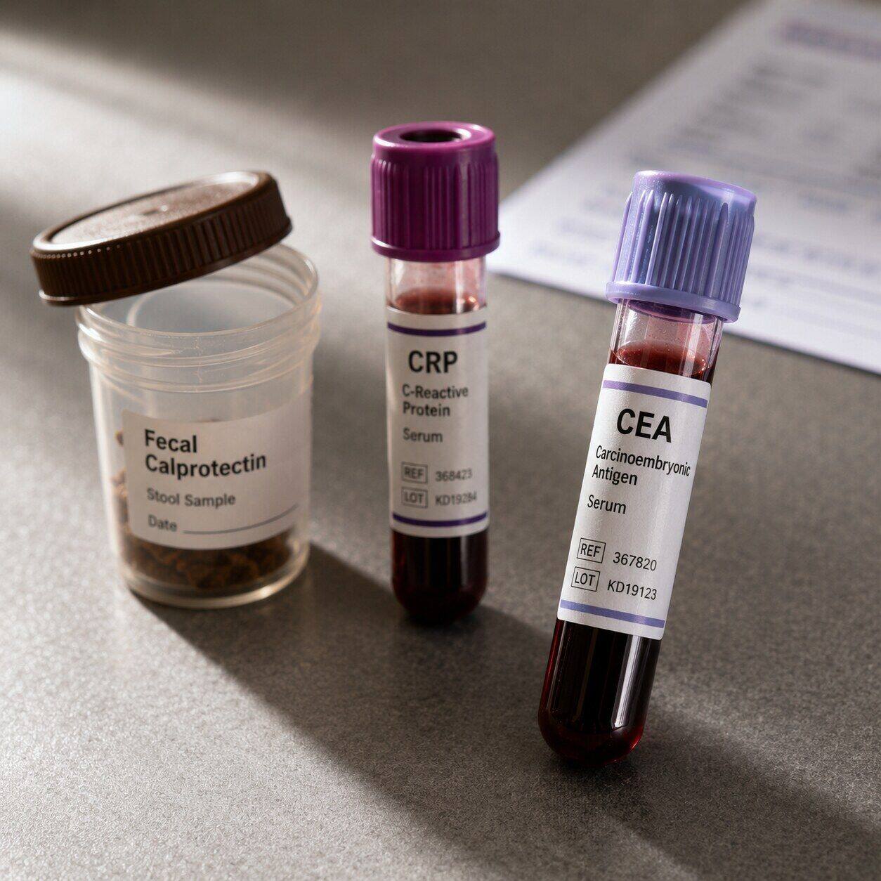
Figure 4. Fecal calprotectin, CRP, and CEA each provide a different layer of information, with CEA serving the narrowest, most cancer-focused role of the three.
Seeing these three specimens side by side is a useful way to internalize why they aren't interchangeable: the stool sample measures inflammation exactly where it's happening, the CRP-drawing tube reflects the body's overall inflammatory broadcast, and the CEA-drawing tube reflects a much narrower signal tied to epithelial turnover and cancer surveillance specifically. A gastroenterologist reading all three together gets a layered picture no single one could provide alone — which is precisely why an elevated CEA, on its own, is rarely enough to change a treatment plan without the other two providing supporting context first. This layered approach is also why patients sometimes leave an appointment having had several different tubes drawn and a stool kit handed to them in the same visit — each one is answering a slightly different question, and CEA's question is the narrowest of the group.
Frequently Asked Questions
Can Crohn's disease or ulcerative colitis alone cause a high CEA without any cancer?
Yes. Active inflammation damages the intestinal lining, loosens the barrier between cells, and increases local blood flow — all of which let more CEA leak into the bloodstream than a healthy gut would release. This is a well-documented, biologically explainable pattern, not a coincidence.
Does CEA go back to normal once an IBD flare is treated?
In many patients, yes — CEA that rose during an active flare often returns toward baseline once inflammation is brought under control, whether through medication adjustment or the flare resolving on its own. A gastroenterologist may recheck CEA after treatment specifically to confirm this pattern.
Should IBD patients get CEA checked regularly, and does it replace colonoscopy for cancer screening?
CEA is sometimes checked periodically in IBD patients as one piece of a broader picture, but it never replaces colonoscopy, which remains the primary surveillance method for colorectal cancer risk in long-standing IBD. CEA is a supporting data point, not a screening tool on its own.
Does it matter whether I have Crohn's disease versus ulcerative colitis for how much CEA rises?
Both can raise CEA, but the pattern can differ. Ulcerative colitis produces continuous surface inflammation across the affected colon, while Crohn's disease often involves deeper, patchier inflammation that in some cases correlates with even more pronounced elevations. Neither pattern is universal, so your own history and current disease activity matter more than which diagnosis you carry.
If I'm on a biologic and my CEA is still high, does that mean the medication isn't working?
Not necessarily on its own. CEA normalization tends to lag behind clinical symptom improvement, often tracking closer to the timeline of deeper tissue healing, which can take several months to become apparent. A single still-elevated value shortly after starting treatment is less informative than a value checked further out, alongside other markers of healing like calprotectin or endoscopic findings.
Conclusion
An elevated CEA in someone with Crohn's disease or ulcerative colitis is far more often a reflection of active gut inflammation than a sign of cancer — the same damaged, leaky, blood-flow-rich tissue that causes IBD symptoms also happens to release more of this particular protein into circulation. That doesn't mean the number should be ignored; it means it needs to be read in context, alongside disease activity, smoking status, prior results, and — when appropriate — direct visualization through colonoscopy, which remains the real gold standard for colorectal cancer surveillance in this population. If your CEA came back high during a flare, the most productive move is usually to bring your bowel disease's current activity into the conversation with whoever ordered the test, rather than assuming the worst from a single lab value. Living with a chronic bowel disease already means learning to hold uncertainty in a way most people never have to — flares come and go, treatments get adjusted, and lab values move around in ways that don't always have an immediate, satisfying explanation. A mildly elevated CEA is one more piece of that same pattern: unsettling to see on paper, but usually explainable once it's placed next to everything else already known about how active your disease is right now, and reassuringly quick to resolve once that activity is brought back under control.
Still Not Sure What Your Results Mean?
Upload your labs and get a complete, visual, plain-language interpretation of every biomarker — delivered to your inbox in under 15 minutes.
Get My ReportThis article is for educational purposes only and does not constitute medical advice. Always consult your healthcare provider regarding your specific lab results.