
Why Is My PSA High With a Normal Prostate Exam?
It feels like a contradiction the moment your doctor says it out loud: your prostate felt completely normal on exam — smooth, evenly sized, no lumps, no hard spots — and yet your PSA blood test came back elevated. If the gland felt fine, shouldn't the blood test agree? The honest answer is that a digital rectal exam and a PSA blood test aren't actually measuring the same thing, and they were never designed to. One is a physical assessment of shape and texture, performed by a finger that can only reach part of the gland. The other is a chemical measurement of a protein leaking into your bloodstream, from a source that finger was never capable of touching in the first place. Understanding exactly where that gap comes from — the real anatomy behind why your prostate can feel entirely normal while still producing an abnormal number — is what turns this confusing mismatch into something genuinely explainable, rather than something that feels like one of the two tests must simply be wrong.
This isn't a rare or unusual scenario either. Given how much of the prostate sits genuinely out of a physical exam's reach, and how many entirely benign, non-cancerous conditions can independently raise PSA, this exact combination shows up in routine urology practice on a regular basis — common enough that most experienced clinicians have a clear, well-rehearsed explanation ready the moment a patient asks why the two results don't seem to match.
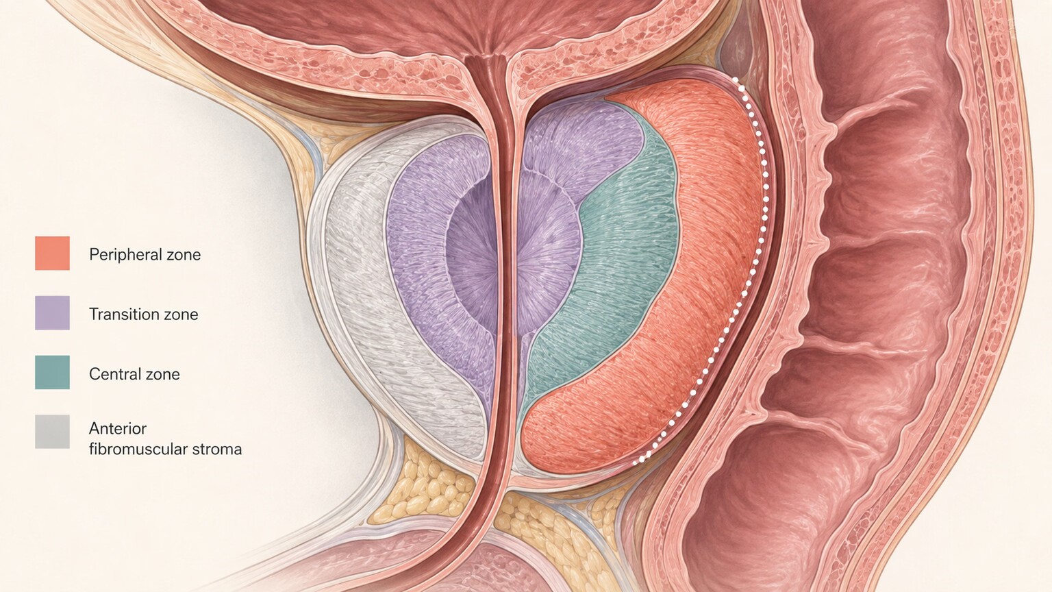
Figure 1. A digital rectal exam can only assess the posterior surface of the prostate's peripheral zone — roughly the back third of the gland — leaving the transition zone, central zone, and anterior fibromuscular stroma entirely outside its reach.
Your Prostate Has Zones, and a Finger Only Reaches One
The prostate isn't a single uniform lump of tissue — anatomists divide it into several distinct regions, each with its own name, location, and tendency toward different kinds of disease. The peripheral zone sits at the back of the gland, closest to the rectal wall, making up roughly 70 percent of the prostate's total tissue and, not coincidentally, the one region a digital rectal exam can actually reach and assess. The transition zone surrounds the urethra near the center of the gland, the central zone sits deeper and more toward the bladder, and the anterior fibromuscular stroma forms a band of mostly non-glandular tissue along the very front of the prostate, closest to the pubic bone and furthest from the rectal wall.
A gloved finger inserted during a digital rectal exam is pressing against the rectal wall, feeling for changes in shape, size, and texture on the posterior surface of the gland — essentially the back of the peripheral zone. It has no physical way to reach the transition zone, the central zone, or the anterior fibromuscular stroma, all of which sit too far forward, too deep, or too shielded by the rest of the gland's own tissue to be assessed by touch through the rectal wall. This isn't a flaw in how the exam is performed — it's simply the physical limit of what a finger, pressing from one specific direction, can ever access.
It helps to picture the prostate's actual size and shape to understand why this limitation is so absolute rather than a minor gap. A typical adult prostate is roughly the size and shape of a walnut, sitting just below the bladder and wrapped around the urethra, with the rectum running directly behind it — which is exactly why a rectal exam can reach it at all in the first place. But "reaching it" only means the examining finger can press against whichever surface happens to face the rectal wall; the front, deeper, and centrally located portions of that same small walnut-sized gland remain on the far side of tissue the finger was never going to press through. Scaling this down to something concrete: on a gland roughly four centimeters across, the accessible posterior surface represents only a thin slice of the total three-dimensional volume, leaving the majority of the gland's actual tissue mass structurally out of reach no matter how thorough or careful the examining clinician is.
Where Prostate Cancer Actually Tends to Start
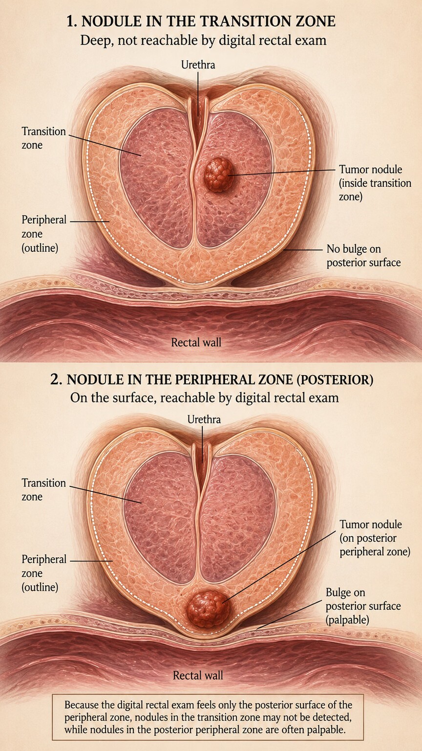
Figure 2. A tumor sitting within the transition zone, deep from the rectal wall, can grow to a meaningful size while producing no detectable change on physical exam, unlike a tumor located on the reachable posterior surface.
Roughly 70 to 80 percent of prostate cancers do arise in the peripheral zone — the exact region a digital rectal exam can assess — which is part of why the exam has real diagnostic value and isn't simply a formality. But that still leaves a genuinely meaningful share of cases, commonly cited in the range of 20 to 30 percent, originating in the transition zone or other harder-to-reach regions instead. A cancer growing in one of these locations can reach a real, biologically significant size — large enough to be actively producing and leaking measurable amounts of PSA into the bloodstream — while remaining completely undetectable by touch, simply because it never grows outward far enough, or in the right direction, to change the shape or texture of the gland's reachable posterior surface.
Interestingly, transition-zone cancers are also somewhat more likely, on average, to be discovered specifically because of an elevated PSA rather than a symptom or an exam finding — a pattern that makes intuitive sense once the underlying anatomy is understood. A peripheral-zone cancer growing toward the rectal wall has a real chance of eventually producing a palpable change an exam can catch, while a transition-zone cancer growing inward, away from the rectal wall and toward the urethra, has essentially no such chance until it becomes large enough to cause urinary symptoms instead — by which point it may already be more advanced than a case caught earlier through PSA screening alone.
This is precisely the mechanism behind the confusing scenario this article opened with. It isn't that the exam was performed poorly, or that the doctor missed something they should have caught — it's that a tumor in the wrong location, relative to where a finger can press, simply has no way of showing up on a physical exam no matter how carefully or skillfully that exam is performed.
It's worth adding one more layer of nuance here, since even a peripheral-zone tumor doesn't automatically guarantee a positive exam finding. Size still matters enormously — a small, early tumor confined to a limited area, even within the reachable zone, may not yet be large enough to create a texture change a finger can distinguish from normal surrounding tissue. This is exactly why physical exam findings tend to correlate more reliably with larger, more advanced tumors than with the smaller, earlier-stage disease that modern PSA-based screening is specifically designed to help catch — a distinction that becomes especially important when thinking about why relying on exam findings alone, even for peripheral-zone cancers, would mean missing exactly the cases where early detection matters the most.
Already have a PSA number from a recent test, but not sure if it's actually good or bad for you? Just plug it in and see instantly.
🧮 Try the Free CalculatorPSA Is a Chemical Signal — It Doesn't Need to Be Touched to Be Detected
The deeper reason this discordance is possible at all comes down to a basic difference in what each test is actually measuring. A digital rectal exam relies entirely on physical deformation — a lump, a hardened patch, an asymmetry the examining finger can feel through the rectal wall. PSA, by contrast, is a protein that the prostate's glandular cells manufacture and normally keep mostly contained within the gland and reproductive tract, with only a small, steady trickle leaking into the surrounding blood vessels under normal circumstances.
Cancerous tissue, wherever it happens to be located within the gland, tends to disrupt the normal architecture that keeps PSA contained — the organized cellular structure and tight junctions that would otherwise hold it back break down, allowing considerably more PSA than usual to leak into the bloodstream. This leakage happens at the cellular and chemical level, entirely independent of whether that disrupted tissue sits somewhere a finger could ever physically reach. A tumor deep in the transition zone, invisible to touch, can be leaking PSA into circulation just as readily as one on the exam-accessible posterior surface — sometimes more readily, depending on the specific tumor's biology — which is exactly why the blood test and the physical exam can tell two genuinely different stories about the same gland.
It helps to understand PSA's actual, everyday job to see why this leakage mechanism makes biological sense rather than feeling like an arbitrary lab quirk. PSA is an enzyme whose normal function is to liquefy semen after ejaculation, breaking down a gel-like protein structure so sperm can move more freely — a purely reproductive function that has nothing to do with cancer detection at all. Nearly all of the PSA a healthy prostate produces is meant to stay within the reproductive tract, doing this job, with only a small, well-regulated amount ever crossing into the bloodstream under normal circumstances. Cancer detection via PSA blood testing is, in a real sense, a side effect of this leakage rather than the protein's intended purpose — we're measuring the amount of a reproductive enzyme spilling into a compartment it doesn't normally belong in, and using the size of that spill as an indirect signal that something has disrupted the tissue architecture responsible for keeping it contained.
This indirect nature is also why PSA, despite being one of the most useful and widely used cancer markers in medicine, was never designed to be perfectly specific to cancer alone — anything that disrupts prostate tissue architecture, cancerous or not, can produce the same basic leakage effect to varying degrees. Understanding PSA as a general signal of "something has changed in how well this tissue keeps its contents contained," rather than a dedicated cancer alarm, is what makes both this article's central discordance and the broader list of non-cancer causes discussed later make complete biological sense together, rather than seeming like separate, unrelated facts about the test.
Exam Sensitivity Has Real, Documented Limits

Figure 3. Beyond anatomical blind spots, published studies show meaningful variation between different examiners assessing the same gland, and the exam's sensitivity for detecting cancer that is present tops out well below 100 percent even under ideal conditions.
Beyond the anatomical blind spots described above, the digital rectal exam carries its own well-documented sensitivity limitations even within the zone it can actually reach. Research comparing exam findings to biopsy-confirmed cancer has found that a meaningful share of cancers located even within the accessible peripheral zone go undetected by touch, particularly when they're small, early-stage, or located toward the edges of the examiner's reach. Studies examining agreement between different examiners assessing the same patient have also found real, measurable variability — one clinician's assessment of "normal" doesn't always match another's for the exact same gland, since the exam depends on subjective tactile interpretation rather than an objective, standardized measurement the way a blood test does.
None of this means the exam is not worth doing — it remains a genuinely useful, low-cost, immediate source of information, and it does catch real cancers, sometimes ones with a normal PSA. But understanding its real sensitivity limits, rather than treating a "normal" result as an absolute all-clear, is part of what allows a normal exam and an elevated PSA to be interpreted correctly together, rather than as one test simply overriding or contradicting the other.
There's also a meaningful learning-curve element to exam accuracy that's rarely discussed openly with patients. Detecting subtle textural differences through a gloved finger and a layer of rectal wall tissue is a genuinely difficult tactile skill, refined over years of examining hundreds or thousands of glands and comparing findings against eventual biopsy or surgical outcomes. A highly experienced urologist who has developed this specific tactile pattern-recognition over decades of practice will, on average, catch subtler abnormalities than a less experienced examiner performing the same physical motion — not because of any difference in effort or care, but because of the sheer accumulated pattern-matching experience involved. This is one more reason a single normal exam finding, especially from a routine primary-care visit rather than a specialist urology exam, shouldn't be treated as the final, definitive word on a gland's condition, particularly when the blood test is telling a different story.
Body habitus adds a further practical complication rarely mentioned outside clinical settings: in patients with significant obesity, the physical distance and tissue thickness a finger must press through can itself reduce how much fine textural detail is transmitted back to the examiner, independent of anything happening within the prostate itself. This is a genuinely under-discussed limitation of the exam's real-world sensitivity, and it's part of why body composition sometimes factors into how much diagnostic weight a clinician places on a "normal" exam finding in a specific individual patient.
What Actually Happens Next: Imaging Fills the Gap
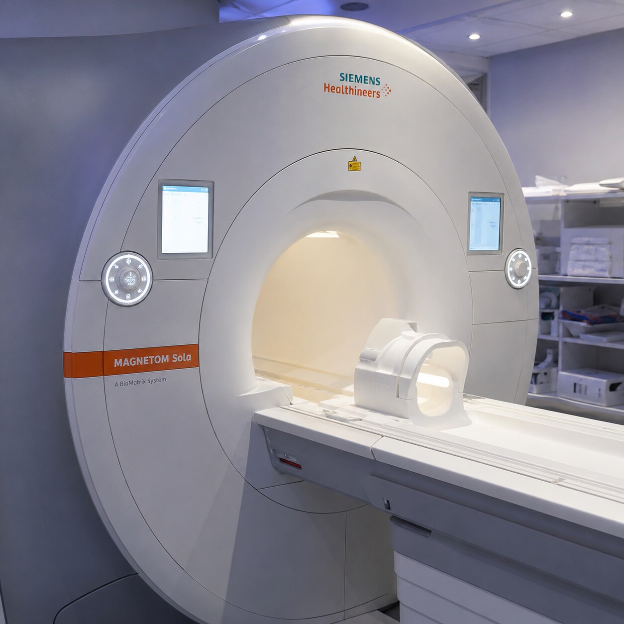
Figure 4. Multiparametric MRI can visualize the transition zone, central zone, and anterior fibromuscular stroma directly, closing exactly the diagnostic gap a physical exam alone cannot cross.
The modern medical answer to exactly this blind spot is imaging, specifically multiparametric MRI of the prostate, which has become an increasingly standard next step when PSA is elevated but the physical exam is reassuringly normal. Unlike a finger pressed against the rectal wall, MRI can visualize the entire gland from the inside out — the transition zone, the central zone, the anterior fibromuscular stroma, all the regions a digital rectal exam simply cannot access — identifying suspicious areas by their distinct appearance on specialized imaging sequences that highlight differences in tissue density, blood flow, and water movement associated with cancerous versus normal tissue.
When a suspicious area is identified on MRI, it can guide a much more precise, targeted biopsy of that exact location, rather than the older approach of taking essentially random samples spread throughout the gland and hoping one happens to land on the abnormal tissue. This targeted approach has meaningfully improved both the detection of clinically significant cancers hiding in hard-to-reach zones and, just as importantly, has reduced unnecessary biopsies of areas that turn out to be entirely normal, making the overall diagnostic pathway considerably more precise than relying on physical exam and PSA alone ever could be.
The scoring system radiologists use to communicate MRI findings, called PI-RADS (Prostate Imaging Reporting and Data System), is worth knowing about by name, since you may see it referenced directly in your own results if you're ever sent for this scan. It rates suspicious areas on a five-point scale, from PI-RADS 1 (very unlikely to be clinically significant cancer) up through PI-RADS 5 (highly likely), giving both the treating physician and the patient a standardized, more objective language for discussing risk than the more subjective descriptions a physical exam alone could ever provide. A PI-RADS 1 or 2 finding on MRI, even alongside an elevated PSA, often supports continued monitoring rather than an immediate biopsy, while a PI-RADS 4 or 5 finding typically prompts a more urgent targeted biopsy of that specific area — precision that simply wasn't available before this imaging technology became widely accessible.
It's worth being upfront that this MRI-first approach, however logical, isn't yet universally available or standard everywhere, since access depends on local equipment, radiologist expertise specific to interpreting prostate MRI, and insurance coverage policies that vary considerably by region and provider. In settings where MRI isn't readily accessible, the traditional approach of monitoring PSA trends over time, sometimes combined with a free PSA percentage, and considering a standard systematic biopsy if the overall picture remains concerning, is still a reasonable and medically sound pathway, just a less precisely targeted one than what MRI can offer where it's available.
Most High-PSA, Normal-Exam Combinations Still Aren't Cancer
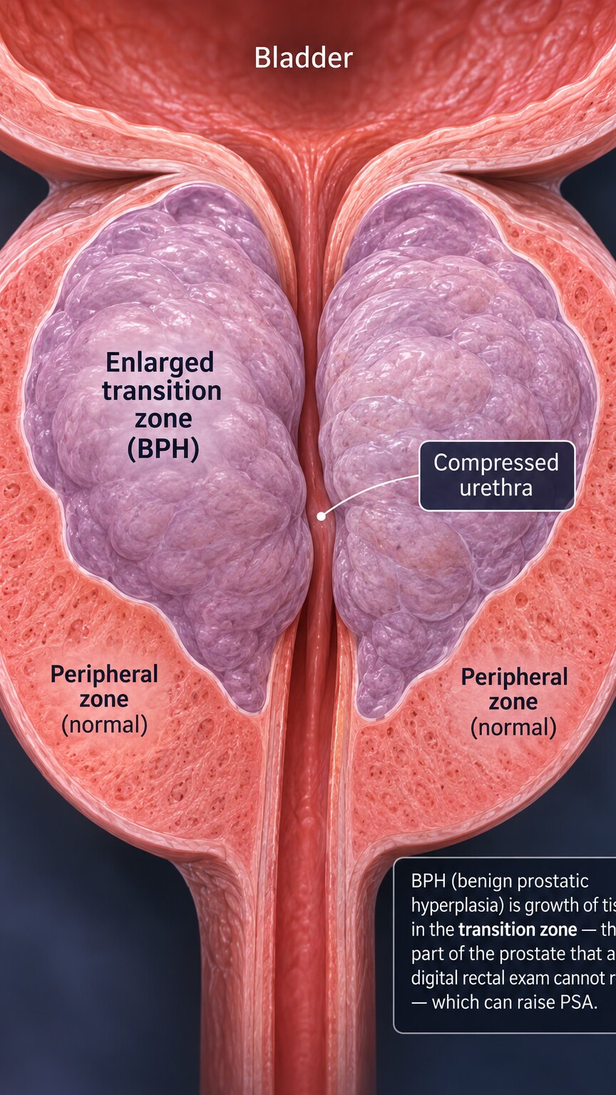
Figure 5. Benign prostatic hyperplasia commonly begins in the transition zone, the exact region a digital rectal exam cannot assess, which is why this extremely common, non-cancerous condition can raise PSA without ever changing how the gland feels.
Before this discordance starts to feel more alarming than it needs to, it's worth stepping back to a broader point: an elevated PSA alongside a normal exam is still, in the majority of cases, explained by something other than cancer entirely. Benign prostatic hyperplasia (BPH), the extremely common, non-cancerous enlargement of prostate tissue that affects most men to some degree as they age, frequently originates specifically in the transition zone — the very region a digital rectal exam cannot assess — and can meaningfully raise PSA simply through the sheer increase in total glandular tissue producing it, entirely independent of any cancer being present.
Prostatitis, whether from an active infection or a related non-bacterial inflammatory process, is another common explanation capable of raising PSA without any palpable change on exam, particularly when the inflammation involves regions of the gland outside the reachable posterior surface. Recent ejaculation, vigorous cycling, or a recent urological procedure can each cause a temporary bump as well, none of which would ever be expected to change how the gland feels to the touch. The point isn't that cancer should be dismissed as a possibility — it's that a normal exam alongside an elevated PSA is a genuinely common, frequently benign combination, and the appropriate next step is thoughtful further evaluation, not immediate alarm.
It's worth appreciating just how common BPH specifically is, since its prevalence is a major reason this discordant combination shows up so often in everyday clinical practice. Some degree of benign prostatic enlargement is present in a majority of men by their sixties, and in the large majority by their eighties, making it one of the most universal age-related changes in male anatomy, comparable in ubiquity to changes like presbyopia (needing reading glasses) or graying hair. Because BPH so often originates in and around the transition zone — precisely because that's the region surrounding the urethra where growth most directly produces the urinary symptoms BPH is known for, like a weaker stream or more frequent nighttime urination — its effect on PSA is, once again, coming from exactly the anatomical region a digital rectal exam cannot evaluate, tying this entire section back to the same core anatomical theme running through this whole article.
Age itself deserves mention as a contributing factor independent of any specific diagnosis, since PSA has a well-documented tendency to drift gradually upward across a normal lifespan even without BPH, prostatitis, or cancer being specifically identified — a reflection of the same slow, cumulative tissue changes responsible for BPH's rising prevalence with age in the first place. This is exactly why age-specific PSA reference ranges exist and why a result that would be flagged as concerning in a 45-year-old might be considered unremarkable, or at least less immediately alarming, in a 75-year-old with the same absolute number.
What to Actually Ask Your Doctor
If you're facing this exact combination — a reassuring exam alongside an unexpectedly elevated PSA — a few specific questions can help you understand where your particular case sits on the spectrum from "very likely benign" to "warrants closer evaluation." Asking directly whether an MRI is being considered, and if not, why, is a reasonable starting point, since this is increasingly the standard next step in exactly this scenario. Asking about your specific PSA trend over time, if you have prior results, matters too — a PSA that's risen quickly carries more weight than one that's been stably, mildly elevated for years, a topic covered in more depth in this site's article on PSA velocity.
Timing questions matter too — asking when a repeat PSA makes sense, and whether it's worth waiting a specific interval versus rechecking sooner, gives you a concrete plan to hold onto rather than an open-ended sense of uncertainty between now and your next appointment.
It's also worth asking whether a free PSA percentage was checked or should be, since that additional measurement — covered in more depth elsewhere on this site — helps refine risk further in borderline cases, and whether any of the temporary, benign explanations described above (recent ejaculation, a recent procedure, an active infection) could plausibly account for your specific result before pursuing more invasive next steps.
Don't hesitate to ask for these answers in plain language either, rather than settling for a quick summary — a genuinely engaged conversation at this stage tends to pay off in how confidently you understand, and follow through on, whatever comes next.
A newer generation of blood-based tests worth knowing the names of, even if not every provider offers them yet, includes the Prostate Health Index (PHI) and the 4Kscore, both of which combine PSA with additional related biomarkers into a single composite risk estimate designed to be more accurate than PSA measured on its own. These tests won't be relevant or available for every patient, and they don't replace imaging when imaging is genuinely warranted, but asking whether one might add useful information in your specific borderline case is a reasonable question, particularly if you're trying to avoid an unnecessary biopsy while still taking a genuinely elevated PSA seriously.
Bring a written list of questions if that helps you stay organized during a short appointment — it's a small preparation step that makes a genuine difference in how much you actually retain and act on afterward.
Finally, it's worth explicitly asking about your individual risk profile beyond the numbers on the page — family history of prostate cancer, particularly in a father or brother, and certain genetic factors (including a meaningfully elevated risk associated with BRCA gene mutations more commonly discussed in the context of breast and ovarian cancer) can shift how aggressively a given PSA and exam combination gets investigated. Two people with the identical PSA value and identical normal exam finding can reasonably receive somewhat different recommendations once their broader individual risk factors are factored into the conversation, which is exactly the kind of nuance a good back-and-forth with your doctor, rather than the raw numbers alone, is meant to capture. None of this needs to happen in a single rushed conversation either — it's entirely reasonable to schedule a dedicated follow-up visit specifically to work through these questions once your results are in hand, rather than trying to absorb every single little detail all at once, at the exact moment the number first shows up unexpectedly on your chart.
A Worked Example: Reading This Combination the Right Way
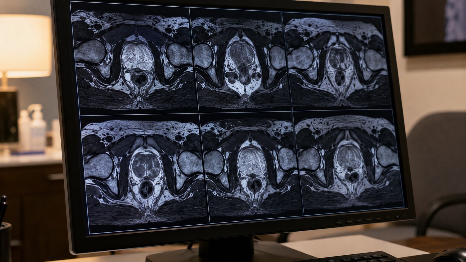
Figure 6. Reviewing MRI images together, rather than relying on the exam and PSA alone, is what actually resolves this discordance into a clear next step.
Walking through a realistic sequence makes this reasoning concrete. Picture a 58-year-old with no urinary symptoms whose routine PSA comes back at 5.8 ng/mL, above the commonly cited general threshold of 4.0, while his digital rectal exam is entirely unremarkable — smooth, symmetric, no nodules. A first, reasonable step is simply repeating the PSA a few weeks later after avoiding ejaculation and strenuous cycling beforehand, since a meaningful share of borderline elevations settle back down once temporary factors are ruled out. If the repeat value stays similarly elevated, a free PSA percentage adds another layer of information — a higher free percentage leans toward a benign explanation like BPH, while a lower one raises relative concern and supports moving forward with imaging.
Suppose the free percentage comes back on the lower, more concerning side. The next reasonable step, rather than an immediate biopsy, is a multiparametric MRI — which in this hypothetical case reveals a small, well-defined area of concern within the transition zone, precisely the region the earlier physical exam could never have assessed. That finding then guides a targeted biopsy of that exact location rather than a broader, less precise sampling approach, arriving at a much more confident answer than the original normal exam and elevated PSA could provide on their own. Whatever that answer turns out to be — benign tissue, or a cancer caught at a genuinely early, highly treatable stage — the path to it ran through recognizing that the normal exam was never capable of ruling anything out in the first place, not through treating it as reassurance the case was closed.
Now imagine a second, alternative branch of this same scenario: the free PSA percentage instead comes back comfortably on the higher, more reassuring side, and the MRI, when obtained, shows a diffusely enlarged gland with no discrete suspicious area at all — a pattern entirely consistent with straightforward BPH rather than anything cancer-related. In this branch, the appropriate path is continued monitoring with periodic PSA rechecks rather than a biopsy at all, since the weight of evidence across every test performed points toward a benign explanation. Both branches start from the exact same starting point — an elevated PSA and a normal exam — and both represent the system working exactly as intended, arriving at two very different, appropriately tailored conclusions based on what the fuller picture actually shows.
Frequently Asked Questions
Does a normal prostate exam mean I don't have prostate cancer?
Not necessarily. A digital rectal exam can only assess the peripheral zone's posterior surface, roughly the back of the gland. Cancers arising in the transition zone, central zone, or anterior fibromuscular stroma — together accounting for a meaningful share of cases — can go undetected by touch even when present.
Why does PSA rise from tissue a doctor's finger can't reach?
PSA leaks into the bloodstream through disrupted cellular architecture, a chemical process that happens regardless of where in the gland it occurs. A tumor deep in the transition zone can leak PSA into circulation just as readily as one on the reachable posterior surface.
What test can check the parts of the prostate an exam can't reach?
Multiparametric MRI can visualize the entire gland, including the transition zone, central zone, and anterior fibromuscular stroma, and is increasingly used as the next step when PSA is elevated but the physical exam is normal.
Is it common for PSA to be elevated with a normal exam?
Yes, and in most cases it isn't cancer. Benign prostatic hyperplasia, which frequently originates in the transition zone outside the exam's reach, along with prostatitis and several temporary factors, commonly explain this exact combination.
How reliable is a digital rectal exam even within the area it can reach?
It has real, documented limits. Studies show meaningful variability between examiners and a notable share of even peripheral-zone cancers going undetected by touch, particularly when small or early-stage, which is why a normal exam alone isn't treated as a definitive all-clear.
What is PI-RADS on an MRI report?
PI-RADS is a standardized five-point scale radiologists use to rate how likely an area seen on prostate MRI is to represent clinically significant cancer, with PI-RADS 1-2 generally supporting continued monitoring and PI-RADS 4-5 typically prompting a targeted biopsy of that specific area.
Does family history change how this discordance should be handled?
Yes. A family history of prostate cancer, particularly in a father or brother, or certain genetic risk factors, can reasonably shift a doctor toward more thorough follow-up for the same PSA and exam combination that might warrant a more conservative approach in someone with no such history.
Conclusion
A normal prostate exam alongside an elevated PSA isn't a contradiction between two tests disagreeing with each other — it's the expected, explainable result of two fundamentally different kinds of assessment, one physical and limited to roughly a third of the gland, the other chemical and blind to location entirely. Understanding the real anatomy behind that gap — the zones a finger can and can't reach, and why PSA doesn't care which zone it came from — turns a confusing, worrying mismatch into a clear next question: not "which test is wrong," but "does this specific result warrant a closer look at the parts of the gland the exam was never able to check in the first place."
The broader takeaway extends well past this one specific combination of tests: no single test in medicine, however well established or widely used, tells the complete story on its own. A physical exam, a blood marker, and modern imaging each illuminate a different slice of the same underlying question, and the real diagnostic power comes from combining them thoughtfully rather than expecting any one of them to serve as a final, standalone verdict. Approaching your own results with that mindset — asking what each test can and cannot see, rather than treating any single number or finding as the whole answer — is a genuinely useful habit that extends well beyond prostate health into how you read almost any medical test result going forward.
If you're currently facing this exact combination, the single most useful step is simply naming the gap out loud in your next conversation with your doctor: which parts of the gland has this exam actually assessed, and what, if anything, is planned to look at the rest. That one question, asked plainly, does more to move a confusing result toward a clear plan than almost anything else you could bring into the room.
Still Not Sure What Your Results Mean?
Upload your labs and get a complete, visual, plain-language interpretation of every biomarker — delivered to your inbox in under 15 minutes.
Get My ReportThis article is for educational purposes only and does not constitute medical advice. Always consult your healthcare provider regarding your specific results.