
Understanding the Difference Between Troponin I and Troponin T
"Troponin" is not actually one single thing your lab measures — it's the name for a small family of proteins, and when your report says "troponin," it almost always means one of two specific members of that family: troponin I or troponin T. These are two different proteins, made from two different genes, measured by two different laboratory tests that are not interchangeable with each other. Most of the time this distinction doesn't change how your result should be interpreted, since both are excellent, highly cardiac-specific markers of heart muscle injury. But the differences between them are real: they behave slightly differently in certain diseases, they can't always be compared numerically between two different hospitals, and one of them has a rare, well-documented blind spot the other doesn't share. Understanding which one your hospital actually tested, and why that even matters, turns a confusing pair of near-identical names into something you can read with real clarity.

Figure 1. The troponin complex sits on the actin thin filament of the cardiac sarcomere, where its three subunits — C, I, and T — jointly control whether the muscle fiber contracts.
What Troponin Actually Is Before It's Ever a Blood Test
To make sense of why there are two different troponin tests, it helps to first understand what troponin does inside a normal, healthy, beating heart — because it isn't floating around in your bloodstream by design. Troponin lives deep inside the machinery of every heart muscle cell, bolted onto a structure called the thin filament, one of the two interlocking protein cables that slide past each other every time your heart contracts. That sliding motion is what a heartbeat physically is at the cellular level, and troponin is the molecular switch that decides, moment to moment, whether that sliding is allowed to happen.
Troponin is actually a team of three separate proteins working together, not a single molecule, and each one has a distinct job. Troponin C is the calcium sensor — when a nerve signal tells your heart to contract, calcium floods into the muscle cell, and troponin C is the piece that physically grabs onto it. Troponin I is the inhibitor — in a resting, relaxed heart muscle, it's the piece actively holding the contraction machinery back, like a hand pressed on a brake. Troponin T is the anchor — it's the piece that physically attaches the whole three-part complex to tropomyosin, a long, rope-like protein that winds around the thin filament and blocks the contraction machinery until troponin tells it to move aside. When calcium binds troponin C, it triggers a shape change that pulls troponin I's "brake" away and lets troponin T drag tropomyosin out of the way, exposing the site where the two filaments can finally grip each other and slide. Multiply that sequence a hundred times a second across billions of cells, and you get a heartbeat.
This matters for understanding the blood test because troponin I and troponin T are only ever released into your bloodstream under one circumstance: when heart muscle cells are damaged badly enough that their outer membrane — the thin wall normally keeping everything inside the cell contained — becomes leaky or breaks open. A lab test for "troponin" is really a test asking one very specific question: has heart muscle tissue been injured recently? The letters I and T simply tell you which one of the two anchor proteins from that same three-part complex the lab chose to measure in order to answer it.
Meet the Two Molecules Behind the Letters
Troponin I and troponin T are not two versions of the same protein — they are genuinely different molecules, built from entirely different genetic blueprints, that happen to work side by side in the same three-part complex. Troponin I is a relatively compact protein, built from instructions carried by a gene called TNNI3, and it weighs in at roughly 24,000 daltons — daltons being the unit scientists use to measure the mass of a molecule, with a single dalton being roughly the mass of one hydrogen atom. Troponin T is a longer, more elongated protein, built from a completely separate gene called TNNT2, and it's noticeably larger, weighing in at roughly 37,000 daltons. Neither protein is simply a longer or shorter copy of the other; they fold into distinct three-dimensional shapes and do distinct structural jobs within the complex, as described above.
This structural difference is not just a biochemistry footnote — it's the reason the two blood tests were engineered so differently from one another. Because troponin I sits more centrally within the complex, tucked against troponin C, an antibody test has to be designed carefully to target a stretch of the molecule that stays exposed and stable even after the protein has been damaged and released into the bloodstream. Troponin T's more elongated shape gives assay developers a longer stretch of exposed, stable amino acid sequence to design an antibody against, which is part of why it lent itself early on to a single, highly reproducible commercial test. Troponin I's more compact shape, and the fact that it can break apart into several different circulating fragments and can also bind to troponin C in ways that partially hide part of its structure, made it a harder target to standardize — different manufacturers ended up choosing different exposed regions of the molecule to build their antibodies against, which is a large part of the root cause behind the standardization gap covered later in this article. In short, the same molecular features that make each protein biologically distinct also explain why one test standardized more easily across the industry than the other.
Here's the detail that makes both of these tests clinically useful in the first place: the heart doesn't use the exact same version of troponin I or troponin T that your skeletal muscles — your biceps, your quadriceps, the muscles that let you walk and lift things — use. Skeletal muscle has its own separate genes for troponin I and troponin T (called TNNI1, TNNI2, TNNT1, and TNNT3, depending on the muscle fiber type), producing versions of these proteins with slightly different amino acid sequences than the heart-specific versions. Modern cardiac troponin tests are deliberately engineered around antibodies that recognize only the unique stretches of amino acids found on the cardiac versions — TNNI3's product and TNNT2's product — and essentially ignore the skeletal muscle versions entirely. This is the entire reason cardiac troponin testing represented such a massive leap forward when it was introduced in the 1990s, replacing older markers like creatine kinase-MB (CK-MB) that couldn't reliably tell the difference between an injured heart and an injured shoulder or leg muscle. Both troponin I and troponin T, in their cardiac forms, share this same core advantage of being genuinely heart-specific — which is exactly why the differences between them, discussed in the rest of this article, are refinements around the edges rather than a fundamental gap in reliability between the two.
Curious what the rest of your labs are telling you beyond troponin? Upload your results and get a complete, plain-language breakdown in under 15 minutes.
Analyze My ResultsWhy Two Separate Tests Exist for the Same Job
The existence of two competing, non-identical tests for essentially the same clinical question traces back to how cardiac troponin testing was actually developed. In the late 1980s and early 1990s, more than one research group and diagnostics company recognized that troponin's cardiac-specific isoforms could make a superior heart-injury marker, and they pursued it somewhat independently, developing antibodies against different subunits of the same complex. Some teams focused on troponin T; others focused on troponin I. Both approaches proved successful, and both were validated in large clinical studies as being highly sensitive and highly specific for heart muscle injury. Rather than one approach winning out and the other disappearing, both went on to become established, guideline-endorsed standards, and the diagnostics industry split along those lines — one major manufacturer built its entire cardiac testing franchise around troponin T, while several other manufacturers each developed their own separate troponin I assays.
That history is the direct reason your hospital's laboratory measures one or the other, rather than always testing both. Which test you get isn't a medical decision made about you personally — it's a decision your hospital's laboratory made years earlier, when it chose which analyzer platform and which manufacturer's testing equipment to install. A hospital running machines from the company that makes the dominant troponin T assay will report troponin T on every patient's chart; a hospital running equipment from any of several other manufacturers will report troponin I. Neither choice reflects a judgment that one test is medically better than the other for a general patient — both are recommended, guideline-endorsed options, and the choice mostly comes down to laboratory contracts, existing equipment, and cost.
Troponin T's One Well-Documented Blind Spot
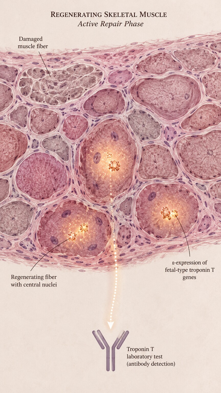
Figure 2. In damaged, regenerating skeletal muscle, fibers can briefly switch back on a fetal-type troponin T gene program, occasionally cross-reacting with troponin T assays.
There is exactly one biological quirk that sets troponin T meaningfully apart from troponin I in clinical practice, and it's worth understanding clearly rather than glossing over, because it's the single most commonly cited reason a doctor might specifically prefer troponin I in certain patients. Skeletal muscle tissue, when it's severely damaged and actively trying to repair itself — think of conditions like inflammatory muscle diseases such as polymyositis or dermatomyositis, advanced muscular dystrophies, or the kind of chronic muscle breakdown and regeneration seen in some patients with long-term kidney failure — can briefly switch back on genetic programs it normally only uses during fetal development. One of the genes that can flicker back to life during this regenerative process produces a troponin T protein that structurally resembles the cardiac version closely enough that some troponin T antibody tests can, in rare cases, mistake it for the real cardiac signal.
The practical result is that a small subset of patients with significant, actively regenerating skeletal muscle disease can show a falsely elevated troponin T result that has nothing to do with their heart at all. This isn't a common event, and newer generations of troponin T testing have substantially reduced how often it happens compared to older assay versions from decades ago — but it remains a recognized limitation that shows up in the medical literature and in clinical guidelines, particularly for patients with known chronic myopathies or advanced kidney disease. Troponin I, built from a genetically distinct molecule with a different fetal-isoform history, is not entirely immune to every form of interference, but this particular skeletal-muscle-regeneration cross-reactivity pattern is far less of a documented concern for troponin I assays. In a patient with a known muscle-wasting condition and an unexpectedly elevated troponin T with no other signs pointing toward the heart, this is often one of the first alternative explanations a cardiologist will consider — and it's a good example of why context always matters more than the number by itself.
Why Your Hospital's Number Might Not Match Another Hospital's
One of the most practically important differences between these two tests has nothing to do with biology at all — it's about how the tests themselves are built and calibrated, and it can genuinely confuse patients who are comparing lab reports from two different facilities. Troponin I assays are manufactured by several different companies, and critically, each company's version uses its own specific antibodies, targeting slightly different regions of the troponin I molecule, calibrated against its own internal reference standard. There has never been a single universal reference material that every troponin I manufacturer calibrates against in exactly the same way, which means a troponin I result of, say, 0.04 nanograms per milliliter from one hospital's testing platform is not guaranteed to mean the exact same thing as a troponin I result of 0.04 nanograms per milliliter from a different hospital using a different manufacturer's equipment — the two numbers can reflect genuinely different scales, even though they're reported using the same units.
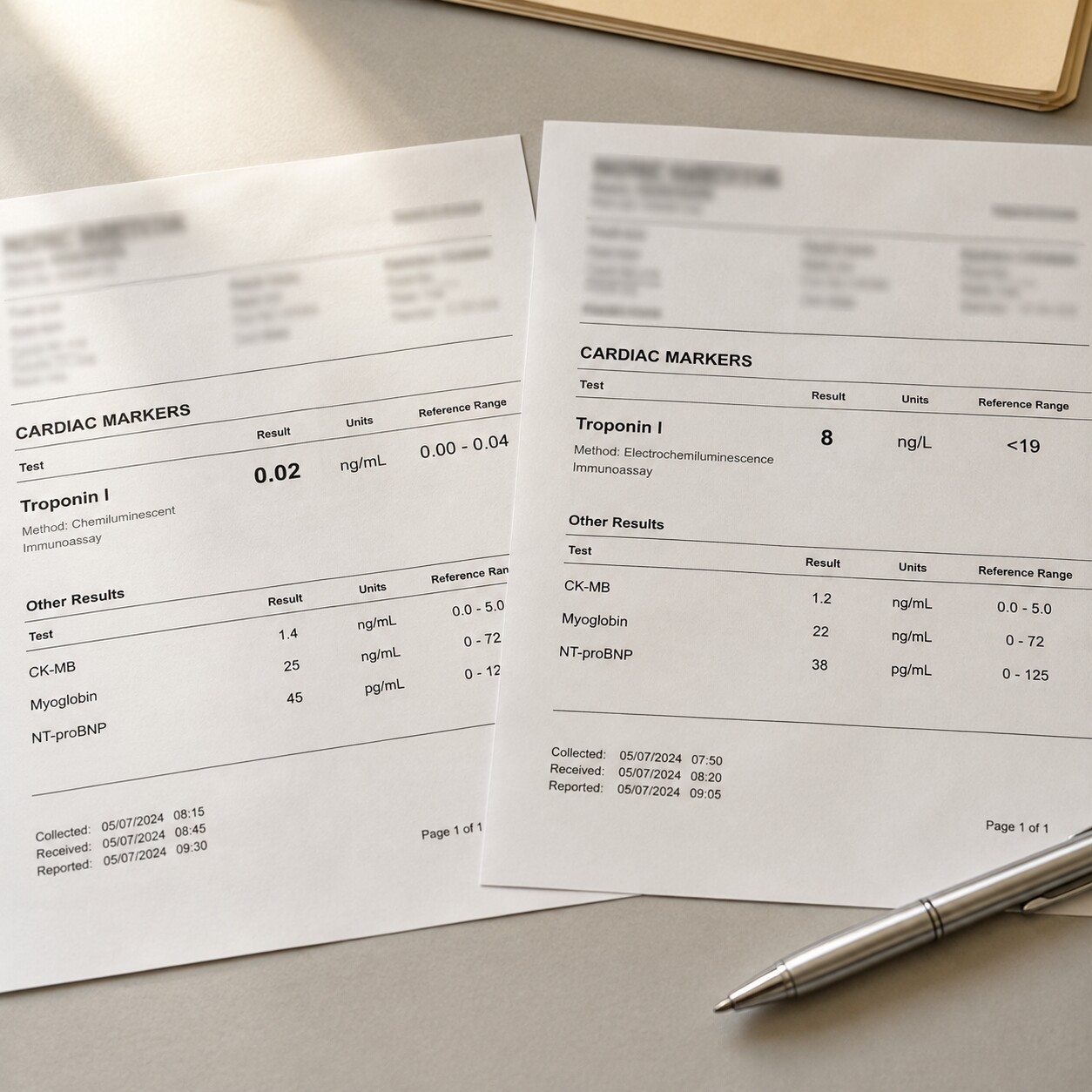
Figure 3. Two hospitals using different troponin I manufacturers can report different reference ranges and numeric scales for what is medically the same underlying result.
Troponin T looks different on this front, mostly because of a historical accident rather than any inherent biological advantage. The dominant high-sensitivity troponin T assay used across most hospitals worldwide is manufactured by a single company, which effectively means that any two hospitals running that same troponin T test are, in practice, using the identical assay, on the identical numeric scale, calibrated the identical way. That gives troponin T a kind of de facto standardization that troponin I has historically lacked — a troponin T number from one hospital using this assay is directly comparable to a troponin T number from another hospital using the same assay, in a way that isn't automatically true for troponin I results from two different manufacturers. This is genuinely useful when a patient is transferred between facilities or has serial testing done at more than one location, though it's worth noting that international standardization efforts have been actively working to bring troponin I assays closer together on this front, and the gap has narrowed over time even if it hasn't fully closed. The practical takeaway for anyone comparing two troponin results from two different hospitals or two different visits is simple: the safest assumption is that the numbers are only directly comparable if you can confirm the same test, from the same manufacturer, was used both times — and if you can't confirm that, the trend and the clinical picture matter far more than comparing the raw numbers to each other.
How High-Sensitivity Testing Changed Both Assays Together
For most of the history of cardiac troponin testing, both troponin I and troponin T assays could only reliably detect relatively large amounts of the protein in blood — enough to confirm a heart attack was already well underway, but not sensitive enough to catch smaller, earlier, or more subtle degrees of heart muscle injury. Starting in the late 2000s in Europe, and reaching routine use in the United States roughly a decade later, both troponin I and troponin T received a major technological upgrade in the form of high-sensitivity assays, often labeled hs-cTnI and hs-cTnT on a lab report. These newer versions can reliably measure troponin concentrations thousands of times smaller than older-generation tests, down into the single-digit nanogram-per-liter range.

Figure 4. High-sensitivity assays use densely packed antibody pairs to detect troponin at concentrations thousands of times smaller than older-generation tests could reach.
This upgrade applied roughly equally to both tests and changed clinical practice for both in the same ways. Emergency departments that once had to wait six to twelve hours between repeat troponin draws to safely rule out a heart attack can now often do so in one to three hours, using rapid rule-out and rule-in protocols built around either hs-cTnI or hs-cTnT — European Society of Cardiology guidelines, for example, publish validated cutoff values for both, using them interchangeably depending on which test a given hospital has available. High-sensitivity testing also introduced sex-specific reference ranges for both troponin I and troponin T, since women generally have a smaller average amount of heart muscle mass than men and, correspondingly, a lower normal upper limit for both tests — a detail that matters because using a single unisex cutoff was shown to under-diagnose heart attacks in women when older, less refined thresholds were used. None of this high-sensitivity upgrade closed the gap between troponin I and troponin T entirely — the standardization issue between manufacturers and the rare skeletal-muscle cross-reactivity issue for troponin T both persisted through the transition — but it did mean both tests became dramatically more useful tools than they were even fifteen years ago, and both remain fully supported, guideline-recommended options today.
Kidney Disease and Chronically Elevated Baseline Troponin
People with significant chronic kidney disease, and especially those on long-term dialysis, present one of the more genuinely complicated scenarios for interpreting either troponin test, and it's worth understanding on its own terms rather than assuming it works the same way it does for someone with normal kidney function. Troponin, like many small proteins, is partly cleared from the bloodstream by the kidneys. When kidney function declines significantly, that clearance slows down, and troponin — even without any new heart injury happening — can build up to a chronically elevated baseline level that simply reflects reduced clearance rather than active cardiac damage. This effect has been observed with both troponin I and troponin T, though it has historically been documented as somewhat more pronounced with troponin T, a pattern researchers attribute partly to differences in how the two proteins and their breakdown fragments are handled by failing kidneys, and possibly to some degree of low-level, ongoing skeletal muscle turnover common in this patient population layering on top of the reduced clearance itself.

Figure 5. Reduced kidney clearance in dialysis patients can raise baseline troponin levels for both tests, complicating the use of a single fixed cutoff to diagnose a new cardiac event.
Because of this, doctors caring for dialysis patients generally don't lean on a single fixed troponin cutoff the way they might for someone with normal kidney function. Instead, they pay much closer attention to the trend — whether a person's troponin, whichever test is being used, is rising meaningfully above their own known baseline, rather than judging a single value against the standard reference range built for the general population. A dialysis patient with a troponin T of, say, three times the general population's upper limit might simply be at their own personal baseline, unchanged from months of prior testing, while a smaller-looking rise from that same baseline could actually represent a real new cardiac event. This is one of the clearest examples in laboratory medicine of why a number alone, divorced from a person's own history and clinical context, can be genuinely misleading — and it applies to some degree to both troponin I and troponin T, even if troponin T's baseline elevation in this population has been more extensively studied and more consistently observed across research.
Does It Actually Matter Which One Your Doctor Used?
For the overwhelming majority of people getting a troponin test — someone in an emergency room with chest pain, someone being monitored after a cardiac procedure, someone whose doctor ordered it as part of a broader workup — it genuinely does not matter whether the result came from a troponin I or troponin T assay. Both are validated, guideline-endorsed, highly cardiac-specific tests, both are interpreted using their own separately established reference ranges and diagnostic cutoffs, and both give a cardiologist essentially equivalent diagnostic information when used correctly and interpreted in the context of symptoms, an electrocardiogram, and, when appropriate, repeat testing over time. Studies comparing head-to-head diagnostic accuracy between modern high-sensitivity versions of both tests have generally found them to perform comparably well at their core job: telling doctors whether a heart attack is happening.
Where it does start to matter is in the more specific situations described throughout this article: a patient with a known chronic muscle-wasting condition where troponin T's rare cross-reactivity issue becomes a relevant consideration; a patient being compared across two different hospitals or health systems where the numeric scale of troponin I results may not line up; and a patient with advanced kidney disease where understanding that either test can run at an elevated personal baseline changes how a single result should be read. None of these scenarios mean one test is broadly "better" than the other — they mean each test has a short list of specific circumstances where its particular quirks are worth a clinician's awareness, and that's precisely the kind of nuance a treating physician, not a patient reading a report at home, is best positioned to apply.
How Troponin Fits Into the Bigger Diagnostic Picture
Neither troponin I nor troponin T is ever meant to be interpreted in isolation, and understanding what surrounds the number on your report goes a long way toward putting a single result in perspective. An electrocardiogram, often shortened to ECG or EKG, records the heart's electrical activity through a set of small sticky patches placed on the chest, and it can show characteristic patterns when a blocked artery is starving a specific region of heart muscle of blood — patterns that look different from the changes sometimes seen with other causes of a troponin rise, such as viral inflammation or strain from a fast heart rate. An echocardiogram, which uses sound waves to build a moving picture of the heart, adds another layer of information by showing whether the heart's pumping motion looks normal everywhere, or whether one specific region isn't contracting as well as the rest — a pattern that points toward a localized blockage rather than a more generalized process affecting the whole heart evenly.
Timing matters just as much as any single test. Because troponin only appears in the blood after heart muscle cells are actually damaged, a result drawn too early after symptoms begin can still look falsely reassuring, which is exactly why doctors so often repeat the test a few hours later rather than relying on one draw alone. The shape of that change over time — whether troponin I or troponin T rises sharply and then gradually falls, stays essentially flat, or climbs steadily with each new sample — often tells a clinician more than the absolute number of either test ever could on its own. In situations where the diagnosis still isn't clear after an ECG, an echocardiogram, and a pattern of repeat troponin measurements, cardiac MRI has become an increasingly useful next step, since it can directly visualize inflammation or scarring within the heart muscle itself and help distinguish a blocked-artery injury from other causes, such as myocarditis, that can elevate either troponin I or troponin T without any coronary blockage at all. Whichever version of the test appears on your report, it's designed to be one piece of this larger puzzle — a highly informative piece, but rarely the entire picture by itself.
Your own personal history feeds into this picture just as directly as any test result does. Age, smoking history, high blood pressure, diabetes, high cholesterol, and a family history of early heart disease all shift how a clinician weighs a given troponin result before any test is even drawn — the same elevated number carries a different level of concern in a 70-year-old with several of those risk factors than it does in a 25-year-old with none of them and a documented viral illness the week before. The specific quality of chest discomfort matters too: pressure or squeezing that spreads to the arm, jaw, or back and comes on with exertion reads very differently to a clinician than a sharp, fleeting pain that changes with breathing or position. None of this replaces the troponin result itself, but it's exactly the kind of surrounding detail that turns a single number into an actual diagnosis, which is part of why the same lab value can lead two different patients toward two entirely different next steps.
Reading the Abbreviations on Your Own Lab Report
Lab reports are not always written with a layperson in mind, and the shorthand used for troponin testing can be genuinely confusing if you don't already know what to look for. If your report lists "cTnI" or "hs-cTnI," that's troponin I — the "c" simply stands for cardiac, distinguishing it from the skeletal muscle version, and the "hs" prefix, when present, means it was measured using a high-sensitivity assay rather than an older-generation test. Likewise, "cTnT" or "hs-cTnT" refers to troponin T, using the same logic. If you see just "troponin" with no letter attached, it's still worth checking the fine print elsewhere on the report — most labs do specify which one was actually run, even if the headline result doesn't spell it out.
Units can add another layer of confusion. Older-generation troponin results were often reported in nanograms per milliliter (ng/mL), while many modern high-sensitivity assays report in nanograms per liter (ng/L) — a unit that's a thousand times smaller, meaning a high-sensitivity result of 14 ng/L is not the same magnitude as an older-style result of 14 ng/mL, even though the two numbers might look superficially similar sitting side by side on a page. This unit shift was intentional on the part of assay manufacturers, partly to help visually signal that a result came from a newer, more sensitive generation of testing, but it does mean that comparing two troponin values without checking both the assay type and the reporting unit can lead to a completely wrong impression of whether a level is rising, falling, or holding steady. When in doubt about what any of these abbreviations or units mean for your own report, the reference range printed directly next to your result — established by your specific lab for its specific assay — is always the most reliable guide, since it was chosen to match exactly what was run on your sample.
What to Do If You're Comparing Results From Two Different Visits
If you've had troponin tested more than once — say, at an urgent care visit and then again at a hospital, or during two separate hospital admissions months apart — and you're trying to make sense of the numbers side by side, the single most useful piece of information to track down is which specific test was used each time, not just whether the report says "troponin." Lab reports don't always spell this out in plain language, but it's usually noted somewhere on the report itself, often abbreviated as "cTnI" or "hs-cTnI" for troponin I, or "cTnT" or "hs-cTnT" for troponin T, sometimes alongside the manufacturer's assay name. If both results came from the same test type, ideally run on the same hospital system's equipment, they can generally be compared directly, and a rising or falling trend between them is meaningful information. If you're not sure, or if the two results came from clearly different facilities, the more reliable approach is to bring both reports to whichever doctor is evaluating you and let them make that comparison — they have access to reference ranges and assay details that aren't always obvious from the printed number alone, and they can determine whether a apparent difference reflects a real clinical change or simply reflects two different measuring sticks.
Frequently Asked Questions
If my hospital only tests troponin T, am I getting a worse test than a hospital that tests troponin I?
No. Both troponin I and troponin T, especially in their modern high-sensitivity forms, are considered equally valid, guideline-endorsed tests for detecting heart muscle injury. Which one your hospital uses reflects which laboratory equipment and manufacturer contract they have in place, not a difference in the quality of care you're receiving.
Can I ask my doctor to run the other test if I already have a troponin T or troponin I result?
You can ask, but it's rarely necessary and isn't standard practice. A single validated troponin test, interpreted alongside your symptoms and, when appropriate, a repeat measurement using that same test, is generally sufficient. Running both tests together doesn't typically add diagnostic value beyond what one properly interpreted test already provides.
Why did my troponin T come back high when I have a muscle disease, but my heart seems fine?
This is a recognized, if uncommon, pattern. Actively regenerating skeletal muscle in certain chronic muscle diseases can occasionally cross-react with troponin T testing, producing an elevated result that isn't coming from the heart. Your doctor will typically look at your overall clinical picture, an ECG, and possibly imaging to determine whether this explanation fits, rather than assuming the elevation is automatically cardiac in origin.
I'm on dialysis and my troponin is always somewhat elevated — does that mean I'm having ongoing heart damage?
Not necessarily. Reduced kidney clearance can raise baseline troponin levels in people with significant chronic kidney disease or on dialysis, for both troponin I and troponin T, independent of any new cardiac event. Doctors managing dialysis patients typically track your own personal trend over time rather than judging a single value against the general population's reference range.
Is one of these tests newer or more advanced than the other?
Not in any meaningful sense. Both troponin I and troponin T were developed around the same general period and have each received the same major technological upgrade — high-sensitivity assay technology — over the past fifteen to twenty years. Neither is considered an outdated or inferior version of the other; they're two parallel, independently developed approaches to the same measurement.
The troponin complex has three parts — why isn't troponin C ever tested?
Troponin C isn't useful as a heart-injury marker because it's essentially the same molecule in heart muscle and skeletal muscle — unlike troponin I and troponin T, it doesn't have a distinct, cardiac-specific version. A test built around troponin C couldn't reliably tell the difference between an injured heart and an injured skeletal muscle, which is exactly the ambiguity troponin I and troponin T were developed to eliminate.
Conclusion
Troponin I and troponin T are two distinct proteins from the same three-part cardiac troponin complex, built from different genes, measured by different tests, and used almost interchangeably in everyday clinical practice because both do their core job — flagging heart muscle injury — extremely well. The real differences between them are narrow but worth knowing: troponin T carries a small, well-documented risk of a false signal in people with certain chronic muscle diseases, troponin I results can't always be compared numerically across different hospitals due to a lack of universal manufacturer standardization, and both can run at an elevated personal baseline in people with significant kidney disease. None of these nuances change the fundamental reliability of either test for the vast majority of people who have it drawn — they simply explain why a cardiologist, not the raw number alone, is the right person to weigh a result that falls into one of these more specific situations.
Still Not Sure What Your Results Mean?
Upload your labs and get a complete, visual, plain-language interpretation of every biomarker — delivered to your inbox in under 15 minutes.
Get My ReportThis article is for educational purposes only and does not constitute medical advice. Always consult your healthcare provider regarding your specific lab results.