
The Cell-Membrane Mechanism Behind a Non-Heart-Attack Troponin Rise
Troponin isn't wired to detect heart attacks specifically — it's wired to detect one physical event: a breach in the outer membrane of a heart muscle cell, releasing the troponin protein normally locked safely inside. A blocked coronary artery is one way to cause that breach, but it's far from the only one, and this article is specifically about the others — the distinct cellular pathways through which kidney disease, severe infection, a blood clot in the lungs, inflammation of the heart muscle itself, a dangerously fast heart rhythm, and even sustained hard exercise each independently compromise that same membrane, through mechanisms that have nothing to do with a blocked artery. Understanding which specific pathway is plausibly at work in a given case is what allows a doctor — and you — to interpret an elevated result correctly instead of jumping straight to the most alarming explanation. That said, an elevated troponin from any cause is still a signal worth having evaluated by a doctor — the point of understanding these other pathways is to inform that evaluation, not to replace it.
What Troponin Actually Is, and Why Any Injury Releases It
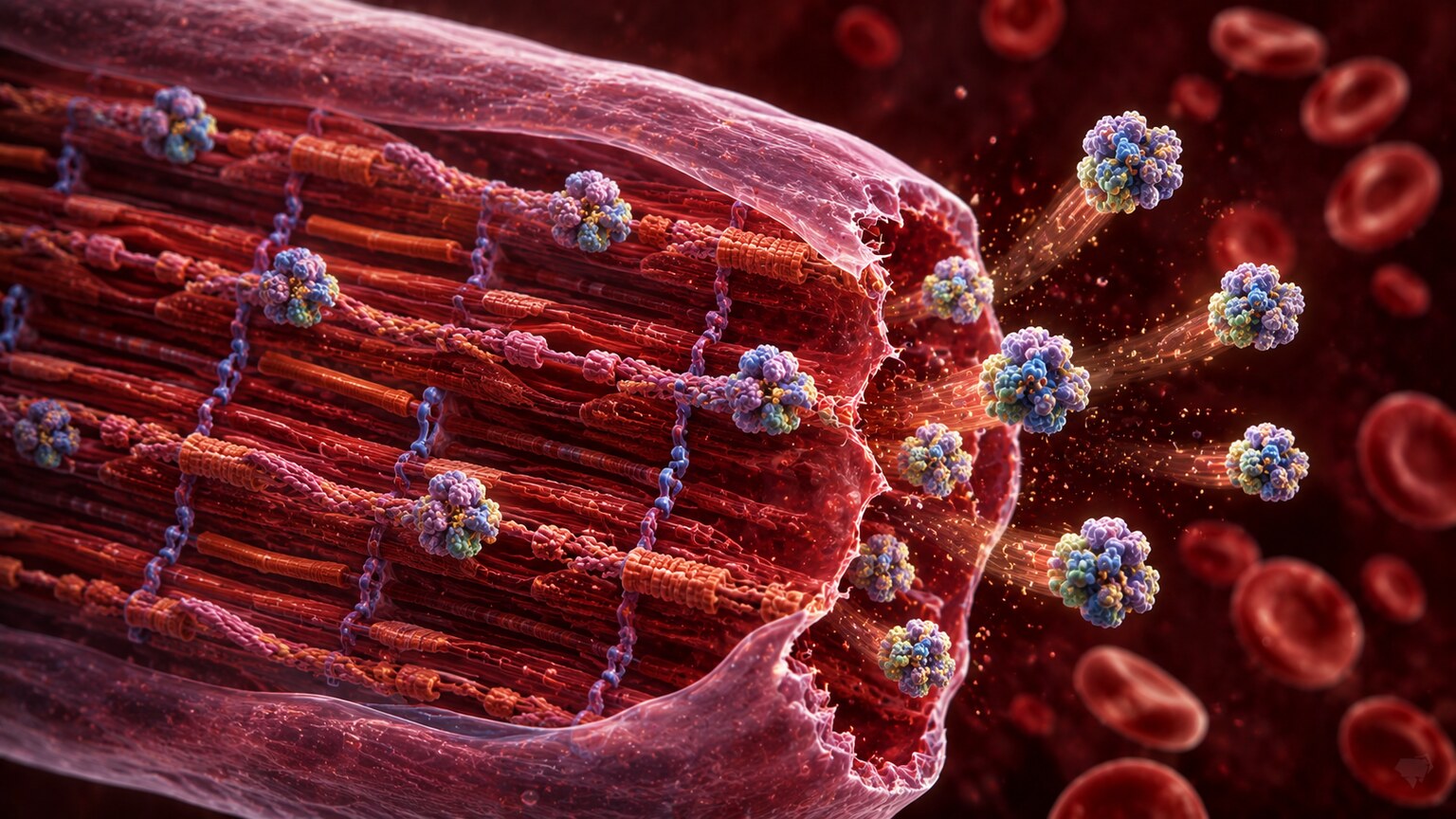
Troponin isn't a substance that floats freely in your blood under normal circumstances — it's a structural protein complex built directly into the contracting machinery inside heart muscle cells, where it helps regulate the interaction between two other proteins, actin and myosin, that slide against each other to generate every heartbeat. Because troponin is normally locked inside intact heart muscle cells rather than circulating in the bloodstream, healthy people have only an extremely small, often undetectable amount in their blood. The moment a heart muscle cell's outer membrane is damaged badly enough to become leaky or to rupture entirely, troponin escapes into the bloodstream, where a blood test can detect it — and modern high-sensitivity troponin assays are remarkably good at picking up even very small amounts of leakage. The critical thing to understand is that the test can't distinguish why the membrane was compromised. It reads the same whether the damage came from a blocked artery cutting off blood supply, or from an entirely different kind of stress that never involved a clot at all.
There are also two distinct forms of troponin measured clinically, troponin I and troponin T, and it's worth knowing they aren't interchangeable numbers with the same reference range — each is produced by a different gene and measured with a different assay, so a result has to be interpreted against the specific reference range for whichever version your lab actually tested. Both forms are considered equally reliable markers of heart muscle injury, and most hospitals settle on using one or the other consistently rather than switching between them, precisely so that serial results drawn from the same patient over hours or days can be compared directly on a stable, consistent scale. What both versions share, and what makes this entire article relevant, is that neither one is exclusive to blocked coronary arteries — both leak out under any of the mechanisms described below, since both are structurally part of the same contractile machinery inside every heart muscle cell, regardless of what specifically damaged it.
Kidney Disease: Reduced Clearance, Not Just Cardiac Injury
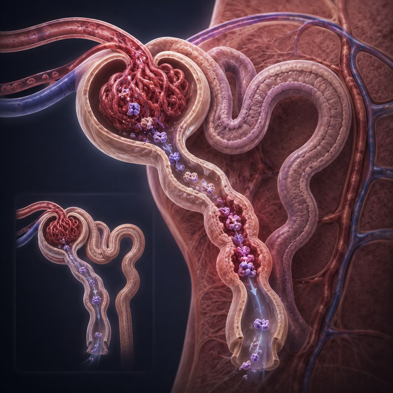
Chronic kidney disease is one of the most common reasons someone walks around with a persistently elevated troponin that has nothing to do with an active cardiac event. Part of how troponin normally gets cleared from the bloodstream after any release, large or small, is through the kidneys, which filter out and break down its smaller protein fragments. When kidney function is significantly reduced, that clearance pathway slows down considerably, and troponin fragments that would normally be filtered out within a day or two instead linger in circulation much longer, allowing baseline levels to sit chronically higher than they would in someone with healthy kidneys. This doesn't mean kidney disease is the only thing happening — people with advanced kidney disease also have real, elevated cardiovascular risk and genuinely can have heart attacks — but it does mean a single elevated troponin in someone with known kidney disease carries different weight than the same number in someone with normal kidney function, which is exactly why doctors interpret this result alongside a patient's known kidney status rather than in isolation.
Curious what the rest of your cardiac panel is telling you? Upload your results and get a complete, plain-language breakdown in under 15 minutes.
Analyze My ResultsSepsis and Critical Illness
Severe infection and sepsis, the body's sometimes life-threatening overreaction to infection, are well-documented causes of elevated troponin even when the heart's arteries are completely clear. Several mechanisms overlap here rather than acting alone. Sepsis frequently causes a mismatch between how much oxygen the heart muscle needs and how much it's actually receiving, since blood pressure often drops and circulation becomes less efficient during a severe infection, straining heart muscle cells even without any physical blockage. The inflammatory chemicals released throughout the body during sepsis, called cytokines, can also directly affect heart muscle cell membranes, making them more permeable and prone to leaking troponin. On top of that, sepsis frequently triggers a fast heart rate as the body tries to compensate for low blood pressure, and that increased workload itself adds further strain. Because sepsis is common in hospitalized patients, and because a troponin elevation in this setting is genuinely associated with worse overall outcomes, this particular cause is taken seriously — not necessarily as a sign of a heart attack requiring emergency cardiac catheterization, but as one more marker of how much physiological stress the body, including the heart, is currently under.
Pulmonary Embolism: When the Right Side of the Heart Is Under Strain
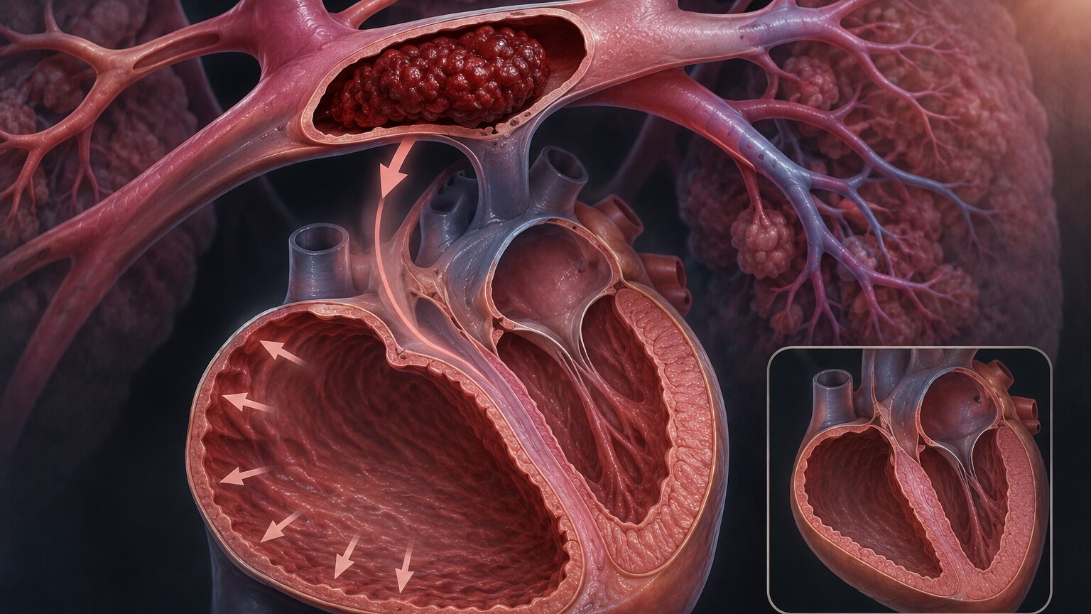
A pulmonary embolism happens when a blood clot, usually formed in a deep vein of the leg, travels through the bloodstream and lodges in one of the arteries supplying the lungs, blocking blood flow through that section of lung tissue. This creates a sudden, significant obstacle for the right side of the heart, whose entire job is pumping blood into the lungs — with a clot suddenly blocking part of that pathway, the right ventricle has to generate far more pressure than it's built for, straining and, in more severe cases, injuring that heart muscle directly. This right-heart strain is a genuinely distinct mechanism from a classic heart attack, which almost always originates on the left side of the heart from a blocked coronary artery, but it produces the same underlying event at the cellular level: stressed, injured heart muscle cells leaking troponin into the blood. In fact, an elevated troponin in someone with a confirmed pulmonary embolism is taken as a meaningful warning sign in its own right, since it indicates the right heart is being significantly strained by the clot, which changes how aggressively that specific case gets treated.
Myocarditis: Direct Inflammation of the Heart Muscle
Myocarditis is inflammation of the heart muscle itself, most often triggered by a viral infection, though it can also stem from autoimmune conditions or, less commonly, certain medications. Unlike a heart attack, where blood flow to a section of heart muscle is physically cut off by a blocked artery, myocarditis involves the immune system's own inflammatory response directly damaging heart muscle cells throughout the affected area, wherever the virus or immune activity happens to be concentrated. That direct cellular injury releases troponin just as effectively as an oxygen-starved heart attack does, and in some cases, myocarditis can produce troponin elevations that are difficult to distinguish from a heart attack based on the number alone. This is exactly why myocarditis is typically confirmed with additional testing beyond troponin — an MRI of the heart can reveal a pattern of inflammation and injury that looks meaningfully different from the pattern left behind by a blocked artery, and this distinction matters enormously for treatment, since myocarditis is managed very differently from a coronary blockage, often with supportive care and monitoring rather than a procedure to open an artery that was never actually blocked.
Fast, Abnormal Heart Rhythms
A sustained rapid heart rhythm, whether from atrial fibrillation, supraventricular tachycardia, or another arrhythmia, can raise troponin through what's sometimes called a supply-demand mismatch, even in someone whose coronary arteries are entirely healthy. When the heart beats extremely fast for a prolonged period, two things work against it simultaneously: the heart muscle's own oxygen demand rises considerably because it's contracting far more often than usual, while at the same time, the heart's own blood supply — which flows into the coronary arteries specifically during the brief relaxation phase between beats — actually decreases, because that relaxation phase gets shorter and shorter as the heart rate climbs. The heart muscle can end up genuinely oxygen-starved during a severe, sustained episode, even without a single blocked vessel anywhere. This is why a person who arrives at an emergency room with a very fast, sustained abnormal heart rhythm sometimes shows a mildly elevated troponin as part of that presentation, and why getting the heart rate back under control is often followed by the troponin gradually settling back down on its own, without any artery ever needing to be addressed.
Strenuous Exercise: A Temporary, Usually Benign Elevation

Perhaps the most counterintuitive cause on this list is intense, prolonged physical exercise. Studies of marathon runners, ultra-endurance athletes, and even healthy recreational athletes doing sustained hard efforts have repeatedly found measurable troponin elevations afterward, sometimes reaching levels that would otherwise prompt real concern in a clinical setting, and this occurs in a meaningful proportion of otherwise healthy competitors, not just a rare outlier or two. The exact mechanism isn't completely settled, but the leading explanations include temporary, reversible increases in the permeability of heart muscle cell membranes under extreme physical stress, small amounts of genuinely reversible microscopic cellular stress, and the sheer mechanical demand placed on the heart during hours of sustained maximal effort. Reassuringly, this kind of exercise-induced elevation typically peaks within a few hours of finishing and returns to baseline within a day or two, without any evidence of lasting heart damage, which is well-documented across a large body of research following recreational and competitive athletes over time. This is exactly why context matters so much when interpreting a troponin result — the same number found in a marathon runner an hour after finishing a race carries a very different meaning than the same number found in someone with chest pain and no recent exercise.
This exercise-related pattern has practical implications beyond athletes curious about their own lab work. Emergency departments occasionally see patients who went for a hard run or played a demanding sport shortly before developing unrelated symptoms, and a treating physician has to weigh how much of a modestly elevated troponin might reasonably be attributed to that recent exertion versus how much genuinely warrants further cardiac workup. Recent exercise history is exactly the kind of detail worth mentioning to a doctor if you're being evaluated for any reason and troponin gets checked — not to talk your way out of appropriate testing, but because it's a real, relevant piece of context that shapes how a borderline result gets interpreted alongside everything else.
Chemotherapy and Certain Cancer Treatments
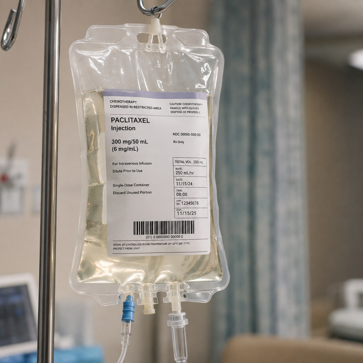
Certain chemotherapy drugs, most notably a class called anthracyclines, along with some targeted cancer therapies, carry a well-documented risk of direct toxicity to heart muscle cells as a side effect of how they work. Because troponin is such a sensitive marker of heart muscle injury, oncology teams often check it before, during, and after these specific treatments as an early warning system, looking for a rising trend that might indicate developing cardiotoxicity well before a person develops noticeable symptoms like shortness of breath or fatigue. Catching this early genuinely changes management, since a treatment plan can sometimes be adjusted, a cardioprotective medication added, or a patient referred to a cardio-oncology specialist for closer monitoring, all aimed at preventing the kind of heart muscle damage that can otherwise become permanent if the toxicity isn't recognized until it's more advanced. This is one of the more proactive, planned uses of troponin testing described in this article — not a value checked in response to symptoms, but one checked routinely as a monitoring tool precisely because the underlying treatment is a known, predictable risk to heart muscle.
Radiation therapy directed at or near the chest, sometimes used for breast cancer, lymphoma, or lung cancer, carries a related but separate risk to heart tissue that oncology teams monitor in much the same way. Unlike the relatively rapid cellular toxicity some chemotherapy drugs cause, radiation-related heart damage tends to develop far more gradually, sometimes not becoming clinically apparent until years after treatment has finished, affecting the heart's blood vessels, valves, and the fibrous sac surrounding the heart in addition to the muscle itself. Because of this delayed timeline, troponin monitoring in this context is often just one part of a broader, long-term cardiac surveillance plan that continues well past the end of active cancer treatment, alongside periodic imaging and cardiology follow-up, reflecting how differently a slow-developing radiation effect needs to be tracked compared to the more immediate cellular toxicity of certain chemotherapy drugs.
Severe Neurological Events
A significant stroke, particularly one affecting certain areas of the brain, or a large hemorrhage inside the skull, can also raise troponin through a genuinely fascinating brain-heart connection called neurogenic myocardial stunning. Severe injury to certain brain regions triggers a sudden, massive surge of stress hormones, including adrenaline, which can directly overstimulate heart muscle cells to the point of temporary dysfunction and injury, even without any blockage in the coronary arteries at all. This same mechanism is closely related to Takotsubo cardiomyopathy, sometimes called broken heart syndrome, where an intense emotional or physical stressor triggers a similar surge and produces a temporary, distinctive pattern of heart muscle weakness, usually with a real but often modest troponin elevation. In both situations, the heart muscle is a genuine bystander reacting to an extreme hormonal surge triggered elsewhere in the body, rather than the primary site of the original problem, which is an important distinction for a care team managing a patient whose main emergency is neurological, not cardiac, even though the troponin result might otherwise suggest a cardiac issue on its own.
Recognizing this connection matters practically, because a stroke or major brain hemorrhage patient with an incidentally elevated troponin doesn't necessarily need the same urgent cardiac catheterization pathway a classic heart attack patient would — pursuing that pathway aggressively in someone whose real emergency is bleeding inside the skull could actually cause harm, particularly since the blood-thinning medications used to treat a heart attack are often the last thing a brain hemorrhage patient should receive. This is a clear, high-stakes example of why understanding the full range of non-cardiac troponin causes isn't just an academic exercise — in situations like this, correctly attributing an elevated troponin to the actual underlying cause can directly shape which treatments are safe to give and which ones could make a life-threatening situation worse.
Why Doctors Focus on the Trend, Not Just One Number
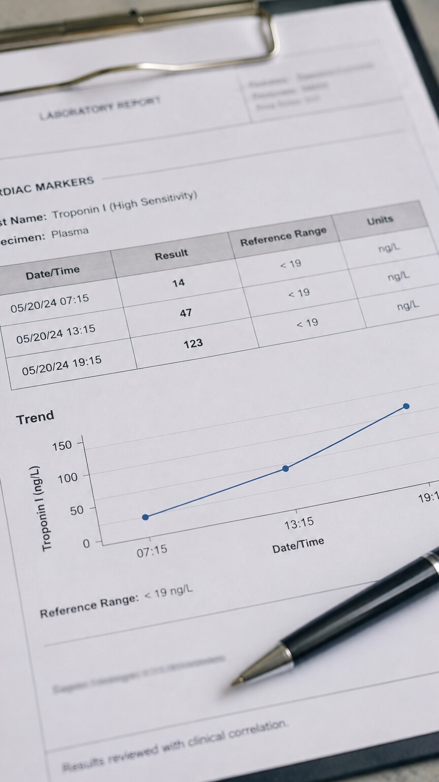
Given how many different processes can raise troponin, a single measurement rarely tells the whole story on its own, which is exactly why serial testing — drawing troponin two or three times over several hours — has become the standard approach in most emergency and hospital settings. A classic heart attack tends to produce a distinctive rise-and-fall pattern over those hours, climbing from an initial value and continuing to climb on a repeat draw before eventually plateauing and slowly declining over the following days. A chronically elevated but stable troponin, like the kind seen in someone with kidney disease, tends to stay relatively flat across repeat measurements, drawn hours apart, without that same dynamic rise. This pattern — rising, falling, or flat — often carries more diagnostic weight than the absolute number itself, since it helps separate an acute, ongoing injury from a chronic, stable elevation that reflects an underlying condition like reduced kidney clearance rather than a new, active event. High-sensitivity troponin assays, now standard in most hospitals, have made this trend-based approach even more precise, since they can detect meaningful changes between two measurements that older, less sensitive tests would have missed entirely.
How a Doctor Actually Sorts Through All of This
Faced with an elevated troponin, a doctor doesn't work through this list of causes at random — the patient's full clinical picture does most of the narrowing before the lab result is even considered in isolation. Someone with crushing chest pain, changes on an EKG consistent with reduced blood flow, and risk factors like smoking or high cholesterol points strongly toward a classic heart attack. Someone with a known history of kidney disease and a stable, unchanging troponin across repeat draws, with no chest pain or EKG changes, points toward chronic reduced clearance instead. Someone recently diagnosed with a blood clot in the leg who suddenly develops shortness of breath and a new troponin elevation raises concern for pulmonary embolism with right heart strain. In nearly every case, troponin is interpreted as one piece of a larger clinical puzzle — symptoms, EKG findings, imaging, and known medical history — rather than a stand-alone verdict, which is exactly why the same elevated number can lead to completely different next steps depending on everything else happening around it.
Age and sex also factor into how a given troponin value gets read, since high-sensitivity assays have revealed genuine, measurable differences in baseline levels across different groups of otherwise healthy people. Men tend to run modestly higher baseline troponin levels than women on average, and baseline levels generally trend upward somewhat with age, likely reflecting the same kind of low-level, cumulative cardiac wear-and-tear that shows up in other markers of cardiovascular health over a lifetime. Some laboratories and hospital systems have begun adopting sex-specific reference ranges for this exact reason, aiming to avoid either under-flagging a genuinely concerning result in a woman whose baseline runs lower, or over-flagging a mildly elevated but ultimately unremarkable result in an older man whose baseline naturally runs a bit higher. This is one more layer of context — alongside kidney function, recent exercise, and known cardiac history — that a lab or clinician reasonably factors in before treating any single troponin number as unambiguous on its own.
Formal clinical guidelines actually codify a version of this reasoning into what's often called the "fourth universal definition of myocardial infarction," a framework cardiology societies use specifically to distinguish a true heart attack from the broader category of troponin elevation from other causes, sometimes labeled "myocardial injury" when no blocked artery is involved. Under this framework, a true heart attack diagnosis requires not just an elevated or rising troponin, but also at least one additional piece of supporting evidence — symptoms consistent with reduced blood flow, specific EKG changes, imaging showing new loss of viable heart muscle, or direct evidence of a clot in a coronary artery found during a procedure. Troponin elevation without any of those additional findings is instead classified as myocardial injury, a broader and less specific category that still deserves attention and often further workup, but doesn't carry the same automatic diagnosis or treatment pathway as a true heart attack. This formal distinction is precisely why a rising troponin alone was never meant to be read as an automatic heart attack diagnosis, even inside a hospital using the most sensitive assays available.
Heart Failure and Chronic Structural Heart Disease
People living with chronic heart failure, where the heart's pumping ability is already reduced, frequently show mildly elevated troponin even at their stable baseline, entirely separate from any new event. Ongoing wall stress on an enlarged, overworked heart chamber, combined with the persistently reduced blood flow that comes with a weaker pump, appears to cause a slow, low-level trickle of troponin release over time rather than the sharp, dramatic spike seen with an acute blockage. This baseline elevation is well recognized in cardiology, and it's actually used constructively: research has found that higher baseline troponin levels in people with chronic heart failure are associated with a greater risk of future hospitalization and worse long-term outcomes, making it a useful piece of risk information for a cardiologist managing that condition over months and years, rather than a signal of a new, acute problem requiring urgent intervention every time it's checked.
Cardiac Procedures and Recent Cardiac Surgery
Any procedure that involves physically manipulating heart tissue is going to cause some degree of troponin release as a direct, expected consequence, not a complication. Cardiac surgery, including bypass grafting and valve repair, inevitably involves handling heart muscle directly, and some cellular injury during that process is unavoidable — troponin levels after these procedures are routinely elevated, sometimes substantially, and interpreting them requires an entirely different reference framework than in someone who hasn't just had heart surgery. Cardiac catheterization procedures, including angioplasty to open a blocked artery, and cardioversion, a controlled electrical shock used to correct certain abnormal heart rhythms, can also cause a modest, expected troponin bump afterward. In every one of these situations, the clinical team already anticipates the rise and interprets any post-procedure troponin against that expected pattern, rather than against the same reference range used for someone walking into an emergency room with new chest pain and no recent cardiac intervention.
Frequently Asked Questions
Can troponin be elevated without any heart attack at all?
Yes. Kidney disease, sepsis, pulmonary embolism, myocarditis, fast abnormal heart rhythms, strenuous exercise, certain chemotherapy drugs, and severe neurological events can all raise troponin through mechanisms that don't involve a blocked coronary artery.
Why does kidney disease cause chronically high troponin?
Troponin is partly cleared from the blood through the kidneys. When kidney function is reduced, that clearance slows down, allowing troponin fragments to linger in circulation longer and baseline levels to sit persistently higher than in someone with healthy kidneys.
Is it dangerous if troponin rises after intense exercise like a marathon?
Research on endurance athletes has generally found these elevations to be temporary and not associated with lasting heart damage, typically returning to baseline within a day or two. Context matters enormously in interpreting this kind of result.
Why do doctors check troponin more than once instead of relying on a single result?
The pattern across repeat measurements — rising, falling, or staying flat — often reveals more than a single number. A classic heart attack typically shows a dynamic rise-and-fall pattern, while a chronic condition like kidney disease tends to produce a stable, unchanging elevation.
How can a doctor tell if an elevated troponin is from a heart attack or something else?
Symptoms, EKG findings, the trend across repeat measurements, and known medical history all factor in together. Troponin is interpreted as one piece of a broader clinical picture rather than a stand-alone diagnosis.
Conclusion
Troponin's sensitivity is exactly what makes it such a valuable test, and exactly what makes it prone to misinterpretation when read in isolation. It detects heart muscle cell injury with remarkable precision, but it was never designed to identify the cause of that injury on its own — a blocked artery, a strained right ventricle from a clot in the lungs, a kidney unable to clear it efficiently, or hours of sustained hard effort during a race can all leave the same fingerprint on this one number. Understanding that range of possibilities doesn't make an elevated troponin any less worth taking seriously — even when the eventual explanation turns out to be non-cardiac, that conclusion should come from a doctor's evaluation, not from self-diagnosis based on this article. It does explain why a doctor gathers symptoms, EKG findings, medical history, and repeat measurements before drawing any conclusion — because the number alone was never meant to tell the whole story.
Still Not Sure What Your Results Mean?
Upload your labs and get a complete, visual, plain-language interpretation of every biomarker — delivered to your inbox in under 15 minutes.
Get My ReportThis article is for educational purposes only and does not constitute medical advice. Always consult your healthcare provider regarding your specific lab results.