
What Does a Very High Troponin Level Indicate?
A very high troponin level means your heart muscle has released a large amount of a specific protein that's only supposed to be locked inside heart muscle cells — and the fact that it's circulating in your blood, especially at a high concentration, means those cells have been significantly damaged or destroyed. Troponin isn't like most lab values, where "high" and "very high" sit on a smooth, gradually more concerning scale. It behaves more like an alarm that's either quiet or extremely loud: a small amount of troponin leaking into the blood can happen for relatively minor, even non-cardiac reasons, but a very high level — many times above the upper limit of normal — almost always reflects a substantial amount of heart muscle injury happening right now or very recently. This article is written for someone looking at a very high number, not a borderline one, and it walks through what actually causes a level that high, why the size of the number matters clinically, and — because this is a topic where timing can be life-saving — why this specific finding is treated as a medical emergency rather than something to research calmly at home before deciding what to do next.
If you are looking at this because you or someone near you currently has chest pain, pressure, shortness of breath, sweating, or sudden severe weakness alongside a very high troponin result, do not wait to finish reading — call 911 or your local emergency number, or go to the nearest emergency department immediately.
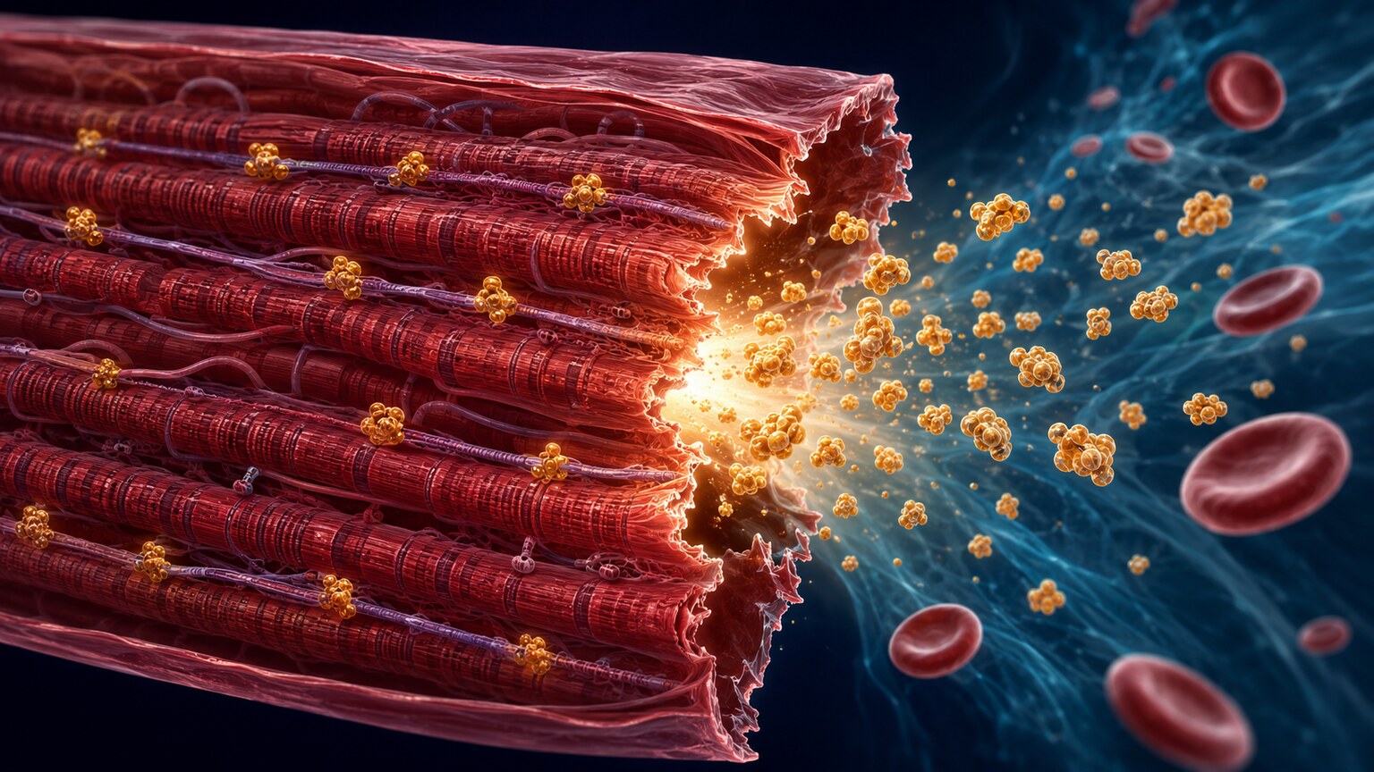
Figure 1. When heart muscle cells are injured badly enough that their outer membrane breaks down, troponin — normally locked inside the cell's contractile machinery — spills directly into the bloodstream.
What Troponin Actually Is and Why It Leaks Into Blood at All
To understand why a very high number is so meaningful, it helps to know what troponin is actually doing inside your heart under normal circumstances. Troponin isn't a hormone or a waste product — it's a structural protein, part of the machinery inside heart muscle cells (cardiac myocytes) that makes them contract. Specifically, it's part of a protein complex sitting along the muscle fibers that controls how calcium triggers each heartbeat's squeeze. Because this protein is so specific to heart muscle — skeletal muscle uses closely related but distinct versions — a lab measuring "cardiac troponin," usually troponin I or troponin T depending on which assay a hospital uses, is measuring something that has essentially no reason to be in your bloodstream unless heart muscle cells have been damaged.
Under healthy conditions, troponin stays locked inside intact heart cells, doing its structural job, invisible to a blood test. It only escapes into circulation when a cell's outer membrane is damaged badly enough to become "leaky" or when the cell dies outright and ruptures, spilling its contents — troponin included — into the surrounding tissue and eventually into the bloodstream, where a blood draw can detect it. Modern lab tests use what's called a high-sensitivity troponin assay, sensitive enough to detect even very small amounts of troponin that a small number of healthy people have circulating at baseline, which is part of why interpreting a troponin result always involves comparing it to a specific reference cutoff (called the 99th percentile upper reference limit) rather than just asking whether any troponin at all is present.
It helps to picture the heart muscle at the microscopic level to understand why this release happens in the first place. Each heart muscle cell is packed with long, repeating protein filaments arranged in a precise, overlapping pattern that slides past itself to generate the mechanical force of a heartbeat. Troponin sits at a specific, critical junction in that filament — it's the molecular switch that responds to calcium and tells the filaments when to grip and slide. This tight structural integration is exactly why troponin makes such a reliable signal of injury: it isn't floating loosely in the cell where minor stress might dislodge a little of it by accident. It's woven into the contractile machinery itself, which means it only escapes in meaningful amounts when that machinery, and the cell membrane holding it in place, has actually been compromised — not simply irritated or mildly stressed. This same tight integration also explains why two closely related versions of the protein, troponin I and troponin T, are both used clinically: they're structurally distinct enough to be measured by separate, highly specific antibody-based assays, but they're released together whenever cardiac muscle cells are damaged, since they're both part of the same troponin complex sitting at the same site inside the same cells.
What Counts as "Very High" — Understanding the Numbers
Troponin results are reported as a concentration, often in nanograms per liter (ng/L) for high-sensitivity assays, and what counts as elevated depends on the specific assay a lab uses — there isn't one universal number that applies to every hospital. What matters more than the raw number is how far above that assay's own upper reference limit the result sits, since that ratio is what actually correlates with the extent of injury. A mildly elevated troponin — say, one to three times the upper limit — can come from a wide range of causes, some cardiac and some not, including things like kidney disease, an irregular heart rhythm, or even intense exercise. A very high troponin, by contrast — commonly described as being ten, twenty, or even fifty times or more above that upper limit — narrows the list of realistic explanations considerably, because reaching that magnitude generally requires a substantial number of heart muscle cells to be damaged or dying at once, not just a handful under mild stress.
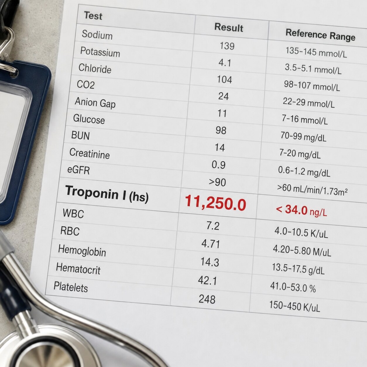
Figure 2. A troponin result reported many times above the assay's upper reference limit reflects the extent of heart muscle injury, not just its presence.
This is one of the reasons troponin is described by clinicians as having a roughly dose-dependent relationship with the size of the injury: in the context of a heart attack, for instance, a small, localized area of damage tends to produce a peak troponin that's meaningfully lower than a large territory of heart muscle affected by a major blockage. That relationship isn't perfectly linear or exact enough to precisely calculate infarct size from a single number, but the general pattern — bigger injury, bigger number — holds up well enough that emergency physicians and cardiologists use the magnitude of the result, alongside symptoms and an ECG, to gauge urgency and severity in real time, well before more detailed imaging is available.
It's also worth understanding why a mildly elevated troponin doesn't automatically mean a milder version of the same problem that causes a very high one. A small rise can come from mechanisms that don't actually involve dying heart cells at all — for example, a heart working under significant strain, such as during a severe infection, a fast abnormal heart rhythm, or advanced kidney disease that slows how quickly troponin is cleared from the blood, can produce a modestly elevated result through cell stress or altered clearance rather than outright cell death. A very high result effectively rules most of those milder mechanisms out on magnitude alone, since none of them tend to push troponin anywhere near the range produced by widespread cell death. This distinction is part of why clinicians resist treating troponin as a single continuous dial from "fine" to "very bad" — it behaves more like two different signals occupying the same scale, one reflecting reversible strain and one reflecting irreversible cellular injury, and the size of the number is one of the clearest clues for telling which one is actually being measured.
The Most Common Cause — A Large Heart Attack
The single most common reason for a very high troponin, particularly in someone presenting with chest pain, is a heart attack (myocardial infarction) involving a substantial amount of heart muscle — most often caused by a blood clot suddenly and completely blocking one of the coronary arteries that supplies oxygen-rich blood to the heart. When that blood supply is cut off, the heart muscle downstream of the blockage begins to die from lack of oxygen within minutes, and the longer the blockage persists, the more cells are affected and the higher the eventual troponin peak tends to be. This is exactly why "time is muscle" is such a central phrase in emergency cardiology — restoring blood flow quickly, through a procedure to open the blocked artery or medication to dissolve the clot, directly limits how much heart muscle ends up permanently damaged, which is reflected in a lower troponin peak in people treated faster.

Figure 3. Crushing chest pressure, pain radiating to the arm or jaw, shortness of breath, and sweating alongside a very high troponin are treated as a heart attack until proven otherwise.
The scale of a coronary blockage generally correlates with how large the affected artery is and how much heart muscle it supplies — a complete blockage near the start of the left anterior descending artery, which feeds a large portion of the heart's main pumping chamber, tends to produce one of the largest and highest-peaking troponin elevations seen in clinical practice, precisely because so much muscle depends on that single vessel. This is also why the classic warning signs — crushing or pressure-like chest pain, pain spreading to the left arm, jaw, or back, shortness of breath, sweating, nausea, or a sense of impending doom — are taken so seriously when they accompany a very high troponin. The combination of severe symptoms and a markedly elevated result is treated as a heart attack until proven otherwise, triggering an urgent pathway toward an ECG and, very often, an emergency procedure to locate and open the blocked artery as quickly as possible.
It's worth noting that not every large heart attack announces itself with textbook symptoms. Women, older adults, and people with diabetes are all somewhat more likely to experience what's sometimes called an atypical presentation — fatigue, nausea, upper abdominal discomfort, or a vague sense of unwellness instead of classic crushing chest pain — even when the underlying blockage and the resulting troponin elevation are just as severe as in a textbook case. This is one of the reasons emergency clinicians take a very high troponin seriously as a standalone finding, even when the accompanying symptoms don't perfectly match the pattern most people associate with a heart attack from television or common knowledge. The lab result itself is treated as strong evidence of significant heart muscle injury regardless of how classic or unusual the symptom picture happens to be, which protects against the specific failure mode of a real heart attack being dismissed simply because it didn't look the way it's "supposed" to.
Curious what the rest of your cardiac panel is telling you? Upload your results and get a complete, plain-language breakdown in under 15 minutes.
Analyze My ResultsMyocarditis — When Very High Troponin Isn't From a Blocked Artery
One of the more surprising facts about troponin is that a heart attack from a blocked artery isn't the only condition capable of producing an extremely high result — in some cases, it isn't even the cause that produces the single highest numbers. Myocarditis, an inflammation of the heart muscle most often triggered by a viral infection (though it can also follow certain autoimmune conditions or, rarely, certain medications), can cause widespread, diffuse damage to heart muscle cells across the heart rather than damage limited to the territory fed by one blocked artery. Because inflammation can affect a broad area simultaneously, some cases of myocarditis produce troponin elevations that rival or even exceed those seen in a large heart attack, despite there being no blockage in the coronary arteries at all.

Figure 4. In myocarditis, immune cells responding to a viral infection infiltrate heart muscle tissue directly, damaging cells across a broad area rather than one blocked artery's territory.
This matters clinically because myocarditis tends to strike a different population than a typical large heart attack — often younger people, sometimes in their teens, twenties, or thirties, frequently in the days to weeks following a viral illness like a cold, flu, or gastrointestinal bug, and sometimes following COVID-19 infection or, in rare cases, certain vaccinations. Someone with myocarditis may describe chest pain that feels different from classic heart attack pain — sometimes sharper, sometimes worsened by lying flat or breathing deeply — alongside fatigue, fever, or a recent history of feeling unwell. Because myocarditis and a large heart attack can look genuinely similar on an initial troponin result, distinguishing between them requires more than the number alone: an ECG, an echocardiogram to look at how the heart is pumping, and often a coronary angiogram to directly check whether the arteries are actually blocked (myocarditis patients typically have clean arteries) are all part of sorting out which of these two very different conditions is actually responsible for the same striking lab result.
Cardiac MRI has become an especially valuable tool in this specific situation, since it can visualize patterns of inflammation and scarring within the heart muscle wall itself with a level of detail an angiogram or standard echocardiogram can't provide. Myocarditis tends to produce a characteristic pattern of injury sitting toward the outer layer of the heart muscle wall, scattered in patches rather than following the clean, wedge-shaped territory that a blocked artery produces — a distinction that can be difficult to appreciate from symptoms or blood work alone but becomes much clearer once the heart muscle itself is directly imaged. This is one of the reasons a young, previously healthy person presenting with a very high troponin and a recent viral illness will often go on to have a cardiac MRI even after a normal angiogram has already ruled out a blocked artery, since confirming myocarditis specifically — rather than simply ruling out a heart attack — changes what kind of monitoring and activity restrictions are recommended during recovery.
Other Serious Causes of an Extremely High Troponin
Beyond a large heart attack and myocarditis, several other conditions are capable of producing a markedly elevated troponin, each through a different mechanism of injuring heart muscle broadly or acutely rather than through a single blocked vessel.

Figure 5. Cardiac arrest and the resuscitation that follows both place major, whole-heart demand on the heart muscle, which is part of why troponin can rise dramatically afterward regardless of the original cause.
Cardiac arrest — the heart suddenly stopping effective pumping — and the resuscitation that follows it can independently drive troponin to very high levels, both because whatever originally caused the arrest may have involved heart muscle injury and because the arrest itself, along with chest compressions and defibrillation shocks used to restart the heart, places significant mechanical and metabolic stress directly on heart muscle cells. Takotsubo cardiomyopathy, sometimes called "broken heart syndrome," is another notable cause: triggered by an intense emotional or physical stressor — the sudden loss of a loved one, a car accident, even a joyful shock — it produces a temporary, severe weakening of part of the heart muscle's pumping ability, along with a troponin elevation that can look strikingly similar to a heart attack despite, again, having no blocked artery behind it. Severe sepsis, a massive pulmonary embolism placing sudden strain on the heart's right side, direct trauma to the chest from an accident, and dangerously fast or irregular heart rhythms sustained for a long period can all independently push troponin into a very high range as well, each by placing the heart under a degree of stress or direct injury severe enough to damage muscle cells broadly.
Takotsubo cardiomyopathy deserves a slightly closer look, since its mechanism is genuinely unusual among the causes on this list. The leading theory is that a sudden, overwhelming surge of stress hormones — adrenaline and related catecholamines — temporarily stuns and weakens heart muscle cells, particularly toward the tip of the heart's main pumping chamber, producing a distinctive ballooned shape on imaging that gives the condition its name (borrowed from a Japanese octopus trap the shape resembles). Unlike a heart attack, where the affected tissue can suffer permanent, irreversible damage if blood flow isn't restored quickly, the heart muscle weakness in Takotsubo cardiomyopathy is usually temporary, often improving substantially over the following weeks as the stunning effect resolves — even though the troponin elevation and the initial symptoms can look every bit as alarming as a major heart attack in the moment. This is precisely why the condition still requires full emergency evaluation despite its generally more favorable long-term trajectory: there is no reliable way to distinguish it from a genuine heart attack based on symptoms and an initial troponin result alone, and the two conditions require different immediate management.
A massive pulmonary embolism — a large blood clot blocking blood flow through the lungs — raises troponin through yet another distinct mechanism worth understanding on its own terms. When a large clot suddenly obstructs blood flow to the lungs, the heart's right side, which normally pumps against relatively low resistance, is forced to work against a sudden, severe increase in pressure. This acute strain can injure the right ventricle's muscle directly, releasing troponin even though the coronary arteries themselves remain completely open and undamaged. A troponin elevation in this context is taken seriously not just as evidence of heart strain but as a marker that helps identify which pulmonary embolism patients are at higher risk of a more complicated course, directly influencing how aggressively the clot itself needs to be treated.
Direct chest trauma and sustained dangerous heart rhythms round out the list through more mechanically straightforward routes. A significant blunt-force injury to the chest — from a car accident, a serious fall, or a direct blow during a contact sport — can bruise or otherwise physically damage heart muscle cells in a condition called cardiac contusion, releasing troponin in a pattern that generally mirrors the location and severity of the impact rather than any pre-existing heart condition. A very fast, sustained abnormal heart rhythm, particularly one that persists for many hours without treatment, can raise troponin by outpacing the heart muscle's oxygen supply during the rhythm disturbance itself, essentially producing a small-scale version of the same oxygen-starvation injury seen in a heart attack, even though no artery is actually blocked. In both situations, the troponin elevation serves the same broad clinical purpose it does throughout this article: flagging that heart muscle cells have been meaningfully injured and that the underlying cause needs urgent identification and treatment, whatever that cause turns out to be.
Why Doctors Repeat the Test — The Trend Matters as Much as the Number
A single troponin result, no matter how high, is rarely the entire story. Because troponin takes time to rise after heart muscle injury begins — typically becoming detectable within a few hours and continuing to climb for many hours afterward — clinicians almost always order repeat troponin measurements spaced a few hours apart, looking specifically at whether the level is still rising, has plateaued, or is already starting to fall.

Figure 6. Repeat troponin draws spaced a few hours apart let clinicians see whether the level is still climbing, has peaked, or is beginning to fall — information a single result can't provide.
A rising trend on repeat testing, even from an already-high starting point, generally indicates that heart muscle injury is still actively progressing — which is part of why timely treatment to restore blood flow, when a blocked artery is the cause, is so urgent even after an initial very high result has already been recorded, since the eventual peak, and the amount of permanent damage it reflects, is still being determined by what happens next. A level that has already peaked and is falling by the time of the second or third measurement generally means the acute injury has stopped progressing and the body has moved into the recovery and healing phase, though "recovering" doesn't mean the damage that already occurred has been undone — troponin measures ongoing cellular injury, not tissue repair. This trend, combined with the ECG pattern, symptoms, and imaging findings, is what ultimately shapes the diagnosis and treatment plan far more precisely than any single number in isolation ever could.
The shape of the curve over time also carries diagnostic information beyond simply "rising" or "falling." A large heart attack tends to produce troponin that rises sharply, peaks within roughly 12 to 24 hours, and then declines gradually over several days as the body clears the released protein from circulation. Myocarditis, by contrast, often produces a somewhat flatter, more prolonged elevation, since ongoing inflammation can continue releasing smaller amounts of troponin over a longer stretch of time rather than everything being released in one concentrated burst. Clinicians familiar with these general patterns sometimes use the overall shape of the troponin trend, not just its peak value, as one more piece of evidence when the rest of the clinical picture is still ambiguous — though, as with the initial number itself, the trend is always interpreted alongside imaging and clinical findings rather than treated as a diagnosis on its own.
Is a Very High Troponin Always a Medical Emergency?
In the overwhelming majority of real-world situations, yes. Because the list of realistic explanations for a very high troponin is dominated by conditions that are actively dangerous — a large heart attack, myocarditis, cardiac arrest, Takotsubo cardiomyopathy, a massive pulmonary embolism — a result in this range discovered in an emergency department or hospital setting is treated as urgent by default, and appropriately so, while the specific cause is being sorted out through ECG, imaging, and clinical evaluation. This is different from a mildly elevated troponin, which sometimes turns out to reflect a chronic, stable condition like longstanding kidney disease and can occasionally be managed with a more measured, outpatient-paced evaluation depending on the full clinical picture. A very high result essentially never falls into that more relaxed category — the magnitude itself is a signal that something significant is actively happening to the heart muscle right now or very recently, and it deserves the same urgency as new, severe chest pain, regardless of how the person happens to be feeling at the exact moment the result comes back.
This is also why a very high troponin discovered incidentally — say, on bloodwork drawn for an unrelated reason, or through a home lab test ordered out of general curiosity rather than in response to symptoms — should never be treated as something to schedule a routine follow-up appointment for later in the week. The absence of dramatic symptoms at the exact moment a very high result is reviewed doesn't lower how urgently it needs to be acted on, since, as covered above, some of the most serious underlying causes can produce a result this high with symptoms that are mild, atypical, or have already partly resolved on their own. Anyone who receives a very high troponin result outside of a hospital setting — through outside lab work, a research study, or any other channel — should treat it the same way as a new episode of severe chest pain: as a reason to seek emergency evaluation immediately, not a reason to wait for a scheduled appointment.
What Happens After a Very High Result — What to Expect
For anyone facing this situation, or trying to understand what a family member is going through, it can help to know the general shape of what typically follows a very high troponin result in a hospital setting. An electrocardiogram (ECG) is usually done immediately, since certain patterns on it can identify a specific type of heart attack (called a STEMI) that requires an emergency procedure within minutes, not hours. An echocardiogram, an ultrasound of the heart, is commonly used to see how well the heart is pumping overall and whether specific areas are moving weakly, which can help distinguish a focal injury (suggesting a blocked artery) from a more diffuse pattern (suggesting myocarditis or Takotsubo). Depending on what the ECG and echocardiogram show, a coronary angiogram — a procedure that directly images the heart's arteries using a thin catheter and contrast dye — is often performed to look for and, if found, immediately treat a blockage. Blood is typically redrawn every few hours to track the troponin trend described earlier, and depending on the eventual diagnosis, treatment may range from medications and close monitoring to an emergency procedure to open a blocked artery, with a hospital stay for observation and recovery almost always part of the picture given the seriousness of the possible underlying causes.
Recovery expectations vary considerably depending on the underlying cause, which is part of why getting a clear diagnosis matters well beyond the initial emergency. Someone recovering from a large heart attack will typically be started on a specific combination of heart-protective medications, referred to a cardiac rehabilitation program to safely rebuild activity tolerance, and given clear guidance on risk-factor changes — blood pressure, cholesterol, blood sugar, smoking — aimed at preventing a second event. Someone diagnosed with myocarditis instead is generally advised to avoid strenuous exercise for a period of weeks to months while the heart muscle heals, since intense exertion during active inflammation has been linked to a higher risk of dangerous heart rhythms, with a repeat echocardiogram or cardiac MRI often used later to confirm the heart's pumping function has returned to normal before activity restrictions are lifted. Someone who experienced Takotsubo cardiomyopathy is usually followed with repeat imaging over the following weeks, since the temporary weakening it causes generally resolves on its own, though a small number of people go on to have recurrent episodes later in life. In every case, the specific cause identified during that initial hospital stay is what determines the entire shape of the recovery plan that follows.
Frequently Asked Questions
How high does troponin need to be before it's considered "very high"?
There's no single universal cutoff, since it depends on the specific assay a lab uses, but clinicians generally consider a result ten times or more above that assay's upper reference limit to be markedly elevated, with results reaching twenty, fifty, or more times the limit reflecting particularly extensive heart muscle injury. The exact ratio matters more than the raw number, since different hospitals and assays report different scales.
Can a very high troponin happen without a blocked artery?
Yes. Myocarditis, Takotsubo cardiomyopathy, cardiac arrest and resuscitation, severe sepsis, a massive pulmonary embolism, and direct chest trauma can all produce very high troponin levels without any coronary artery blockage at all. Sorting out which of these is responsible generally requires an ECG, echocardiogram, and often a coronary angiogram to directly check the arteries.
Does a higher troponin number mean a worse long-term outcome?
Generally, yes, though it isn't the only factor. Higher peak troponin levels are statistically associated with larger amounts of heart muscle injury and, on average, with a somewhat higher risk of complications like heart failure afterward. That said, outcomes also depend heavily on how quickly treatment was received, the specific underlying cause, and a person's overall health, so the number alone doesn't determine the outcome for any individual person.
Why do doctors keep drawing blood for troponin instead of relying on the first result?
Troponin rises over hours after heart muscle injury begins, so a single measurement only captures one point on a curve that may still be climbing. Repeat measurements a few hours apart show whether the injury is still actively progressing, has peaked, or is already resolving — information that shapes both the diagnosis and how urgently further treatment is needed.
If troponin comes back very high but symptoms have already gone away, is it still an emergency?
Yes. A very high troponin reflects heart muscle injury that has already happened or is actively happening, regardless of how someone feels at the exact moment the result comes back — some serious causes, including Takotsubo cardiomyopathy and certain heart rhythm disturbances, can produce symptoms that ease on their own even while significant injury is still present. A very high result should always prompt emergency evaluation, not just a response to symptoms in the moment.
Conclusion
A very high troponin level is one of the clearer, less ambiguous signals a lab test can send: it means a substantial number of heart muscle cells have been damaged or destroyed, and the most common explanations — a large heart attack, myocarditis, cardiac arrest, and a handful of other serious acute conditions — are all things that require prompt medical evaluation, not a wait-and-see approach. Unlike many lab values where a high number simply means "worth discussing at your next appointment," a very high troponin reflects an active, ongoing process in heart muscle that clinicians treat with urgency by default, using serial measurements, an ECG, and imaging to figure out exactly what's happening and how to treat it as quickly as possible. If you or someone you're with is facing this situation with any accompanying symptoms, the right move is always emergency care first — understanding the details of what caused it can come afterward, once the immediate danger has been addressed.
Once the acute situation has been stabilized and a specific diagnosis has been reached, the same lab value that first signaled an emergency becomes a useful tool for something calmer: tracking recovery and understanding what a follow-up conversation with a cardiologist is actually about. Knowing the difference between a blocked artery, an inflamed heart muscle, a stunned but structurally intact one, and a right ventricle strained by a clot in the lungs turns what first arrived as an alarming, unexplained number into a specific, well-understood diagnosis with its own clear path forward — which is ultimately the most reassuring thing a very high troponin result can become, once the emergency itself has passed.
Still Not Sure What Your Results Mean?
Upload your labs and get a complete, visual, plain-language interpretation of every biomarker — delivered to your inbox in under 15 minutes.
Get My ReportThis article is for educational purposes only and does not constitute medical advice. If you are experiencing chest pain, shortness of breath, or other symptoms of a possible heart emergency, call 911 or your local emergency number immediately. Always consult your healthcare provider regarding your specific lab results.