
Understanding What Mosaicism Means in a Chromosomal Result
Most people assume every cell in their body carries an identical set of chromosomes, copied faithfully from the very first cell formed at conception, and passed down unchanged through every subsequent round of cell division for the rest of a person's life. For most people, that assumption is true. But in a meaningful share of pregnancies and in some people throughout their lives, it isn't — a genetic error partway through early cell division can leave a person with two, or occasionally more, genetically distinct populations of cells coexisting in the same body, a condition called mosaicism. Chromosomal mosaicism shows up in roughly 1% to 2% of samples tested with standard karyotyping and in up to 4% of samples tested with more sensitive chromosomal microarray technology, making it far from a rare curiosity in genetic testing and a term genetic counselors encounter regularly, in prenatal clinics and adult genetics practices alike, rather than only in unusual, one-off cases. This article explains exactly how mosaicism happens at the cellular level, why the percentage of affected cells matters so much for interpreting a result, how the specific laboratory method used to detect it shapes what actually gets reported, how this same phenomenon can occasionally even become visible on the skin, and what a mosaic finding typically means depending on when and how it was discovered.
What Mosaicism Actually Means
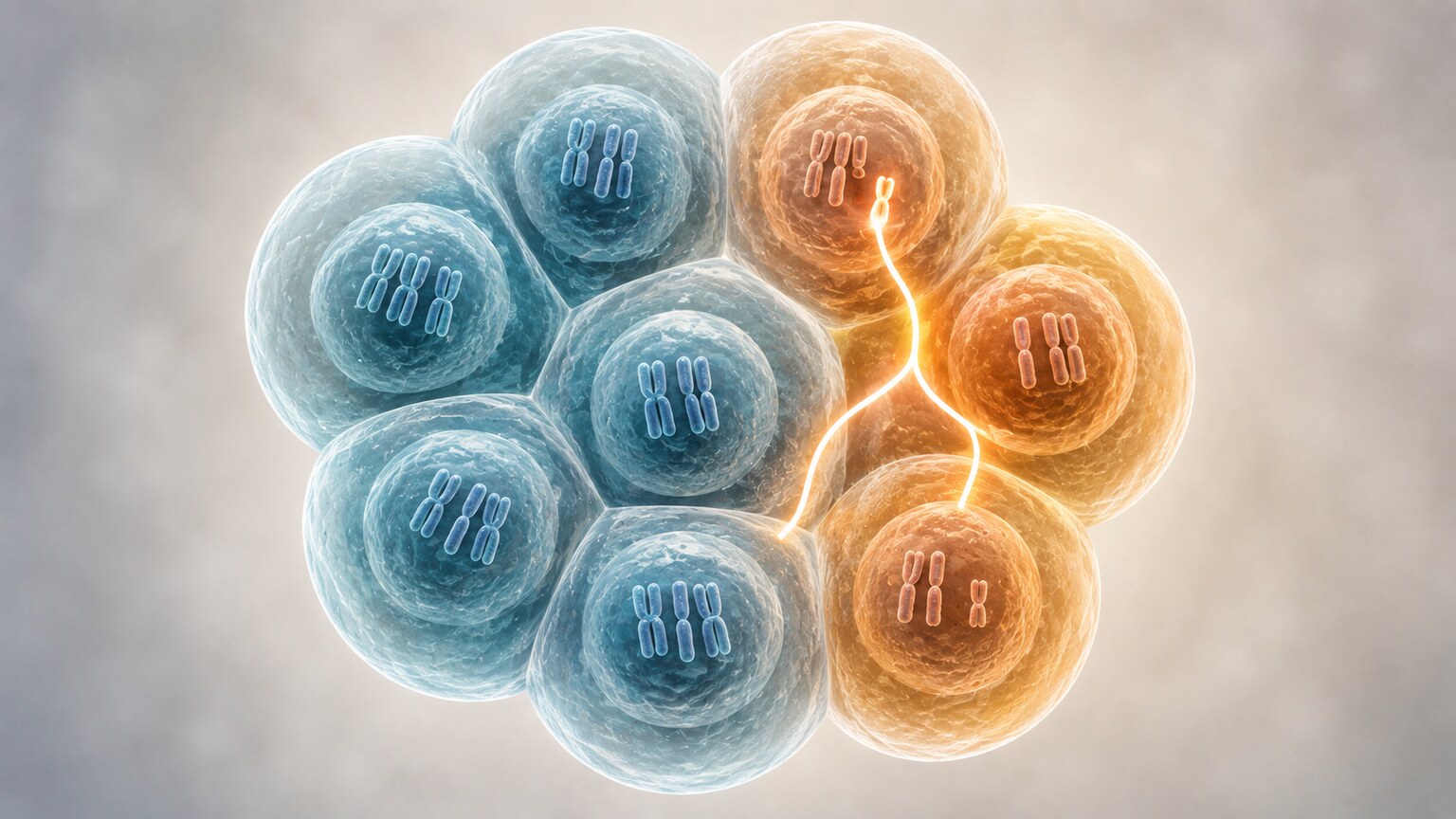
In genetics, mosaicism refers to the presence of two or more genetically different cell lines within a single individual, all of whom originated from the exact same fertilized egg — this shared origin is actually the key detail that separates mosaicism from a related but distinct phenomenon called chimerism, where genetically different cell lines come from two separate fertilized eggs that fused early in development instead of just one splitting apart. This shared cellular ancestry, despite the eventual genetic divergence between the two populations, is what makes mosaicism a fundamentally different phenomenon from an inherited condition passed down through egg or sperm. It is an important distinction from a condition present in every single cell of the body from the very start — in mosaicism, some fraction of the body's cells carry one chromosome pattern, typically the normal one, while another fraction carries a different pattern, often involving an extra or missing chromosome. The proportion between these two groups can range enormously, from a tiny minority of affected cells detectable only with the most sensitive testing methods to something close to a fifty-fifty split, and that proportion turns out to be one of the single most important pieces of information for understanding what a mosaic result actually means for a given person or pregnancy, shaping everything from how the finding is initially interpreted to what kind of follow-up, if any, gets recommended.
How Mosaicism Actually Happens
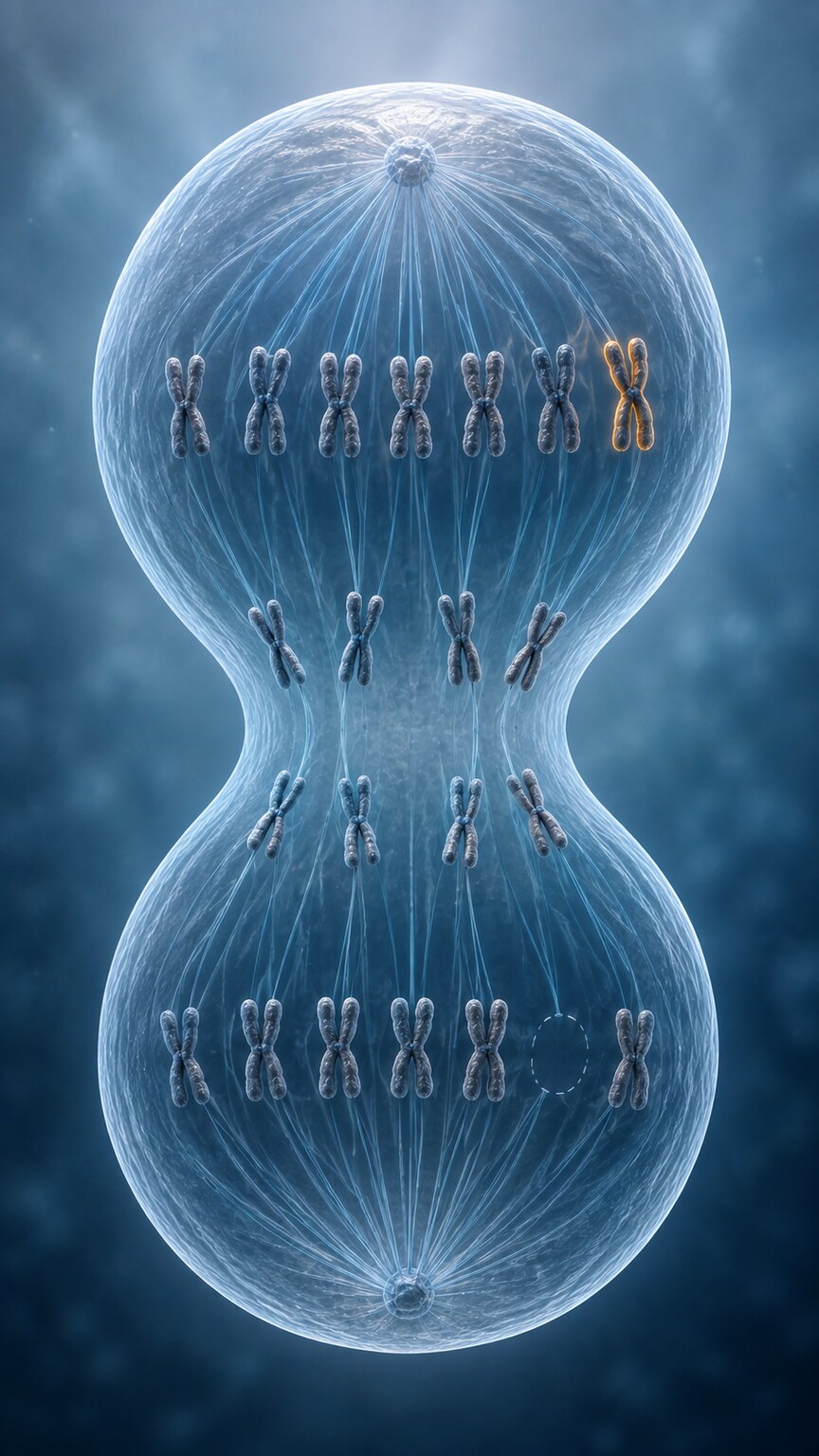
Researchers have identified two primary mechanisms behind mosaic aneuploidy, and both trace back to an error that happens after fertilization, once the embryo has already begun dividing — this "after fertilization" timing is itself an important distinguishing feature, since it means the original fertilized egg started out with a completely normal, uniform chromosome count before the error introduced any variation. The first is called postzygotic mitotic nondisjunction, a mistake that occurs when a single cell within the developing embryo divides and fails to properly separate its chromosomes, producing two unequal daughter cells — one ending up with an extra chromosome (47 instead of the usual 46) and the other missing one entirely (45 instead of 46). Because a completely missing chromosome is almost always lethal to that particular cell line early in development, the surviving lineages typically settle into a two-cell-line pattern: some descendants carrying the normal chromosome count and others carrying the extra one, while the monosomic line is naturally eliminated through ordinary developmental attrition before it can contribute meaningfully to the finished body. The second mechanism, called trisomy rescue, works somewhat in reverse. It begins with an embryo that started out with three copies of a chromosome in every single cell, a full trisomy present from conception, and then one of those cells manages to correct the error partway through development by discarding the extra copy — effectively "rescuing" itself back toward a normal chromosome count in a process that essentially fixes the problem in some cellular descendants while leaving it uncorrected in others. When this correction happens in only some cells rather than all of them, the result is a mosaic mixture of normal and trisomic cell lines coexisting in the same embryo, with the exact ratio between the two depending on precisely when during development the correcting cell divided relative to the rest of the embryo. Both mechanisms produce the same basic outcome — two genetically distinct populations within one individual — but they arise from opposite starting points, one moving from normal toward abnormal and the other moving from abnormal back toward normal, only for the correction to remain incomplete, leaving behind a mosaic footprint of the original error even after most of the embryo's cells have returned to a typical chromosome count.
Trying to make sense of a genetic or chromosomal result of your own? Upload your labs and get a complete, plain-language breakdown in under 15 minutes.
Analyze My ResultsWhy the Percentage of Affected Cells Matters So Much
Once a mosaic result is identified, the single most clinically important number that follows is the percentage of cells carrying the abnormal chromosome pattern, because this percentage correlates directly with how likely the finding is to actually affect the person's health or development. A result showing only a small minority of abnormal cells — say, 5% or 10% — behaves very differently in practice than a result showing that abnormal cells make up half or more of the sampled tissue, and genetic counselors will typically frame the discussion around this number specifically rather than treating "mosaic" as a single, uniform category of finding. This isn't just a theoretical distinction: research on prenatal chromosomal mosaicism has generally found that cases where the fetus was genuinely affected tend to show a notably higher average mosaic level than cases where the abnormality turned out to be confined to the placenta and didn't involve the fetus at all — illustrative figures cited in some studies put that difference at roughly double, though exact percentages vary between studies and shouldn't be applied directly to an individual result without a genetic counselor's interpretation. That said, percentage alone isn't a perfect predictor either — which chromosome is involved, which tissue was actually sampled, and how the abnormal cells are distributed throughout the body all factor into the final clinical picture alongside the raw percentage, meaning two people with an identical mosaic percentage but a different chromosome involved can end up with very different outlooks entirely, since not every chromosome carries the same functional consequences when present in an extra or missing copy.
Confined Placental Mosaicism: When the Finding Isn't Really About the Baby
One of the most important concepts to understand about prenatal mosaicism is that a mosaic result found on a chorionic villus sampling (CVS) test doesn't necessarily mean the fetus itself is affected. CVS samples tissue from the placenta, which develops from the same original fertilized egg as the fetus but can, through the exact same postzygotic errors described above, end up with a chromosomally different cell line that stays confined entirely to placental tissue while the fetus itself develops with a completely normal chromosome count — a divergence that becomes possible because the cells destined to form the placenta and the cells destined to form the fetus itself actually separate into distinct developmental lineages quite early on, meaning an error occurring after that split can affect one lineage without ever touching the other. This phenomenon, called confined placental mosaicism, occurs in roughly 1% to 2% of CVS samples, and research following large numbers of these cases has found that true fetal involvement — meaning the abnormal cell line genuinely made it into the fetus and not just the placenta — is confirmed in only a small minority of pregnancies where placental mosaicism was initially detected. Large published case series, involving tens of thousands of CVS samples, have found only a small handful of mosaic rare autosomal trisomies ultimately confirmed in the fetus itself through follow-up testing — a reassuringly low confirmation rate given how frequently the underlying placental finding itself occurs, though the exact figures should be discussed with a genetic counselor rather than applied directly from these illustrative numbers. In other words, the overwhelming majority of mosaic findings on a CVS test turn out to be a placental phenomenon that never touched the developing baby at all, which is genuinely reassuring information once a family understands the distinction, even though the initial report language alone can sound alarming without that context attached, which is exactly why genetic counseling is typically offered alongside any mosaic CVS result rather than the finding being communicated as a bare number on its own.
Why a Mosaic Finding Often Leads to a Follow-Up Test
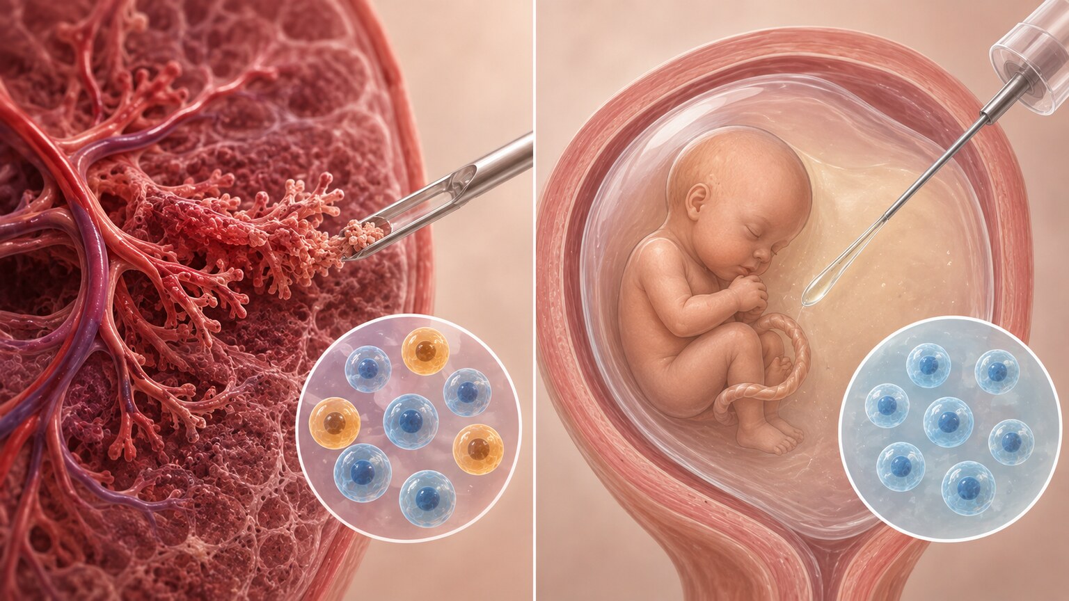
Because a mosaic CVS result can't, on its own, distinguish between a placenta-only finding and one that genuinely involves the fetus, the standard next step is a follow-up amniocentesis, which samples amniotic fluid containing fetal cells shed directly from the baby rather than placental tissue, typically performed a few weeks after the original CVS to allow enough time for a technically adequate amniotic fluid sample to be collected safely. If the amniocentesis comes back with a completely normal chromosome count, that strongly supports a diagnosis of confined placental mosaicism, and the pregnancy is typically managed with reassurance, though some studies suggest even confined placental mosaicism can carry a modestly elevated risk of complications like preterm birth or lower birth weight depending on which chromosome was involved, which is why continued monitoring — sometimes including additional growth ultrasounds later in pregnancy — is still often recommended even after the fetus itself is confirmed to be chromosomally normal. If the amniocentesis also shows the abnormal cell line, that confirms true fetal mosaicism, and genetic counseling then focuses on what's known about that specific chromosome abnormality, the percentage detected, and how it's likely to affect development going forward, often drawing on published case series of similar mosaic findings to give families the most concrete outlook available. This two-step testing approach — an initial screening or diagnostic test followed by a confirmatory test using a different tissue source — is specifically designed to resolve exactly the kind of ambiguity that a mosaic result on its own can't answer, and it reflects a broader principle in prenatal genetics that no single test result is interpreted in isolation when a more definitive answer is available through a reasonable follow-up step.
Mosaic Turner Syndrome: A Real-World Example of How Much Percentage Matters
Turner syndrome, caused by a completely or partially missing X chromosome in someone assigned female at birth, offers one of the clearest real-world illustrations of how dramatically mosaicism can soften a condition's typical presentation. In its non-mosaic form, every cell carries only a single X chromosome instead of the usual two, and the condition is associated with short stature, ovarian insufficiency affecting fertility, and an elevated risk of specific heart and blood vessel abnormalities that require lifelong monitoring. In mosaic Turner syndrome, some cells carry the standard two X chromosomes (46,XX) while others carry only a single X (45,X), and research has found that the percentage of affected 45,X cells across different individuals with this mosaic pattern ranges anywhere from about 15% to 79% of sampled cells — a wide enough range that two people both labeled "mosaic Turner syndrome" on paper can have meaningfully different actual cellular compositions underlying that shared label. That wide range in cellular composition maps onto an equally wide range in outcomes: women with a 45,X/46,XX mosaic pattern were found, on average, to be less short in stature, to have a normal reproductive lifespan and birth rate, and to show none of the cardiovascular complications commonly associated with non-mosaic Turner syndrome — a dramatically milder overall picture than what's typically seen when every single cell carries the missing X chromosome, and one that has led researchers to describe mosaic Turner syndrome specifically as showing reduced penetrance compared to the classic, non-mosaic form of the condition.
Mosaic Down Syndrome: A Second Example
Down syndrome offers a second useful example of the same underlying principle. The great majority of Down syndrome cases involve a full trisomy 21 present in every cell from conception, but less than 1% of people with Down syndrome have the mosaic form instead, where only some of their cells carry the extra copy of chromosome 21 while the rest carry a completely typical chromosome count. Research on mosaic Down syndrome has found that the resulting presentation varies enormously from person to person, ranging from very mild features to a presentation nearly indistinguishable from classic, non-mosaic Down syndrome — and notably, the percentage of mosaicism measured in a blood sample doesn't reliably predict where along that spectrum a given person will fall, which can be genuinely difficult for families to reconcile when a lab report gives a specific, seemingly precise percentage that ultimately doesn't map onto a specific, predictable outcome. Part of the explanation lies in the fact that different organs and tissues develop from different embryonic layers, so the actual percentage of trisomic cells can vary meaningfully between blood, skin, muscle, and brain tissue in the same individual, meaning a blood test alone captures only a partial, tissue-specific snapshot of the body's true overall mosaic composition rather than a single number that applies uniformly everywhere.
Why Different Tissues in the Same Body Can Show Different Percentages
This tissue-to-tissue variability is a genuinely important concept for interpreting any mosaic result, prenatal or otherwise. Because the postzygotic error that creates mosaicism can occur at different points during early development, the resulting abnormal cell line may end up distributed unevenly across the body depending on which embryonic structures descended from the specific cell where the error first occurred — an error happening very early, before the embryo has differentiated into distinct tissue layers, tends to produce a more evenly distributed mosaic pattern, while an error happening later, after some tissue-layer separation has already occurred, can produce a pattern confined largely to just one or two tissue types. A blood sample, a skin biopsy, and a tissue sample from an internal organ can each show meaningfully different percentages of the same abnormal cell line in the very same person, which is why a single blood-based mosaic percentage should generally be understood as a useful estimate rather than a precise, universal number applicable to every tissue in the body. This is also part of why genetic counselors are often cautious about making firm predictions based purely on a percentage measured from whichever tissue happened to be the most convenient or least invasive to sample, and why, in cases where the stakes of a prediction are especially high, doctors will sometimes recommend sampling more than one tissue type specifically to build a fuller, more representative picture before drawing firm conclusions.
When Mosaicism Is Actually Visible on the Skin
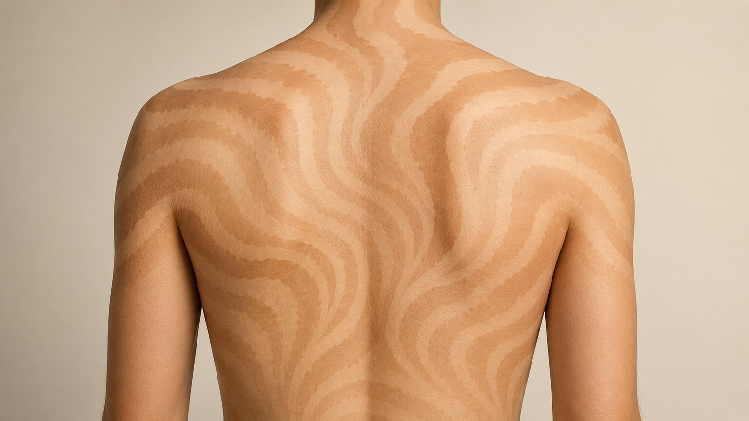
Most mosaicism is entirely invisible, detectable only through laboratory testing, but there's one striking exception worth knowing about: mosaicism can occasionally be seen directly on the skin, in a pattern called the lines of Blaschko. These lines trace a distinctive whorled or S-shaped pattern on the trunk and a more linear pattern along the arms and legs, and they represent the literal migration paths taken by two genetically different populations of skin cells — most often melanocytes, the pigment-producing cells of the skin — as they proliferated and spread outward from their point of origin while the embryo developed. Despite following such a consistent, well-documented pattern across different people and different underlying genetic causes, these lines are entirely undetectable on genetically uniform skin and only become visible at all when two distinct cell populations happen to exist side by side. When one of these two cell populations produces a different amount of melanin than the other, the boundary between them becomes visible as a pattern of lighter or darker skin following these developmental lines exactly, essentially making the underlying cellular mosaicism visible to the naked eye without requiring any laboratory test at all to first suspect it. Research has found that mosaic autosomal trisomies specifically account for roughly 30% of pigmentary mosaicism cases following this pattern, and cases have been documented involving trisomy 7 mosaicism and mosaic Turner syndrome, among other chromosome abnormalities, underscoring that this visible skin pattern isn't tied to any single specific chromosome abnormality but can arise from a range of different underlying mosaic conditions. Because these lines follow the exact developmental migration routes of embryonic skin cells rather than any nerve or blood vessel pathway, doctors and dermatologists specifically look for this Blaschko-line pattern as a visible clue that mosaicism may be present, sometimes prompting genetic testing that wouldn't otherwise have been pursued, turning a subtle skin finding into the starting point for a broader genetic workup that might otherwise never have been considered.
What a Mosaic Result Means Outside of Pregnancy
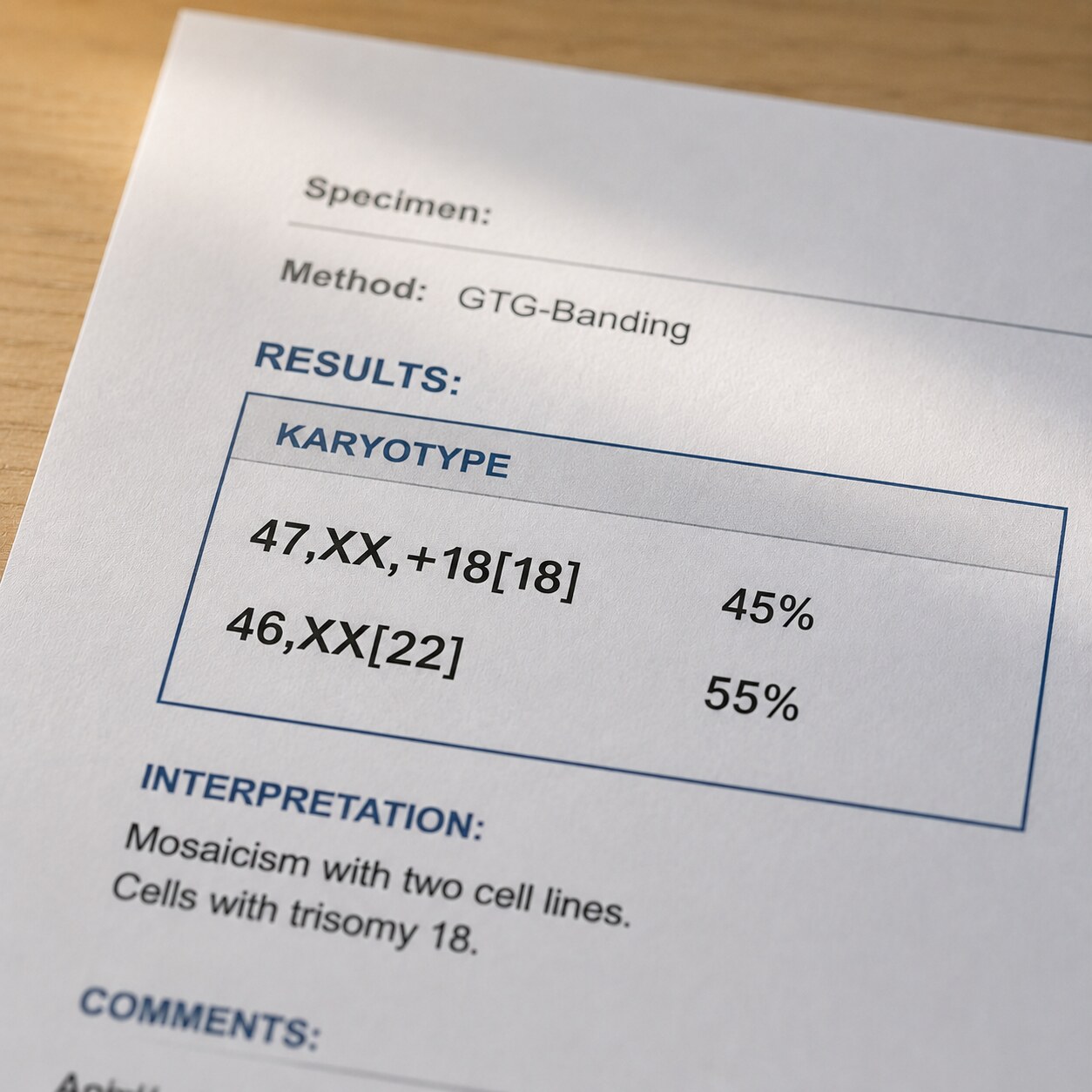
While mosaicism is most commonly discussed in a prenatal testing context, it can also be identified later in life — sometimes incidentally during genetic testing pursued for an entirely different reason, such as fertility evaluation, a workup for an unrelated medical condition, or even routine genetic screening, and sometimes specifically because a person's symptoms or physical features didn't fully match what would be expected from a non-mosaic version of a given chromosomal condition, prompting a doctor to specifically request mosaicism analysis rather than a standard single-result chromosome count. In these cases, the same core principles apply: the percentage of abnormal cells found, the specific chromosome involved, and which tissue was tested all shape how the result is interpreted and what it might mean going forward, exactly as they do in the prenatal context described throughout this article. A mosaic finding identified in adulthood is typically followed by genetic counseling to review what's specifically known about that chromosome abnormality's mosaic form, drawing on published case reports and research cohorts of similar findings where available, and sometimes by additional testing on a second tissue type — often a skin biopsy alongside the original blood sample — to build a more complete picture of how the abnormal cell line is distributed throughout the body before finalizing any conclusions about what the finding actually means for that person's health.
How Labs Actually Detect and Report Mosaicism
Understanding a mosaic result also benefits from knowing, at least broadly, how the lab arrived at that percentage in the first place. Traditional karyotyping works by growing cells from the sample in a culture dish, then physically examining a set number of individual cells under a microscope — often 20 or more separate cells — and directly counting how many show the normal chromosome pattern versus the abnormal one, with the reported percentage simply reflecting that count. Chromosomal microarray testing, a newer and more sensitive technology, works differently: rather than examining individual cells one at a time under a microscope, it measures the overall balance of genetic material across the entire sample at once, which is part of why it can detect lower levels of mosaicism that might be missed if only a small handful of individual cells happened to be examined under traditional karyotyping. This methodological difference is a meaningful part of why chromosomal microarray testing detects mosaicism in up to roughly 4% of samples compared to the 1% to 2% detected by standard karyotyping — it isn't that microarray testing is finding something that wasn't there before, but that it's more sensitive to smaller-scale mosaic patterns that older methods could reasonably miss entirely, given how few individual cells get directly examined in a standard karyotype count. Neither method is simply "better" in every situation; karyotyping still offers a direct, cell-by-cell visual confirmation that some clinical scenarios specifically call for, while microarray testing offers broader sensitivity across the whole sample, and the two are sometimes used together specifically to take advantage of each method's particular strengths, giving both a precise cell-level count and a comprehensive whole-sample estimate within the same overall workup.
Frequently Asked Questions
What does it mean if my chromosomal test result shows mosaicism?
It means two or more genetically different cell lines were detected in the tissue sampled, typically one with a normal chromosome count and one with an extra or missing chromosome, both originating from the same fertilized egg.
How common is chromosomal mosaicism?
It's detected in roughly 1% to 2% of samples using standard karyotyping and in up to 4% of samples using more sensitive chromosomal microarray testing, making it a genuinely common finding rather than a rare one.
Does a mosaic result on a prenatal test always mean the baby is affected?
No. A large share of mosaic findings on CVS testing are confined to placental tissue and never involve the fetus, which is why a follow-up amniocentesis is typically recommended to clarify the situation.
Does the percentage of abnormal cells predict how severe a condition will be?
It's a meaningful factor, but not a perfect predictor. Which tissue was sampled, which chromosome is involved, and how the abnormal cells are distributed throughout the body all matter alongside the raw percentage.
Can mosaicism be found outside of pregnancy?
Yes. It can be identified incidentally during genetic testing done for other reasons, or specifically pursued when a person's features don't fully match what's expected from a non-mosaic version of a chromosomal condition.
Can mosaicism actually be seen on the skin?
Yes, in some cases. A pattern called the lines of Blaschko can make mosaicism visible as whorled or linear areas of lighter or darker skin, tracing the migration paths of two genetically different cell populations during development.
Why does chromosomal microarray testing detect more mosaicism than standard karyotyping?
Karyotyping examines a limited number of individual cells directly under a microscope, while microarray testing measures the overall genetic balance across the whole sample at once, making it more sensitive to smaller-scale mosaic patterns.
Conclusion
Chromosomal mosaicism can sound alarming the first time it appears on a lab report, but understanding the biology behind it — two genetically distinct cell lines arising from a single postzygotic error, whether through mitotic nondisjunction or trisomy rescue — turns an unfamiliar term into something genuinely explainable rather than a vague, frightening label. The percentage of affected cells, which tissue was tested, and which chromosome is involved all shape what a specific mosaic finding actually means, and real-world examples like mosaic Turner syndrome and mosaic Down syndrome show just how dramatically these factors can soften a condition's typical presentation compared to its non-mosaic form, sometimes producing an outcome close to unaffected despite an abnormal chromosome count technically being present in some fraction of the body's cells. Even the rare cases where mosaicism becomes visible directly on the skin, tracing the lines of Blaschko, reflect the exact same underlying cellular principle described throughout this article, just made unusually tangible. Whether a mosaic result surfaces during pregnancy or later in life, the same underlying framework applies: it's a snapshot of genetic variation within one body, not a single fixed diagnosis, and it's a finding that genetic counseling and appropriate follow-up testing are specifically designed to help interpret with real clarity, rather than something meant to be understood from a single percentage on a page in isolation, disconnected from the tissue it came from and the specific chromosome it involves.
Still Not Sure What Your Results Mean?
Upload your labs and get a complete, visual, plain-language interpretation of every biomarker — delivered to your inbox in under 15 minutes.
Get My ReportThis article is for educational purposes only and does not constitute medical advice. Always consult your healthcare provider or a genetic counselor regarding your specific results.