
What Does a Chromosomal Deletion or Duplication Indicate?
A chromosomal deletion or duplication, described on paper, sounds like a fairly simple accounting problem — a piece of DNA missing somewhere, or an extra copy of something sitting where it shouldn't be. But the actual consequence has almost nothing to do with the DNA itself being gone or extra; it has everything to do with dosage. In reality, your cells are built and calibrated to run on a precise, carefully balanced two-copy amount of most individual genes, and both losing a single copy entirely and gaining one single extra copy beyond the normal two can genuinely throw that finely tuned balance off in ways that produce meaningfully different, and sometimes genuinely opposite, effects depending on exactly which specific genes happen to sit inside the particular affected region in question. This article walks through the actual underlying biology behind why size and precise genomic location decide almost everything about the real-world consequences of a given deletion or duplication, using several well-documented, extensively studied named syndromes as concrete, specific illustrations of a broader dosage-sensitivity principle that genuinely applies to nearly every single copy number finding a modern genetics report is capable of returning.
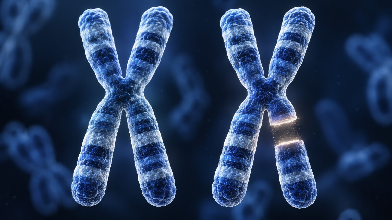
Figure 1. A deletion removes a segment from one copy of a chromosome pair, leaving only a single working copy of every gene inside that missing stretch instead of the usual two.
Why Your Cells Care About Having Exactly Two Copies
Nearly every single gene contained within your genome exists in exactly two functional copies — one copy inherited directly from each parent — and for a large share of genes, your cells have evolved to depend on getting roughly double the output that a single copy alone would produce. This precise dependence genuinely isn't arbitrary or purely incidental in any meaningful way: many individual proteins only ever function correctly within a fairly narrow, specific concentration range, and the many cellular processes that rely on those particular proteins have been calibrated, over the course of evolution, specifically around that expected two-copy baseline output. A specific gene where a single remaining working copy genuinely isn't enough on its own to maintain fully normal cellular function is formally described as "haploinsufficient" — lose the other copy, and the one copy that remains, even functioning entirely perfectly in every other respect, simply cannot manufacture a sufficient quantity of the needed protein to keep every downstream process running exactly as it normally would. Certain other genes show essentially the mirror-image sensitivity in the opposite direction instead: having an extra, third functional copy present pushes that gene's total output high enough to meaningfully disrupt the exact same finely tuned biological balance, a distinct phenomenon researchers refer to specifically as triplosensitivity. Whether any given deletion or duplication ultimately causes a noticeable, clinically meaningful effect depends very heavily on precisely how many haploinsufficient or triplosensitive genes happen to sit inside that specific affected stretch of chromosome in question.
It helps to picture why this dosage sensitivity exists at all, rather than treating it as an arbitrary biological quirk. Genes don't function in isolation — they're part of interconnected regulatory networks, often controlling the production of enzymes or structural proteins that other cellular processes depend on running at a specific, predictable rate. A protein produced at half its normal amount can, for some processes, simply mean things happen a bit more slowly with no meaningful downstream consequence; for others, particularly during the tightly timed, sequential stages of early embryonic development, a protein arriving at half-strength at a critical window can mean a structure forms incompletely or a signal never reaches the threshold needed to trigger the next developmental step at all. This is a large part of why so many dosage-sensitive genes turn out to be genes active specifically during early development — the tight timing and threshold-dependent nature of that process leaves comparatively little room for a reduced or excessive dose to be absorbed without consequence, compared to a gene whose job continues steadily throughout adult life.
Genome-wide studies cataloging which specific genes are haploinsufficient versus dosage-tolerant have become an increasingly central resource in modern clinical genetics, since knowing in advance which genes fall into which category lets a lab predict, before any single patient's result is even reviewed, how concerning a deletion or duplication overlapping a specific gene is likely to be. Databases maintained by international genetics consortia assign a probability score to most human genes reflecting how intolerant that gene appears to be to a single-copy loss, based on how rarely such losses are observed in large, healthy population samples — a gene where losing one copy is never observed in tens of thousands of healthy sequenced individuals is treated as a strong haploinsufficiency candidate, while a gene where single-copy loss turns up regularly in people with no related symptoms is treated as considerably more dosage-tolerant.
Why Size Alone Doesn't Predict Severity
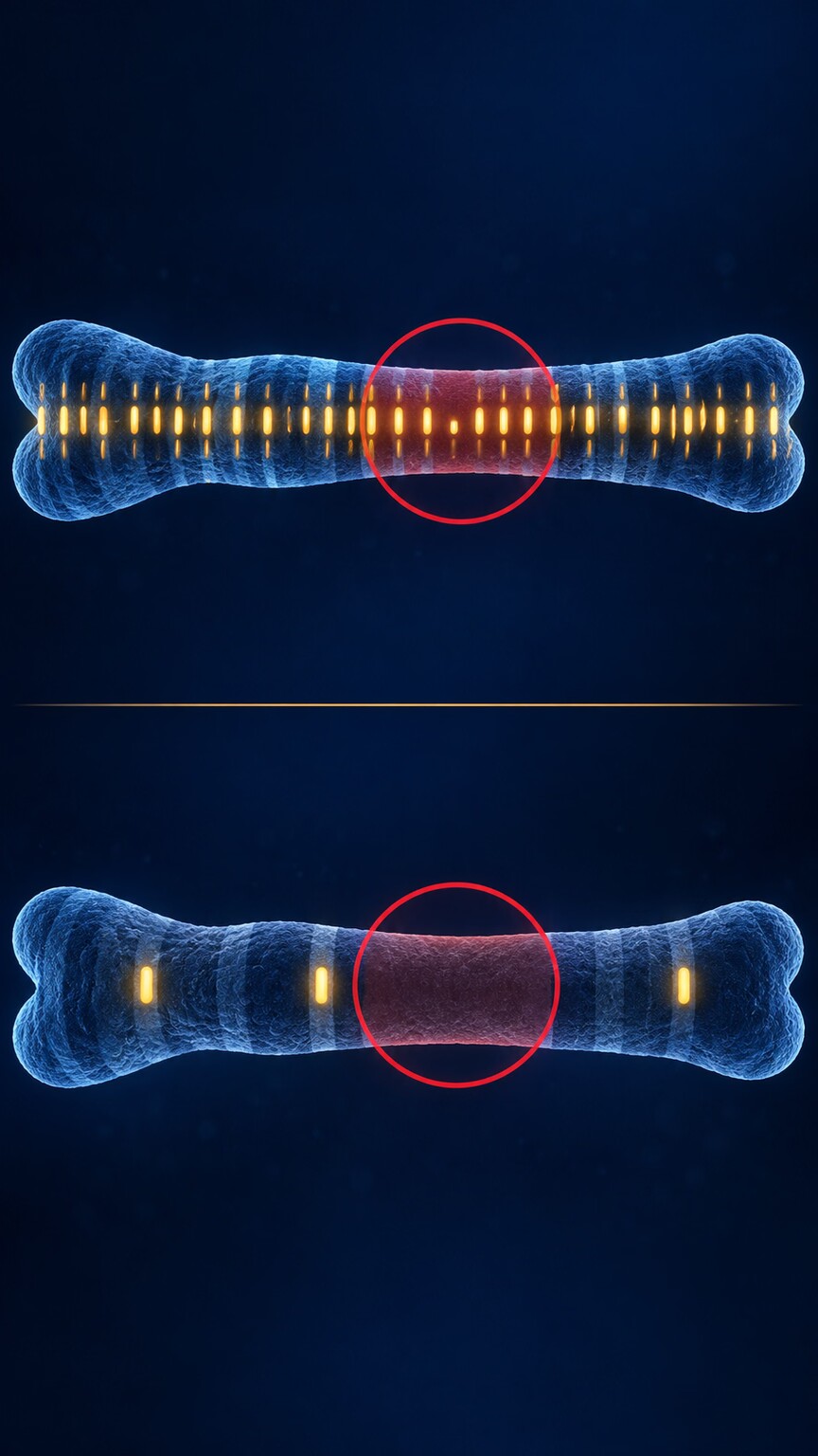
Figure 2. Two deletions of the exact same physical size can have completely different consequences depending on whether the missing stretch is gene-dense or largely empty of functional genes.
A genuinely counterintuitive but critically important fact is that a larger deletion isn't automatically worse than a smaller one, and a tiny deletion isn't automatically safer than a large one. What actually matters is gene density — how many functional genes happen to sit inside the specific stretch that's missing or duplicated. A relatively large deletion spanning several million base pairs, sitting entirely within a region of the genome that's mostly non-coding "junk" DNA of no known function, with only one or two genes scattered thinly across that whole large span, can turn out to be entirely harmless in practice. A much smaller deletion, only a fraction of that overall size, but landing squarely on a stretch densely packed with several critical, individually haploinsufficient genes, can instead produce a serious, wide-reaching, multi-system condition. This is exactly why a lab report listing the size of a deletion or duplication in base pairs doesn't, by itself, tell you anything about severity — the specific genes contained within that span are what actually determine the outcome, which is why every copy number finding gets individually annotated against gene databases rather than judged by size category alone.
This same principle explains a related pattern worth understanding: two genetically unrelated conditions can sometimes be caused by deletions of wildly different sizes, simply because the actual functionally important gene content overlaps despite the surrounding span being different. A very targeted, gene-focused deletion affecting only the specific dosage-sensitive gene responsible for a condition can produce the exact same clinical picture as a much larger deletion that happens to include that same gene along with a great deal of surrounding, functionally unimportant DNA. Clinicians and researchers sometimes describe this by identifying a "critical region" within a larger, more variably-sized deletion — the specific minimal stretch of DNA that, when missing, is sufficient on its own to produce the associated condition, distinct from the sometimes much larger total deleted region an individual patient's specific test result might show.
This critical-region concept also explains why some patients with what looks like "the same syndrome" on paper can present with meaningfully different severity or slightly different combinations of features from each other. If two patients each have a deletion overlapping the identical critical region, but one patient's deletion extends further in one direction, capturing one or two additional genes beyond the core critical region, that patient may show additional features beyond the syndrome's typical presentation, reflecting the extra genes caught up in their specific, slightly larger deletion. This is part of why genetic counseling for a named microdeletion syndrome typically still involves reviewing the individual patient's exact breakpoints — the specific start and end coordinates of their deletion — rather than assuming every patient with the same syndrome name has an identical genetic finding down to the base pair.
Trying to make sense of a genetics report or lab result on your own? Upload your results and get a complete, plain-language breakdown in under 15 minutes.
Analyze My Results22q11.2 Deletion Syndrome: One Region, Many Systems Affected
One of the most well-studied microdeletion syndromes, 22q11.2 deletion syndrome (sometimes still called DiGeorge syndrome), illustrates the gene-dosage principle concretely. A small missing segment on the long arm of chromosome 22 removes roughly 30 to 40 genes at once, several of which are individually haploinsufficient and each contribute to a different piece of early development — heart formation, the immune system's thymus gland, palate development, and specific brain circuits all draw on genes within this one region. This is exactly why 22q11.2 deletion syndrome produces such a wide, seemingly unrelated combination of features in different individuals — heart defects, immune deficiency, cleft palate, and learning differences — despite tracing back to a single genetic cause: it isn't one gene malfunctioning in one system, it's several independently haploinsufficient genes, each affecting its own separate developmental process, all missing from the same small deleted stretch at once.
One specific gene within this region deserves particular mention, since it illustrates the haploinsufficiency concept especially concretely: a gene involved in early heart and facial development is widely considered a major contributor to several of 22q11.2 deletion syndrome's most consistent features. Research comparing patients whose deletions happen to spare this specific gene against patients whose deletions include it has found a meaningfully different rate of heart defects between the two groups, offering fairly direct evidence that this one gene, among the thirty-odd genes removed by the full deletion, carries an outsized share of responsibility for that particular feature. This kind of gene-by-gene dissection — identifying which specific genes within a larger deleted region drive which specific clinical features — is an active, ongoing area of research for essentially every microdeletion syndrome discussed in this article, since fully understanding it opens the door to potential future treatments targeting the specific downstream consequence of losing that one gene's function, rather than treating the syndrome as an undifferentiated whole.
It's also worth noting that 22q11.2 deletion syndrome shows considerable variability in which features actually appear in a given individual, even though the deletion itself, in the majority of cases, removes essentially the same set of genes. Some individuals with the deletion have significant heart defects requiring surgery in infancy; others have no cardiac involvement detected at all, despite carrying an identical or near-identical deletion. This phenomenon, called variable expressivity, appears throughout the microdeletion and microduplication syndromes discussed in this article, and reflects the reality that other genetic background factors, and likely some degree of chance during development, influence whether a given dosage imbalance actually crosses the threshold into a clinically noticeable effect for each specific affected system in each specific individual.
Williams Syndrome: A Duplication Producing the Nearly Opposite Picture
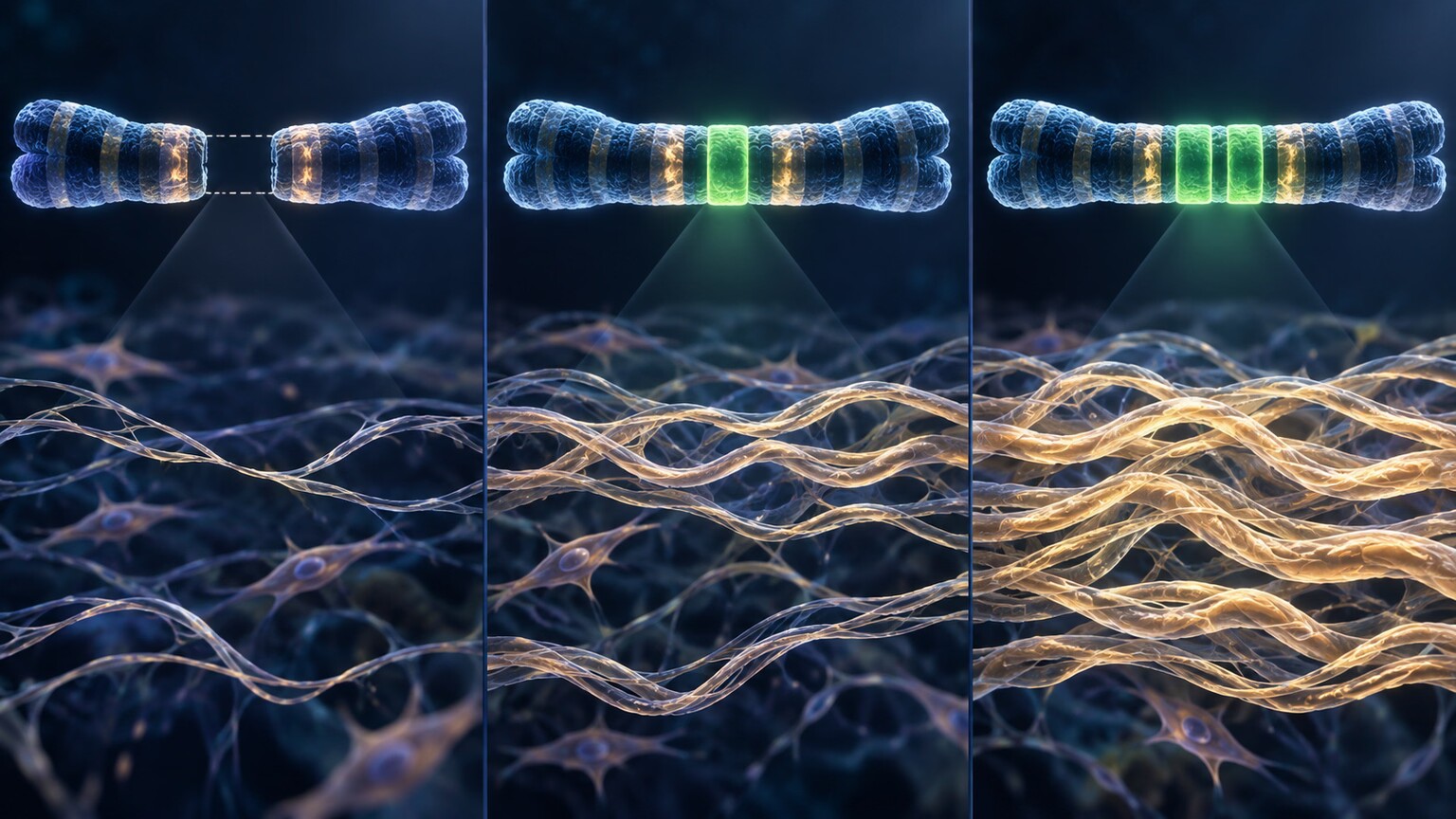
Figure 3. The same chromosome 7 region containing the elastin gene produces genuinely opposite tissue effects depending on whether it's deleted (Williams syndrome) or duplicated (7q11.23 duplication syndrome).
A different region, on chromosome 7, demonstrates the deletion-versus-duplication contrast especially clearly. Deleting this stretch — which includes the gene for elastin, a protein that gives connective tissue like blood vessel walls their stretch and recoil — produces Williams syndrome, characterized in part by a distinctive cardiovascular finding called supravalvular aortic stenosis, a narrowing that results directly from connective tissue lacking adequate elastin. Duplicating the same exact region produces a genuinely different, sometimes nearly opposite-leaning condition, 7q11.23 duplication syndrome, associated with its own distinct pattern of features including speech delay and, in some studies, a tendency toward different cardiovascular findings than the deletion produces. The same genomic address, one copy too few versus one copy too many, and two clinically distinguishable conditions — about as clean a real-world demonstration of gene-dosage sensitivity as exists in modern clinical genetics.
Beyond the specific cardiovascular contrast, Williams syndrome is also well known for a distinctive cognitive and personality profile — a characteristic combination of relative strength in language and social engagement alongside significant difficulty with visual-spatial tasks, paired with an unusually warm, highly sociable personality style that researchers have studied specifically in connection with this deletion. The 7q11.23 duplication syndrome, by contrast, is more often associated with speech and language delay rather than the relative language strength seen in the deletion, along with a higher reported rate of certain anxiety-related features — differences plausibly connected to the same genes being pushed in opposite dosage directions affecting the same neurodevelopmental pathways differently depending on whether they're under- or over-expressed.
Researchers studying this specific chromosome 7 region have used it as something of a natural experiment for understanding how gene dosage shapes brain development and behavior more broadly, precisely because it offers a rare, clean comparison: the same genes, the same chromosomal address, examined at both a reduced and an elevated dosage in different groups of patients. Findings from this line of research have informed broader thinking about dosage-sensitive genes elsewhere in the genome that may contribute to more common, non-syndromic neurodevelopmental and psychiatric conditions, even in the absence of an identifiable, large chromosomal deletion or duplication — suggesting that the dosage-sensitivity principle demonstrated so clearly in these rarer, well-defined syndromes likely operates on a smaller, harder-to-detect scale throughout the broader population as well.
Prader-Willi and Angelman: When Which Parent Contributed the Deletion Matters
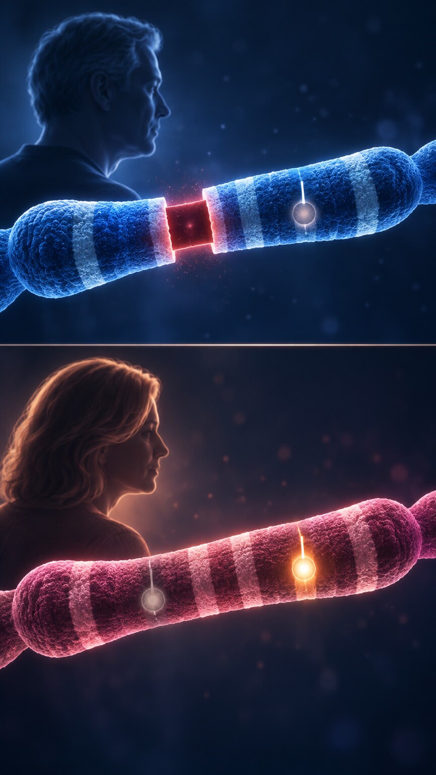
Figure 4. The identical chromosome 15 deletion produces Prader-Willi syndrome when the missing copy came from the father, but Angelman syndrome when it came from the mother — a phenomenon called genomic imprinting.
A third region, on chromosome 15, adds a layer of complexity beyond simple gene dosage: parent-of-origin effects, a phenomenon called genomic imprinting. Certain genes are chemically marked, or "imprinted," so that only the copy from one specific parent is normally active, while the copy from the other parent is naturally switched off regardless of whether it's structurally intact. In this particular chromosome 15 region, genes are normally active only on the father's copy; the same identical deletion, if inherited on the chromosome that came from the father, removes the only active copy and produces Prader-Willi syndrome, characterized by low muscle tone in infancy and, later, an insatiable appetite and risk of severe obesity. The same deletion inherited on the chromosome from the mother instead removes a copy that would have been silenced anyway, leaving the region's mother-specific active genes intact — but a deletion occurring on that same region of the maternal chromosome instead specifically removes a different, separately imprinted gene that's normally active only on the maternal copy, producing the entirely clinically distinct Angelman syndrome as a result, marked by severe developmental delay, a distinctive happy demeanor, and often a near-complete absence of expressive speech throughout life. This is the clearest possible illustration that a deletion's consequence depends not just on which genes are missing, but sometimes on which parent's copy was lost.
Genomic imprinting itself is worth understanding at a slightly deeper level, since it's a genuinely unusual departure from how most of the genome behaves. For the vast majority of genes throughout the genome, both the maternal and paternal copies are equally active, and losing either one has an identical consequence regardless of which parent it came from — this is exactly why, for the 22q11.2 and chromosome 7 regions discussed earlier in this article, parent-of-origin generally doesn't change the outcome at all. Imprinted regions are the exception, not the rule, estimated to represent well under one percent of all genes in the human genome, each one specifically marked early in egg or sperm formation through a chemical modification that silences one parent's copy without altering the underlying DNA sequence itself. This modification is what actually determines whether losing the maternal or paternal copy of a specific imprinted gene matters — the DNA sequence lost is identical either way, but which copy was already switched off before the deletion ever happened is what changes the result.
A related, rarer mechanism can also produce Prader-Willi or Angelman syndrome without any deletion at all, worth mentioning briefly since it reinforces how central parent-of-origin is to this specific region's biology: a phenomenon called uniparental disomy, where a child inherits both copies of chromosome 15 from the same parent rather than one from each. If both copies come from the mother, the region behaves as though the paternal, normally-active genes are entirely absent — despite no physical deletion having occurred at all — producing the same Prader-Willi syndrome phenotype through an entirely different underlying mechanism. This is exactly why comprehensive testing for these two conditions typically includes methods specifically designed to detect both deletions and uniparental disomy, rather than relying on a standard microarray alone, which can sometimes miss a uniparental disomy case since the total amount of DNA present is technically normal — it's simply all inherited from one parent instead of the usual one-from-each pattern.
Why Some Deletions and Duplications Cause No Symptoms at All
Not every deletion or duplication a genetic test identifies causes any noticeable effect, and understanding why explains a genuinely common, often reassuring scenario in modern genetic testing. Regions of the genome vary enormously in how tolerant they are of copy number changes — some stretches contain no genes at all, or only genes with enough built-in redundancy elsewhere in the genome that losing one copy locally has no measurable consequence. Large population databases built specifically from testing hundreds of thousands of otherwise healthy people worldwide have carefully cataloged many of these dosage-tolerant regions over time, allowing labs to recognize a newly identified deletion or duplication as one that's been seen repeatedly in people with no related health effects, and classify it confidently as benign.
Some regions of the genome are actually structured in a way that makes copy number changes considerably more likely to occur there in the first place, an important piece of context for understanding why certain deletions and duplications, including several of the specific syndromes discussed in this article, recur independently in unrelated families with strikingly similar breakpoints each time, rather than each case being a unique, one-off event. These regions typically contain repeated DNA sequences flanking a central segment, and during egg or sperm formation, the cellular machinery responsible for correctly pairing and separating chromosomes can occasionally misalign these repeated flanking sequences, causing an uneven exchange that deletes the segment between them on one resulting chromosome while duplicating that same segment on the other. This specific mechanism, sometimes called non-allelic homologous recombination, is the actual reason 22q11.2 deletion syndrome and the chromosome 7 region discussed earlier both occur repeatedly, independently, across many unrelated families with such consistent breakpoints — the underlying DNA architecture at these specific locations essentially predisposes that exact stretch to this kind of uneven exchange, rather than each family's case arising through pure chance at a random location.
Understanding this mechanism also clarifies why these specific microdeletion and microduplication syndromes, despite being individually rare, are collectively common enough to be well-characterized in the medical literature: rather than requiring a random, one-in-a-billion mutation at any arbitrary genomic location, they occur at a modest but measurable, predictable rate specifically because certain locations in the genome are structurally prone to this exact kind of recombination error. This is also exactly why, once a family has one child with a de novo occurrence of this kind of recombination-driven deletion, the recurrence risk for future pregnancies generally remains low — the event happened during that one specific instance of egg or sperm formation, and the same random misalignment isn't meaningfully more likely to recur in a future pregnancy just because it happened once, unless a parent is found to actually carry the finding themselves.
This is exactly why parental testing, mentioned in the imprinting discussion above, carries such weight in interpretation more broadly: a deletion or duplication found in an apparently healthy parent who passed it to their child is meaningful evidence, though not absolute proof, that the same finding in the child is less likely to be the explanation for whatever symptom prompted testing in the first place.How a Suspected Deletion or Duplication Actually Gets Confirmed
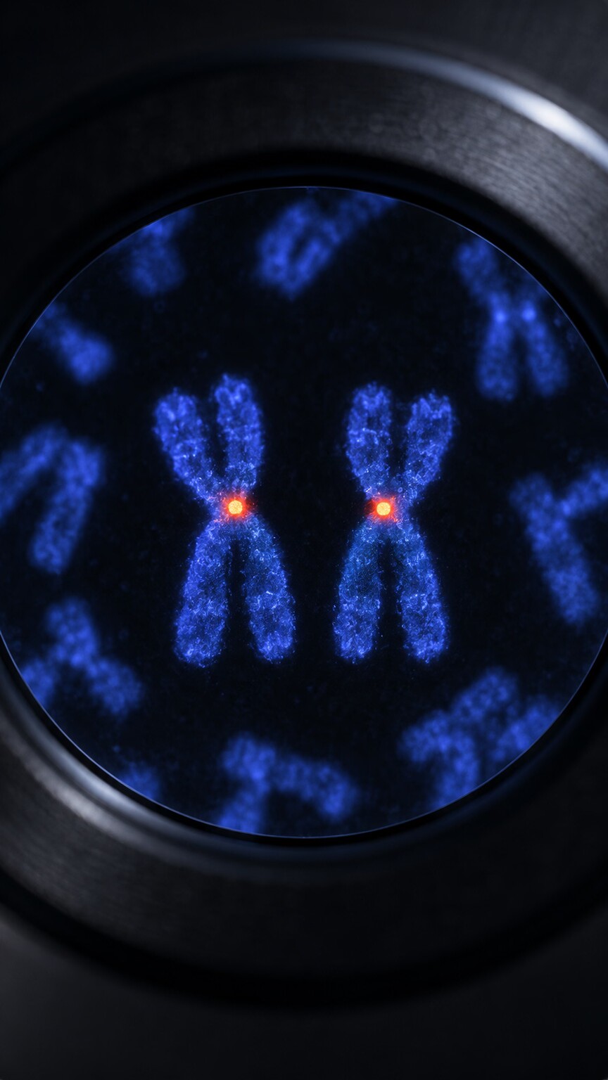
Figure 5. A FISH probe is engineered to bind only to a specific chromosome region, lighting it up under fluorescent microscopy — one glowing signal confirms a normal copy is present, while a missing signal confirms a deletion.
Once a chromosomal microarray flags a specific deletion or duplication, that finding is often confirmed using a targeted technique called fluorescence in situ hybridization, or FISH — a genuinely different technology from the microarray itself, worth understanding since both terms appear together on many genetics reports. A FISH probe is a short, custom-built piece of DNA chemically tagged with a fluorescent marker and engineered to bind only to one specific region of one specific chromosome, matching its sequence precisely. When applied to a sample of the patient's cells and viewed under a fluorescence microscope, each normal copy of that targeted region lights up as a distinct glowing dot; a person with two normal copies shows two dots, while someone with a deletion of that specific region shows only one, and someone with a duplication can show an extra signal or an unusually large, split signal, depending on the specific technique used.
FISH serves two distinct clinical purposes worth distinguishing. First, it directly confirms a deletion or duplication that a microarray already flagged, providing a visual, independent verification using a completely different underlying method rather than simply rerunning the same test. Second, and just as importantly, it's used to test other family members — most commonly parents — quickly and specifically for that one exact region, without needing to run a full microarray on each person, which is exactly the practical tool behind the parental testing discussed earlier in this article, letting a lab efficiently answer the specific question of whether a parent carries the identical finding.
Recurrence Risk: Why Some Findings Change Future Family Planning and Others Don't

Figure 6. Whether a deletion or duplication arose new in a child or was inherited from an unaffected parent meaningfully changes the estimated recurrence risk discussed during genetic counseling for future pregnancies.
Whether a specific deletion or duplication arose fresh in a child (de novo, as described earlier) or was inherited from a parent carrying the identical finding has direct, practical implications for genetic counseling around future pregnancies, which is part of why this distinction receives so much attention during result interpretation. A de novo finding, arising newly rather than being inherited, generally carries a low recurrence risk for future pregnancies from the same parents, since it reflects a one-time event during egg or sperm formation rather than something either parent is consistently passing down. A finding inherited from a parent carrying the same deletion or duplication, particularly one large enough to be reliably passed through standard inheritance patterns, carries a meaningfully higher recurrence risk, generally estimated around fifty percent for each future pregnancy, mirroring the same basic inheritance math that applies to many single-gene genetic conditions.
This distinction becomes considerably more nuanced for the imprinted regions discussed earlier, where recurrence risk also depends on which parent is the carrier and which specific mechanism produced the original finding, sometimes requiring more specialized counseling than the general de novo-versus-inherited framework alone can provide. This is exactly why genetic counselors, rather than a lab report in isolation, are typically the ones translating a specific finding into a specific, personalized recurrence risk estimate for a given family — the underlying math genuinely does depend on the specific region, specific mechanism, and specific inheritance pattern involved, not a single universal rule that applies identically to every deletion or duplication a report might list.
A Worked Example: Same Category of Finding, Three Different Outcomes
Consider three separate genetic test results, each reporting a copy number variant of a similar physical size, somewhere in the range of one to two million base pairs. The first, a deletion on chromosome 22 overlapping the region discussed above, is classified pathogenic and explains a child's combination of a heart defect and immune concerns — multiple haploinsufficient genes, multiple affected systems, exactly as the gene-dosage principle predicts. The second, a duplication on a different chromosome entirely, sits in a well-characterized gene desert with no genes of note inside it and a long track record in population databases of appearing in healthy people; it's classified benign and dismissed as an incidental, unrelated finding. The third, a deletion overlapping a single, poorly characterized gene that current research hasn't yet linked definitively to any clinical picture, is classified a variant of uncertain significance — not dismissed, but not confidently explanatory either, prompting a recommendation for parental testing and periodic reclassification as research continues to catch up with what that specific gene actually does.
This third case is worth sitting with a little longer, since it's genuinely the most common real-world outcome families encounter and often the hardest to receive emotionally. Unlike the first two scenarios, which resolve cleanly into "this explains it" or "this is unrelated," a variant of uncertain significance leaves a family in a state of genuine, honest ambiguity — not a diagnosis, but not full reassurance either. Genetic counselors generally frame this outcome carefully, emphasizing that "uncertain" reflects the current limits of scientific knowledge about that specific gene or region, not an indication that something more concerning is being withheld or downplayed. Many variants initially classified this way are reclassified within a few years as more data accumulates from other families tested for the same finding, more research is published on the specific gene involved, or larger population databases grow large enough to reveal whether that exact variant turns up in healthy people or not — which is exactly why labs generally recommend rechecking in periodically, sometimes every one to two years, on any result that returns in this uncertain middle category, rather than treating the initial classification as necessarily final.
Frequently Asked Questions
Is a duplication generally less serious than a deletion?
Not as a general rule. Some genes are more sensitive to losing a copy (haploinsufficient), others to gaining one (triplosensitive), and some regions like the elastin-containing area on chromosome 7 show that a deletion and duplication of the identical region can each cause distinct, comparably significant conditions.
Can the same deletion cause different severity in different people?
Yes, a pattern called variable expressivity. Even within the same family carrying an identical deletion, other genetic and environmental factors can influence how severely, or whether at all, the associated features actually appear.
Why does it matter which parent a deletion came from?
For a small number of chromosome regions subject to genomic imprinting, only one parent's copy of certain genes is normally active. The same deletion can therefore produce different conditions depending on whether it removed the active copy or the already-silenced one.
Does a large deletion always cause more problems than a small one?
No. What matters most is how many functional, dosage-sensitive genes fall within the deleted or duplicated region, not its physical size — a small deletion in a gene-dense area can be more significant than a much larger one in a gene-sparse region.
What happens if my result comes back as a variant of uncertain significance?
It means current research hasn't yet definitively linked that specific finding to a known outcome. Parental testing and periodic reclassification over time, as more data accumulates, often eventually resolve these findings toward benign or pathogenic.
Conclusion
A chromosomal deletion or duplication is never just a matter of DNA present or absent — it's a change in dosage, and the actual consequence depends on precisely which genes sit inside the affected region, how sensitive those specific genes are to having one copy instead of two (or three instead of two), and, in certain regions, which parent contributed the affected copy in the first place. The same category of finding, the same rough size, can mean anything from a serious multi-system condition to a completely harmless variant carried silently through generations — which is exactly why every deletion or duplication gets evaluated on its own specific genetic content, never judged by size or category alone.
For anyone reading their own genetics report with this framework in mind, a few questions are worth bringing directly to whoever ordered the test or to a genetic counselor: which specific genes fall within the reported region, and is the finding classified as pathogenic, benign, or uncertain; was parental testing performed, and if so, did either parent carry the identical finding; and, if the region happens to be one of the small number known to be subject to genomic imprinting, which parent's copy was actually affected. These aren't just academic details — as this article has shown through several concrete, well-documented examples, each one can genuinely change what a given deletion or duplication actually means for the specific person who received that result, sometimes dramatically so.
The broader lesson underlying every example in this article is that chromosomal copy number changes are never simply "present" or "absent" in a meaningful clinical sense — they exist on a spectrum shaped by which genes are involved, how sensitive those particular genes are to dosage changes in either direction, and occasionally by inheritance patterns unique to a small number of specially regulated genomic regions. A report listing a deletion or duplication is the beginning of an interpretive process, not the end of one, and that process is exactly what turns an otherwise abstract finding into something a family can actually act on with confidence.
Still Not Sure What Your Results Mean?
Upload your labs and get a complete, visual, plain-language interpretation of every biomarker — delivered to your inbox in under 15 minutes.
Get My ReportThis article is for educational purposes only and does not constitute medical advice. Always consult your healthcare provider regarding your specific lab results.