
What Is the Difference Between Numerical and Structural Chromosomal Abnormalities?
When a genetic report comes back describing a "chromosomal abnormality," that single phrase is actually standing in for two entirely different kinds of biological event, and knowing which one you're dealing with changes almost everything about what the finding actually means. Chromosomes carry your genetic instructions in tightly organized packages, and something can go wrong with them in essentially two distinct ways: either the wrong number of packages ends up in the cell, or the right number of packages is present but one of them has been rearranged, broken, or reassembled incorrectly on the inside. The first category is called a numerical abnormality; the second is called a structural abnormality. These aren't just two labels for the same underlying problem — they arise through completely different biological mechanisms, get detected by different kinds of tests, carry different implications for whether a condition can be passed down to future children, and are read completely differently by the specialists interpreting a report. This article walks through exactly what a chromosome is and why its exact count matters so much, what a numerical abnormality is and the real-world conditions it produces, what a structural abnormality is and how it differs mechanically from a numerical one, the important distinction between balanced and unbalanced structural changes, how different genetic tests are actually built to catch one type versus the other, why this classification changes how doctors think about recurrence risk for future pregnancies, and how to actually read which category a specific finding belongs to on your own report.
What a Chromosome Actually Is, Before Splitting Into Types of Abnormality
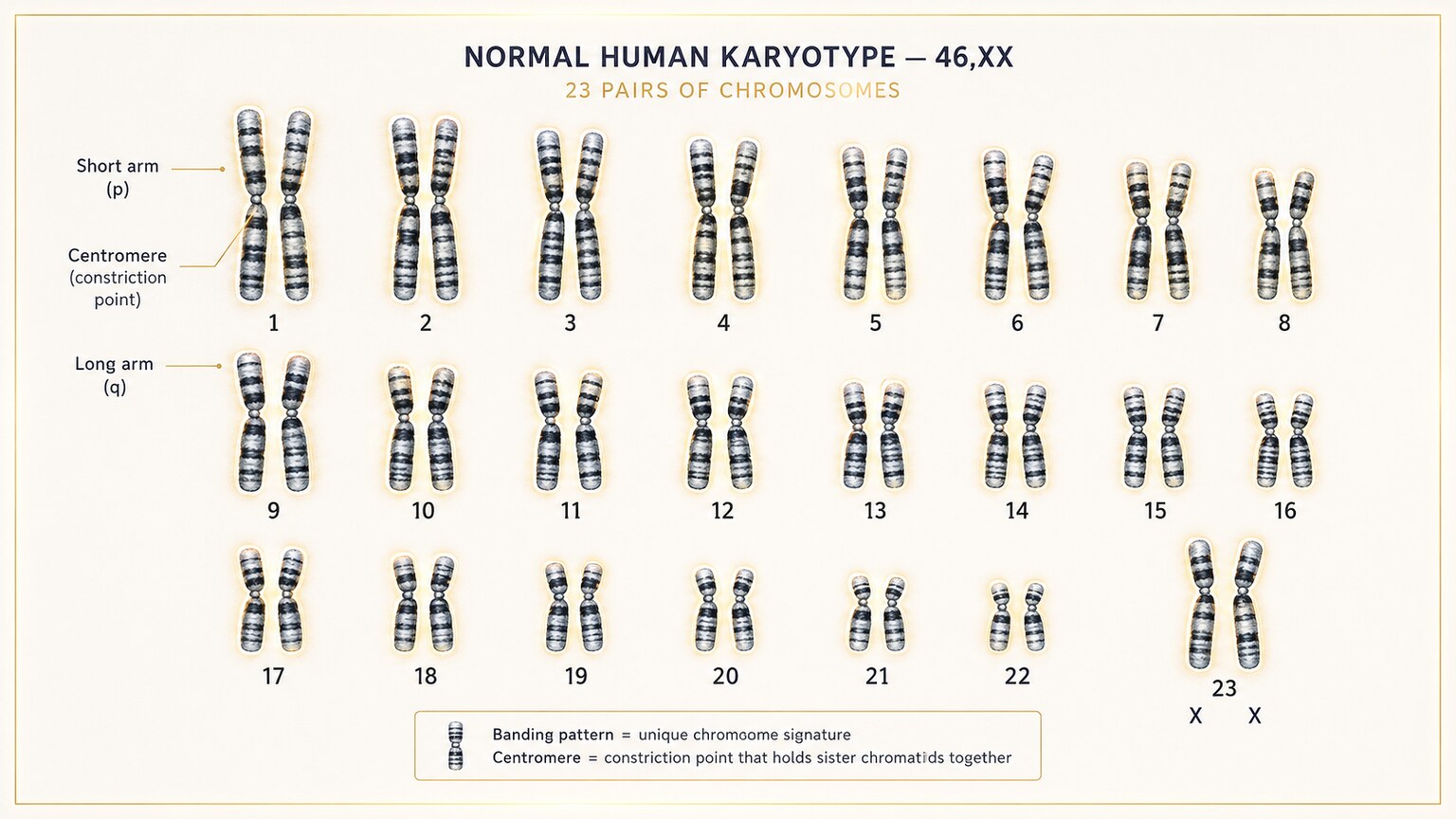
Figure 1. A typical human cell contains exactly 46 chromosomes, organized into 23 matched pairs — the baseline that both numerical and structural abnormalities are defined against.
Every cell in your body (with the exception of eggs and sperm) is built to carry exactly 46 chromosomes, organized into 23 matched pairs — one member of each pair inherited from your mother, the other from your father. Twenty-two of those pairs are called autosomes, and they're numbered roughly from largest to smallest (chromosome 1 down to chromosome 22); the 23rd pair is the sex chromosomes, either XX or XY. Each chromosome is essentially a tightly coiled, organized package of DNA, and the specific genes located along its length are arranged in a consistent order and location, generation after generation, the same way a book's chapters stay in the same order every time it's printed.
This dual requirement — the right number of chromosomes, each one internally intact and correctly organized — is exactly where the numerical/structural distinction comes from. A numerical abnormality is a problem with the count: too many or too few whole chromosomes present in the cell. A structural abnormality is a problem with the architecture: the correct number of chromosomes is present, but the internal arrangement of one or more of them has been altered — a piece is missing, a piece is duplicated, a piece has broken off and reattached somewhere else, or a segment has been flipped end to end. Both categories are genuinely "chromosomal abnormalities" in the broadest sense, but they represent almost entirely different biological events, which is why lumping them together under one umbrella term can be genuinely confusing for anyone trying to understand their own or a family member's specific result.
Numerical Abnormalities: When the Count Itself Is Wrong
A numerical abnormality — also called aneuploidy in medical terminology — means a cell ends up with an extra whole chromosome, a missing whole chromosome, or in rarer cases, an extra full set of chromosomes altogether. The most common mechanism behind this is something called nondisjunction, an error that happens during the specialized cell division that creates eggs and sperm. Normally, each matched pair of chromosomes is supposed to separate cleanly, sending exactly one copy into each resulting reproductive cell. Nondisjunction happens when a pair fails to separate properly, so one resulting egg or sperm cell ends up with two copies of that chromosome instead of one, while the other ends up with zero copies instead of one.
When an egg or sperm carrying this kind of numerical error combines with a normal partner cell at fertilization, the resulting embryo ends up with either three copies of that chromosome (called trisomy, since the normal partner cell still contributes its own single copy) or just one copy (called monosomy). Because this error happens once, essentially by chance, during the formation of a single egg or sperm cell, it typically isn't something inherited from a parent's own chromosomal makeup — the vast majority of numerical abnormalities occur in parents whose own chromosomes are completely normal, with the risk of this kind of error increasing measurably with maternal age due to changes in how egg cells are stored and matured over decades, a pattern explored in more detail later in this article.
Real-World Examples of Numerical Abnormalities You May Have Heard Of
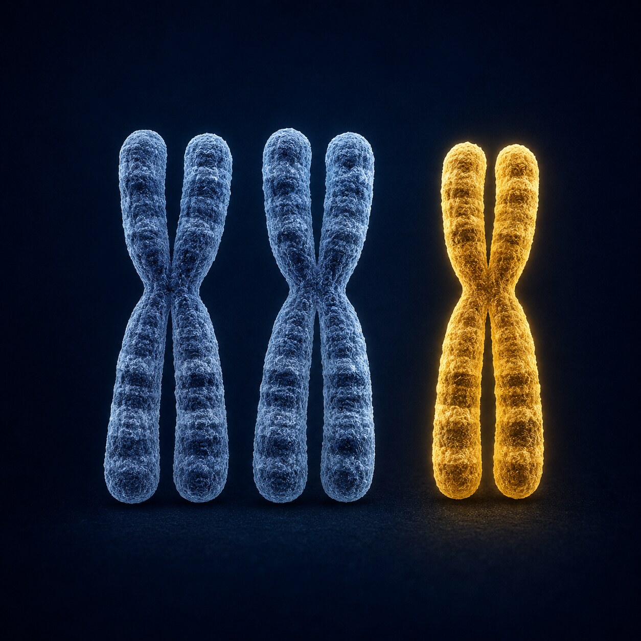
Figure 2. Trisomy 21, three copies of chromosome 21 instead of the usual two, is the most common numerical chromosomal abnormality compatible with survival to birth.
The best-known numerical abnormality is trisomy 21, an extra full copy of chromosome 21, which causes Down syndrome. It's the most frequently occurring numerical abnormality among liveborn babies, and it's a textbook example of the mechanism described above: a nondisjunction error during egg or sperm formation, with no structural rearrangement of any chromosome involved at all — chromosome 21 itself is completely normal in its internal structure, there's simply one extra whole copy of it present. Trisomy 18 (Edwards syndrome) and trisomy 13 (Patau syndrome) work through the identical mechanism, an extra whole copy of a different chromosome, and are both associated with more significant medical complexity.
Numerical abnormalities involving the sex chromosomes follow the same basic principle. Turner syndrome results from monosomy of the X chromosome — a person with only a single X chromosome instead of the usual XX or XY pairing. Klinefelter syndrome results from an extra X chromosome in a male, producing an XXY pattern instead of the typical XY, and is itself one of the more commonly occurring numerical abnormalities involving the sex chromosomes, though it often goes undiagnosed until later in life since its physical effects can be relatively subtle. In every one of these examples, the defining feature is purely a count problem: the internal genetic material on each individual chromosome involved is structurally normal, there is simply the wrong number of whole chromosomes present in the cell.
Structural Abnormalities: When the Count Is Right but the Architecture Is Wrong
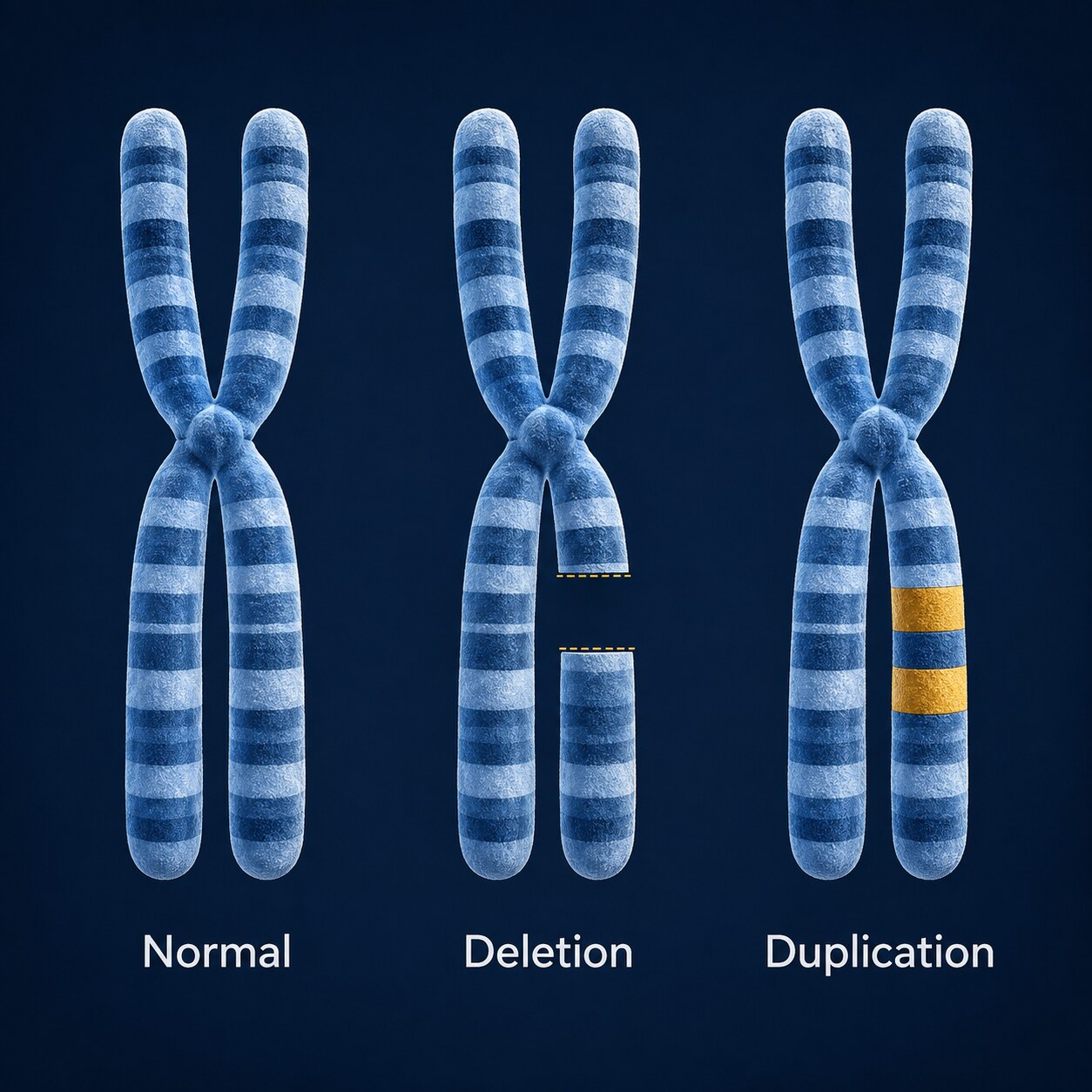
Figure 3. A structural abnormality changes the internal architecture of a single chromosome — a missing segment (deletion) or an extra copy of a segment (duplication) — without necessarily changing the total chromosome count.
Structural abnormalities work through a completely different mechanism than numerical ones. Rather than an entire chromosome going missing or being duplicated, a structural abnormality happens when a chromosome breaks at one or more points and then gets repaired incorrectly, or when genetic material gets exchanged between two different chromosomes during the normal process of genetic recombination. The total chromosome count can, in many cases, remain exactly 46 — the problem isn't how many packages are present, it's that the internal contents of one specific package have been rearranged.
Several distinct types of structural change exist, each with its own name depending on exactly what happened at the break point. A deletion means a segment of a chromosome is missing entirely — that stretch of genetic material, and every gene located on it, simply isn't there anymore. A duplication is the reverse: an extra copy of a segment is present, so genes in that region are represented more times than they should be. A translocation happens when a broken piece of one chromosome reattaches to a different, unrelated chromosome instead of repairing back onto its own. An inversion happens when a chromosome segment breaks at two points, flips completely end to end, and reattaches backwards. A ring chromosome, a rarer structural change, happens when a chromosome loses material at both of its ends and the two remaining ends fuse together into a closed loop. An isochromosome, another distinct structural pattern, happens when a chromosome divides incorrectly along the wrong axis during cell division, producing a chromosome made of two mirror-image copies of just one arm instead of the normal two genetically distinct arms.
Curious what your own lab results actually mean for your specific situation? Upload your results and get a complete, plain-language breakdown in under 15 minutes.
Analyze My ResultsBalanced vs. Unbalanced: The Critical Sub-Distinction Within Structural Changes
Within the structural category specifically, there's one more distinction that matters enormously for how a finding is interpreted: whether the rearrangement is balanced or unbalanced. A balanced structural rearrangement means all the genetic material that was originally present is still present somewhere in the cell — it's simply been reorganized, like a translocation where a piece from chromosome 5 swapped places with a piece from chromosome 12, with nothing actually lost or gained overall. An unbalanced structural rearrangement means genetic material has genuinely been lost or gained in the process — a deletion, a duplication, or a translocation where the exchange between chromosomes wasn't equal on both sides.
This balanced-versus-unbalanced distinction is often more clinically important than the numerical-versus-structural distinction itself, because a person carrying a perfectly balanced structural rearrangement typically has no health effects from it at all — every gene they need is present in the correct total dose, just relocated. The real significance of a balanced rearrangement usually only becomes apparent when that person has children, since their eggs or sperm can end up with an unbalanced version of the same rearrangement, passing on a genuine deletion or duplication to the next generation even though the parent carrying the balanced version was never affected. This is also why balanced rearrangements are sometimes discovered only after a couple experiences recurrent pregnancy loss or difficulty conceiving, since testing prompted by those specific concerns can be what first reveals a rearrangement that had been present, silently, in one parent's chromosomes their entire life, with no earlier symptom, illness, or health effect of any kind ever hinting at its existence beforehand.
Mosaic Forms: When Only Some Cells in the Body Carry the Change
Both numerical and structural abnormalities can also occur in what's called a mosaic form, meaning the abnormality is present in only some of a person's cells rather than every single one, existing side by side with a separate population of completely normal cells within the same body. Mosaicism happens when the underlying error — whether a nondisjunction event or a structural breakage — occurs not during egg or sperm formation, but slightly later, after fertilization, during one of the very first rounds of cell division as the embryo begins to grow. Because every future cell descends from whichever cell divided at that point, only the specific lineage of cells descending from the one affected division carries the abnormality, while cells descending from other, unaffected divisions remain completely normal.
Mosaic forms exist for both categories discussed in this article — mosaic trisomy 21 (some cells with the standard two copies of chromosome 21, others with three) and mosaic structural rearrangements both occur, and in both cases, the proportion of affected cells, and specifically which tissues in the body those affected cells happen to be concentrated in, can meaningfully influence how a person is affected. A relatively low percentage of affected cells, particularly if concentrated in tissues with less overall functional impact, can sometimes correspond to a considerably milder presentation than the same abnormality would produce if present in every single cell of the body — one more layer of nuance that sits on top of the basic numerical-versus-structural classification this article has focused on.
How Different Genetic Tests Are Built to Catch One Type but Not Always the Other
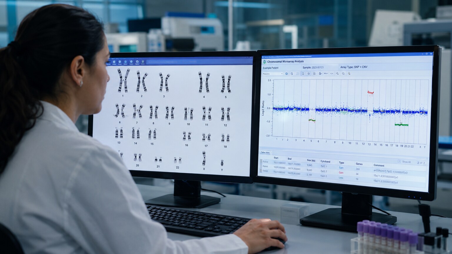
Figure 4. A karyotype test visually counts and inspects whole chromosomes, while a chromosomal microarray measures DNA dosage at thousands of points — each method is suited to catching a different category of abnormality.
This distinction isn't just academic — it directly determines which laboratory test is actually capable of detecting a given problem. A karyotype test, the classic method where a lab technician photographs and visually arranges all 46 chromosomes under a microscope, is excellent at catching numerical abnormalities (an extra or missing whole chromosome is easy to count) and large structural rearrangements big enough to see directly, like a major translocation between two large chromosomes. What a karyotype often cannot catch is a very small structural change — a deletion or duplication involving only a tiny segment of a chromosome, far too small to see even under high magnification.
A chromosomal microarray, a newer and more sensitive technology, works completely differently: instead of visually counting and inspecting chromosomes, it measures the relative dosage of DNA at thousands of specific points along the genome, comparing a person's sample against a reference standard to detect regions with too much or too little genetic material. This makes it dramatically better than a karyotype at catching small deletions and duplications, the kind of unbalanced structural change that might involve only a handful of genes. What a microarray generally cannot catch, however, is a balanced rearrangement — since a balanced translocation or inversion doesn't change the total amount of genetic material at any point along the chromosome, there's no dosage difference for the microarray to detect, even though the rearrangement is real and could still matter for that person's future children.
Because each method has this genuine, well-defined blind spot, specialists often order both tests together rather than choosing one over the other, particularly when the clinical picture doesn't clearly point toward one specific category of finding in advance. A karyotype and a microarray run side by side cover almost the entire range of possible chromosomal findings between them — the karyotype catching numerical changes and large structural rearrangements including balanced ones, the microarray catching small unbalanced structural changes the karyotype's resolution would miss entirely. Newer sequencing-based technologies are increasingly able to bridge even this remaining gap, offering the potential to detect both dosage changes and balanced rearrangements from a single test, though karyotype and microarray remain the most widely available, well-validated combination in routine clinical use as of this writing.
Why This Distinction Changes How Doctors Think About Recurrence Risk

Figure 5. Whether a finding is numerical or structural directly changes the recurrence-risk conversation a genetic counselor has with a family about future pregnancies.
Because numerical and structural abnormalities arise through such different mechanisms, they carry very different implications for a couple planning future pregnancies. A numerical abnormality like trisomy 21 is, in the large majority of cases, a one-time, sporadic event tied to an error in the formation of a single egg or sperm cell, not a reflection of an underlying, permanent chromosomal pattern in either parent. This means the recurrence risk for a future pregnancy — while somewhat elevated compared to the general population, and rising further with maternal age — is still relatively low in most individual cases, and testing the parents' own chromosomes typically shows both of them to be completely normal.
A structural abnormality found in a child, by contrast, often prompts testing of both parents specifically to check whether one of them silently carries a balanced version of that same rearrangement. If a parent is found to carry a balanced translocation, for example, the recurrence risk for future pregnancies can be substantially higher than the general population risk, because that parent has a structural chromosomal pattern built into every one of their own reproductive cells, not a one-time random event. This is precisely why a structural finding in a child so often leads to parental testing, genetic counseling, and a much more detailed conversation about future family planning than a numerical finding typically requires.
When a parent is confirmed to carry a balanced structural rearrangement, genetic counselors can typically calculate a more specific recurrence risk estimate for that family than a generic population-wide figure, based on exactly which chromosomes are involved and precisely where the break points sit. Some balanced rearrangements carry a meaningfully elevated risk of producing an unbalanced version in a future pregnancy, while others, depending on the specific chromosomes and segments involved, carry a risk closer to the general population baseline — which is exactly why identifying the parent as a carrier is only the first step, with the specific rearrangement's own risk profile determining what comes next. Reproductive options in these situations can include closer prenatal monitoring in a future pregnancy, or for some families, exploring assisted reproductive approaches that allow embryos to be screened for the specific rearrangement before a pregnancy begins at all.
Prenatal Versus Postnatal Testing: Why the Same Classification Applies at Every Life Stage
The numerical-versus-structural framework described throughout this article applies identically whether testing happens before birth, in a newborn, in a child being evaluated for developmental differences, or in an adult referred for testing decades later — the underlying biology of what makes something numerical or structural doesn't change depending on when in life it's discovered. What does change across these different contexts is which testing method is typically used first and why. Prenatal testing often starts with a less invasive screening approach analyzing fetal DNA fragments circulating in a pregnant person's blood, which is particularly well-suited to flagging likely numerical abnormalities like trisomy 21, 18, and 13, before a more definitive diagnostic test (typically amniocentesis or chorionic villus sampling, followed by a full karyotype or microarray) confirms the specific finding and clarifies whether it's numerical, structural, or occasionally both.
Postnatal testing in a child or adult, by contrast, is usually prompted by a specific clinical question — developmental differences never fully explained, recurrent pregnancy loss, unexplained infertility, or a family history of a known rearrangement — and the choice between karyotype and microarray (or sometimes both together) is guided by which specific category of finding is suspected based on that clinical picture. A family history of a known balanced translocation, for instance, points a specialist toward karyotype testing specifically, since that's the method capable of detecting a balanced rearrangement; a child with unexplained developmental delay and no specific family history more often starts with a microarray, since it's better positioned to catch the kind of small, previously unknown structural change most likely to be relevant in that particular clinical scenario.
Can a mosaic chromosomal finding be either numerical or structural?
Yes. Mosaicism describes what proportion of a person's cells carry an abnormality, not which category that abnormality belongs to — both numerical mosaicism (like mosaic trisomy 21) and structural mosaicism can occur, existing alongside a separate population of the person's own normal cells.
Does the testing method change depending on whether testing happens before or after birth?
The initial screening approach often differs — prenatal testing frequently starts with a blood-based screen well-suited to flagging likely numerical findings — but the same karyotype and microarray methods used to confirm a diagnosis, and the same numerical-versus-structural classification framework, apply at every life stage.
How to Read Which Category a Finding Belongs to on an Actual Report
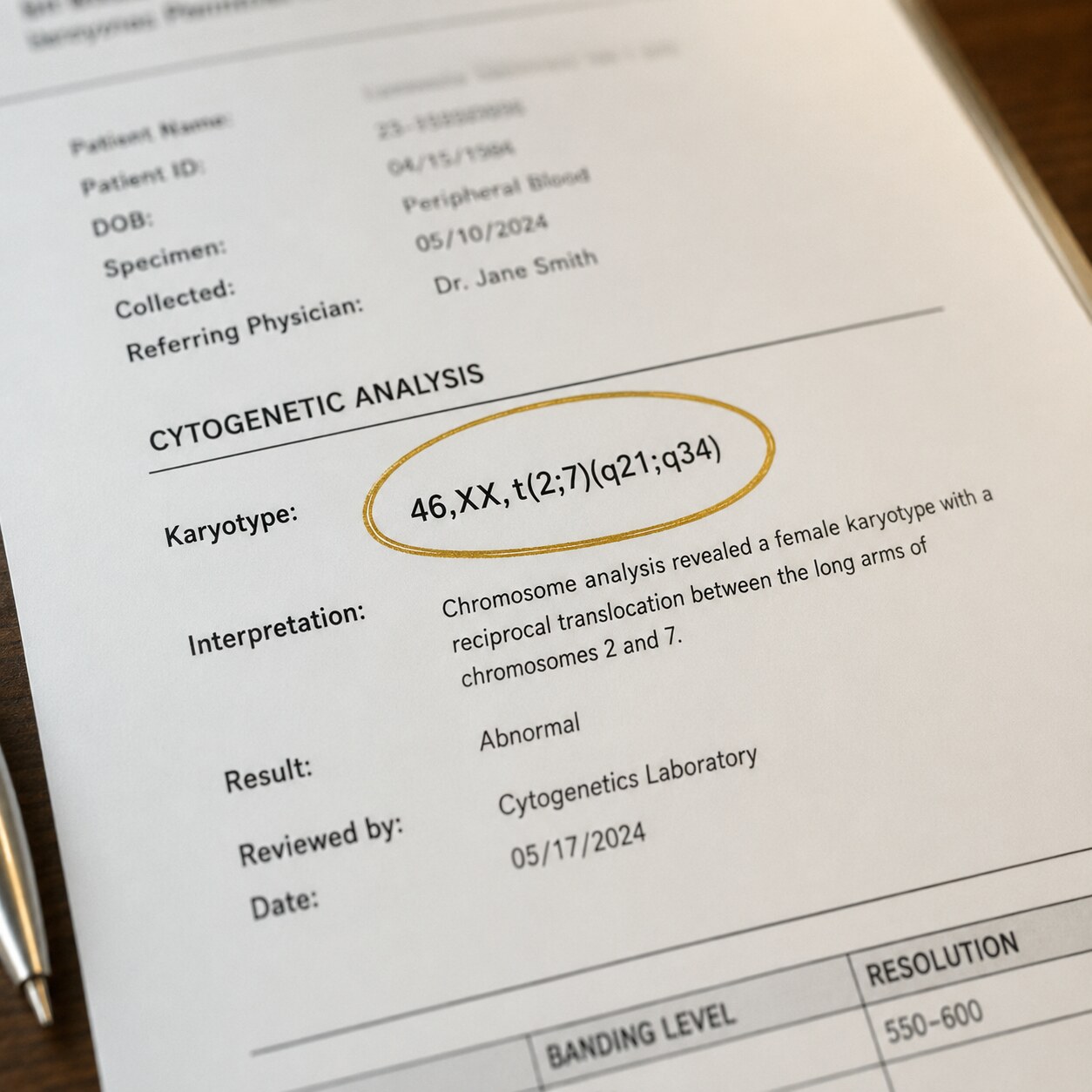
Figure 6. Standard karyotype notation on a report — such as "47,XY,+21" for trisomy 21 or "46,XX,t(2;7)" for a balanced translocation — encodes exactly which category of abnormality was found.
Chromosomal reports use a standardized shorthand notation that, once decoded, tells you directly which category a finding belongs to. The notation always starts with the total chromosome count, followed by the sex chromosomes, followed by a description of anything abnormal. A result written as "47,XY,+21" tells you the total count is 47 (one more than the normal 46), followed by the sex chromosomes XY, followed by "+21," meaning the extra chromosome is a third copy of chromosome 21 — a numerical abnormality, specifically trisomy 21.
A result written as "46,XX,t(2;7)(q21;q34)" tells a very different story: the total count is a completely normal 46, followed by XX, followed by "t(2;7)," which stands for a translocation between chromosomes 2 and 7, with the specific break points noted in parentheses. Because the total count here is normal 46, this is immediately identifiable as a structural finding, not a numerical one — the count is right, but the internal arrangement of two chromosomes has been altered. Learning to recognize this pattern — whether the leading number differs from 46 (numerical) or stays at 46 while a specific rearrangement is spelled out afterward (structural) — is one of the most useful skills for making sense of a genetic report before ever discussing it with a specialist, and it works consistently across essentially every laboratory report using this standardized international notation system.
What Happens to a Cell Biologically When Each Type of Error Occurs
It's worth walking through the underlying cell biology a little more closely, since understanding exactly when and how each error occurs helps explain why they behave so differently afterward. Numerical errors happen during a specific, tightly choreographed stage of cell division called meiosis, which is the process that produces eggs and sperm with exactly half the normal number of chromosomes, so that when two of these reproductive cells combine at fertilization, the resulting embryo ends up back at the correct total of 46. During meiosis, matched chromosome pairs are pulled apart by a network of molecular fibers, and nondisjunction is essentially a mechanical failure in that pulling-apart process — the fibers fail to separate one particular pair cleanly, and the error is locked in for every single cell that grows from that one affected egg or sperm going forward, since every subsequent cell division simply copies whatever chromosome count was present at the very start.
Structural errors happen through an entirely different biological process: DNA breakage and repair. Chromosomes are, under completely normal circumstances, subject to a huge number of tiny breaks and repairs constantly, as part of ordinary cellular maintenance and the routine process of genetic recombination that shuffles genetic material between matched chromosome pairs during egg and sperm formation. The overwhelming majority of these breaks are repaired with complete accuracy, restoring the chromosome to its original, correct sequence. A structural abnormality happens on the rare occasion when this repair process goes wrong — when a break is repaired using the wrong piece of DNA as a template, or when two separate breaks on two different chromosomes get repaired by mistakenly swapping their loose ends with each other instead of with their own original partner. This is a fundamentally different kind of biological accident than a failed separation of matched pairs, which is part of why the resulting abnormalities behave so differently in terms of inheritance and recurrence.
Why Numerical Abnormalities Become More Common With Advancing Maternal Age
One of the most well-documented patterns in reproductive genetics is the steadily rising risk of numerical chromosomal abnormalities as maternal age increases, and understanding why this happens specifically (rather than just knowing that it happens) helps clarify why this age effect applies so much more to numerical abnormalities than to structural ones. Unlike sperm, which are produced continuously throughout a man's reproductive life, all of the egg cells a woman will ever have are actually formed before she is even born, and then held in a paused, dormant state within the ovaries for decades, sometimes for 40 years or more, before any individual egg is finally matured and released during ovulation.
The molecular machinery responsible for correctly separating chromosome pairs during that final maturation step is thought to gradually degrade the longer an egg cell has been sitting in this dormant state, making the mechanical failure behind nondisjunction measurably more likely to occur in eggs that have been waiting the longest. This is a plausible, well-supported explanation for why the risk of numerical abnormalities like trisomy 21 rises noticeably after a woman's mid-30s and continues climbing through her 40s — it isn't a reflection of anything a person did or didn't do, or any lifestyle factor within their control, but a consequence of the decades-long biological storage period unique to how human egg cells are formed. Sperm, by contrast, are freshly produced on an ongoing basis, which is part of why paternal age shows a much weaker and less consistent association with numerical abnormalities specifically, even though advanced paternal age does carry its own separate, smaller genetic risks related to a different category of error entirely (new mutations within individual genes, rather than whole-chromosome count problems) — a distinction worth keeping in mind since the two are often, mistakenly, conflated in casual conversation.
Why Severity Doesn't Track Neatly With Either Category Alone
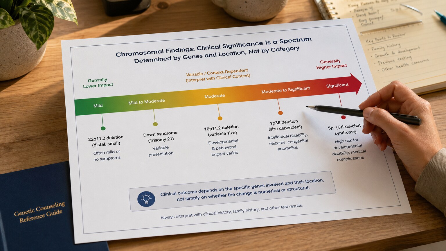
Figure 7. Neither category is inherently "more severe" than the other — a tiny structural deletion can carry more significant effects than a well-tolerated numerical variation, and the reverse is equally true.
It's tempting to assume that one category is automatically more serious than the other — that a missing or extra whole chromosome must be worse than a small internal rearrangement, simply because it sounds like a bigger event. In reality, severity has far more to do with which specific genes are affected and how many of them, not which broad category the change falls into. A numerical abnormality involving a chromosome that's relatively gene-sparse can sometimes be compatible with a fuller range of health outcomes than a tiny structural deletion that happens to remove a segment packed with genes essential for early development — size and category tell you relatively little on their own; location and gene content tell you most of what actually matters.
This is exactly why genetic counselors and laboratory geneticists spend so much of their interpretation effort not on the broad numerical-versus-structural label itself, but on precisely which genes fall within the affected region, what's already known from published case reports about similar findings, and whether population databases show the same change occurring in people with no health effects at all. The numerical/structural distinction is the essential first branch in understanding a finding's mechanism and inheritance pattern — but it is deliberately not, by itself, a reliable predictor of how significant that finding's health impact will turn out to be for any one specific person.
What This Means the Next Time You See These Terms on a Report
Armed with this framework, a genetic report no longer needs to read as an intimidating block of unfamiliar shorthand. The first, most useful question to ask of any chromosomal finding is simply which of the two broad categories it falls into: does the leading number in the notation differ from the expected 46, signaling a numerical change, or does it stay at 46 while a specific rearrangement is spelled out afterward, signaling a structural one. From there, the second most useful question is whether a structural finding has been described as balanced or unbalanced, since that single word carries enormous weight for understanding both the finding's likely health impact and its implications for future family planning.
Neither of these two initial questions requires a genetics degree to ask, and both give a genuine, substantive head start on understanding a result well before a formal consultation with a genetic counselor or specialist ever takes place. That head start doesn't replace the value of that expert conversation — the specific genes involved, the relevant published literature, and the individualized recurrence-risk calculation still require professional expertise — but walking into that conversation already understanding the basic numerical-versus-structural, balanced-versus-unbalanced framework this article has covered allows for a far more informed, far less overwhelming discussion from the very first question asked.
Frequently Asked Questions
Can a person have both a numerical and a structural chromosomal abnormality at the same time?
Yes, though it's less common than having just one. Some conditions involve an extra copy of a rearranged chromosome, combining both a count problem and an architecture problem in the same finding, which a specialist would describe using both parts of the standard notation together.
Is a structural abnormality always inherited from a parent?
No. While parental testing is commonly done to check, many structural rearrangements arise new, for the first time, in a single egg or sperm cell or very early in embryonic development, with both parents' own chromosomes testing completely normal.
Which type is more common overall, numerical or structural?
Numerical abnormalities are considerably more common among detected chromosomal findings overall, largely because nondisjunction errors during egg and sperm formation happen relatively frequently, especially as maternal age increases, compared to the specific breakage-and-repair events that produce structural changes.
Does a balanced structural rearrangement ever need treatment?
Typically not for the person carrying it, since all their genetic material is present in the correct amount. The relevant conversation is usually about future family planning and the chance of passing an unbalanced version to a child, not about treating the carrier themselves.
Conclusion
Numerical and structural chromosomal abnormalities represent two fundamentally different biological events sharing the same broad label: one is a problem with how many whole chromosomes are present, and the other is a problem with the internal architecture of an otherwise correctly counted chromosome. That distinction shapes which laboratory test can actually detect a given finding, how a specialist interprets its significance, and — perhaps most importantly for families — how the recurrence risk conversation unfolds for future pregnancies. Learning to recognize which category a specific finding belongs to, including the important balanced-versus-unbalanced sub-distinction within structural changes, turns an intimidating block of genetic notation into something considerably more understandable and actionable in conversation with a genetics specialist.
Beyond the mechanics of classification itself, the broader lesson worth carrying forward is that neither category is inherently more alarming than the other, and neither by itself predicts severity, treatment needs, or long-term outlook — those answers depend on the specific genes and chromosomes involved in each individual case. What the numerical-versus-structural distinction reliably does provide is the correct starting framework for asking the right follow-up questions: how the finding likely arose, whether it's likely to recur in a future pregnancy, and which additional testing, if any, might add useful clarity. Approached with that framework in hand, a genetic report that once looked like an intimidating wall of unfamiliar notation becomes a considerably more navigable, understandable piece of one's own or one's family's health story.
Still Not Sure What Your Results Mean?
Upload your labs and get a complete, visual, plain-language interpretation of every biomarker — delivered to your inbox in under 15 minutes.
Get My ReportThis article is for educational purposes only and does not constitute medical advice. Always consult your healthcare provider regarding your specific lab results.