
Why Is Chromosomal Testing Done During Pregnancy?
Chromosomal testing is offered during pregnancy to check whether the developing baby has the correct number and structure of chromosomes — the 46 tightly-packaged bundles of DNA that carry every instruction needed to build a person — because certain chromosome differences, like an extra copy of chromosome 21 in Down syndrome, are common enough and clinically significant enough that most obstetric guidelines now recommend offering some form of this testing to every pregnant patient, not just those considered higher risk. What often confuses expecting parents is that "chromosomal testing" during pregnancy isn't one single test — it's a menu of several very different options, ranging from a simple blood draw with zero risk to the pregnancy, to a diagnostic procedure that samples cells directly from the placenta or the amniotic fluid. This article walks through exactly what each option actually checks, why your OB might recommend one over another, and — critically — the difference between a screening result and a diagnosis, since mixing those two up is the single most common source of unnecessary panic in prenatal care.
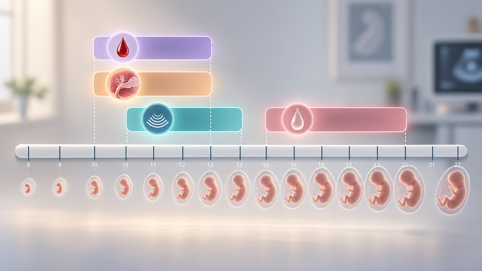
Figure 1. Different prenatal chromosomal tests become available at different points in pregnancy, from cell-free DNA screening as early as week 10 to amniocentesis starting around week 15.
What "Chromosomal Testing" Actually Means in Pregnancy
Every cell in your body — and every cell in a developing fetus — normally contains 46 chromosomes, arranged in 23 matched pairs, one member of each pair inherited from each parent. Think of chromosomes as 23 volumes of an instruction manual, each volume holding a different, non-overlapping set of chapters (genes) needed to build and run a human body. Prenatal chromosomal testing exists to answer one core question: does this particular set of 23 volumes have the right number of volumes, and are they structurally intact, or is a volume missing, duplicated, or rearranged in a way that changes the instructions the body receives?
The most common chromosome differences tested for during pregnancy are numerical — an extra or missing whole chromosome, like trisomy 21 (Down syndrome, an extra chromosome 21), trisomy 18 (Edwards syndrome), trisomy 13 (Patau syndrome), or an atypical number of the X and Y sex chromosomes. These conditions occur at a rate strongly correlated with maternal age but can happen in a pregnancy of any age, which is exactly why most professional guidelines — including those from the American College of Obstetricians and Gynecologists — now recommend offering some form of chromosomal screening to every pregnant patient, regardless of age or risk factors, rather than reserving it for those over 35.
It helps to understand why these particular chromosomes — 21, 18, and 13 — are the ones most commonly discussed, rather than any of the other 20 numbered chromosome pairs. Chromosomes vary enormously in how much genetic material they carry, and chromosome 21 happens to be the smallest of all 23, carrying relatively few genes compared to its neighbors. An extra copy of a gene-dense chromosome, like chromosome 1 or chromosome 3, is essentially incompatible with a pregnancy continuing at all and almost always results in an early miscarriage, often before a person even knows they're pregnant — which is exactly why those extra-chromosome conditions are rarely discussed in prenatal testing conversations at all: they're simply not compatible with a pregnancy reaching a stage where testing would catch them. Chromosomes 21, 18, and 13, being smaller and carrying comparatively less genetic material, are among the few whose extra copy allows a pregnancy to continue to term often enough that it became clinically important to develop reliable ways of detecting them.
This same logic extends to differences in the number of sex chromosomes — X and Y — which tend to be more compatible with ongoing pregnancy and life after birth than an extra autosome (a non-sex chromosome) typically is, producing conditions like Turner syndrome (a single X chromosome instead of the usual pair) or Klinefelter syndrome (an extra X chromosome in a male fetus). Most current-generation NIPT panels screen for these sex chromosome differences as a standard part of the same blood draw, alongside the more commonly discussed trisomies, though the pregnant patient can typically choose in advance whether they want this additional category of information included in their results.
Cell-Free DNA Screening (NIPT): The Blood Test Option
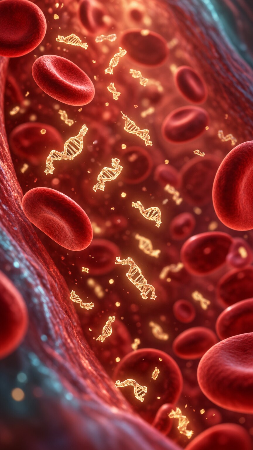
Figure 2. Cell-free DNA screening works by detecting small fragments of placental DNA that circulate freely in the pregnant person's own bloodstream from around the tenth week of pregnancy onward.
Non-invasive prenatal testing, almost universally shortened to NIPT, is a simple blood draw from the pregnant person's arm — nothing touches the baby or the placenta directly. It works because of a genuinely elegant biological quirk: throughout pregnancy, small fragments of DNA from the placenta break off and circulate freely in the mother's bloodstream, mixed in among her own DNA fragments. Because the placenta develops from the same fertilized egg as the fetus, this placental DNA is, in the overwhelming majority of pregnancies, an accurate stand-in for the fetus's own chromosomes. A specialized lab sorts through this mixed pool of DNA fragments and counts how much comes from each chromosome — too much material from chromosome 21 relative to the rest suggests a possible extra copy, exactly the way finding twice as many pages torn from chapter 21 of a shredded book would suggest an extra copy of that chapter existed somewhere in the pile.
NIPT can typically be performed starting around week 10 of pregnancy, since enough placental DNA needs to have accumulated in the maternal bloodstream by that point for reliable detection, and results usually take about one to two weeks to come back. Its major strength is accuracy for the conditions it's actually designed to detect: for trisomy 21 specifically, NIPT correctly flags the vast majority of affected pregnancies while producing very few false alarms in the general population, a performance level meaningfully better than the older screening methods it has largely replaced as a front-line option. It's important to understand that NIPT is a screening test, not a diagnostic one — a "high risk" result means the pregnancy needs a diagnostic test to confirm the finding, not that the baby definitely has the condition, a distinction covered in detail further down.
NIPT can occasionally come back with a result labeled "no call" or "test failed," most often because the sample simply didn't contain enough placental DNA relative to the mother's own DNA — a proportion referred to as the fetal fraction — to produce a reliable answer either way. This happens more frequently earlier in pregnancy, in patients with a higher body weight (since fetal fraction tends to run lower in that circumstance for reasons not fully understood), or in pregnancies with certain underlying chromosomal differences, which is itself sometimes a subtle clue worth following up on rather than simply repeating the blood draw and moving on. When this happens, the usual next step is either a repeat blood draw a week or two later, once more placental DNA has typically accumulated, or a conversation about proceeding directly to a different screening or diagnostic option instead.
It's also worth knowing that NIPT can occasionally detect something entirely unrelated to the fetus's own chromosomes: a small percentage of unusual or discordant results eventually trace back to an undiagnosed chromosomal change in the pregnant person themselves, or, more rarely, to an early, undetected vanishing twin whose placental DNA is still circulating in the maternal bloodstream even though only one fetus continues developing. These scenarios are uncommon, but they're part of why an unexpected or hard-to-interpret NIPT result is always worked through with a genetic counselor rather than taken at face value, since the range of possible explanations is genuinely broader than "the baby does or doesn't have the condition being screened for."
Trying to make sense of a genetics or prenatal lab report on your own? Upload your results and get a complete, plain-language breakdown in under 15 minutes.
Analyze My ResultsFirst-Trimester Combined Screening: The Older Blood-and-Ultrasound Approach
Before cell-free DNA screening became widely available, and still used today in some practices or alongside NIPT, first-trimester combined screening pairs a maternal blood test — measuring two specific pregnancy-associated proteins — with a specialized ultrasound measurement called nuchal translucency, which measures the thickness of a small, normally fluid-filled space at the back of the developing baby's neck. An unusually thick nuchal translucency measurement, combined with certain patterns in the blood protein levels, statistically raises the estimated chance of a chromosomal difference, and a software algorithm combines the maternal age, blood results, and ultrasound measurement into a single personalized risk estimate, typically expressed as an odds ratio like "1 in 250."
Combined screening detects meaningfully fewer cases of Down syndrome than NIPT does and produces more false-positive results, which is exactly why most practices that have access to cell-free DNA screening have shifted toward offering NIPT as the primary front-line option. Combined screening still has a genuinely useful role, though: because it happens earlier in some clinical workflows and includes an anatomy-focused ultrasound component, it can pick up on structural findings — like an increased nuchal translucency associated with a heart defect — that a blood-only test like NIPT was never designed to catch in the first place, since NIPT only reads chromosome material and has no way to see physical structure at all.
Chorionic Villus Sampling: The Earlier Diagnostic Option
Chorionic villus sampling, almost always shortened to CVS, is the first of the two diagnostic (not screening) options, and it's the one performed earlier in pregnancy, typically between weeks 10 and 13. A diagnostic test, unlike a screening test, directly examines actual fetal-origin cells rather than statistically estimating risk, which is why it can give a definitive yes-or-no answer rather than a probability.

Figure 3. Chorionic villus sampling collects a small tissue sample directly from the chorionic villi — finger-like projections of the placenta — allowing fetal chromosomes to be examined directly rather than estimated statistically.
CVS collects a tiny sample of the chorionic villi, small finger-like projections of placental tissue, either through a thin catheter passed through the cervix or a needle inserted through the abdomen, guided continuously by real-time ultrasound to keep the sampling instrument well clear of the fetus itself. Because the villi share the fetus's own chromosome makeup in the vast majority of cases, examining them under a microscope allows lab specialists to directly count and examine the chromosomes rather than infer them statistically. The tradeoff for this earlier timing and diagnostic certainty is a small but real procedure-related pregnancy loss risk, generally cited in modern published data as somewhere in the range of 1 in 300 to 1 in 500 procedures when performed by an experienced specialist, a figure worth discussing directly with a maternal-fetal medicine specialist rather than a generic online source, since it varies with the specific technique used and the individual clinical situation.
Two different access routes exist for CVS — transcervical, where a thin, flexible catheter is passed through the cervix, and transabdominal, where a needle is inserted through the lower abdomen, similar in feel to the needle used in amniocentesis. Which route is used depends mainly on where the placenta has attached inside the uterus, something the ultrasound performed just before the procedure identifies; a placenta attached toward the back of the uterus is often more easily reached transcervically, while one attached toward the front more often favors the transabdominal route, and the specialist performing the procedure makes this call in real time based on the clearest, safest path to the tissue being sampled. Most patients describe the sensation as similar to a Pap smear for the transcervical route, or a sensation similar to an amniocentesis needle for the transabdominal route, with the entire sample collection itself typically taking well under a minute once the needle or catheter is correctly positioned.
Because CVS is performed earlier than amniocentesis, choosing it can mean having a definitive answer several weeks sooner in the pregnancy — a meaningful practical difference for families for whom that additional time matters, whether for medical decision-making, emotional preparation, or simply reducing the length of time spent waiting on uncertain information. This earlier timing is, for many families who already know they want diagnostic certainty, the single deciding factor in choosing CVS over waiting for amniocentesis instead.
Amniocentesis: The Second-Trimester Diagnostic Option
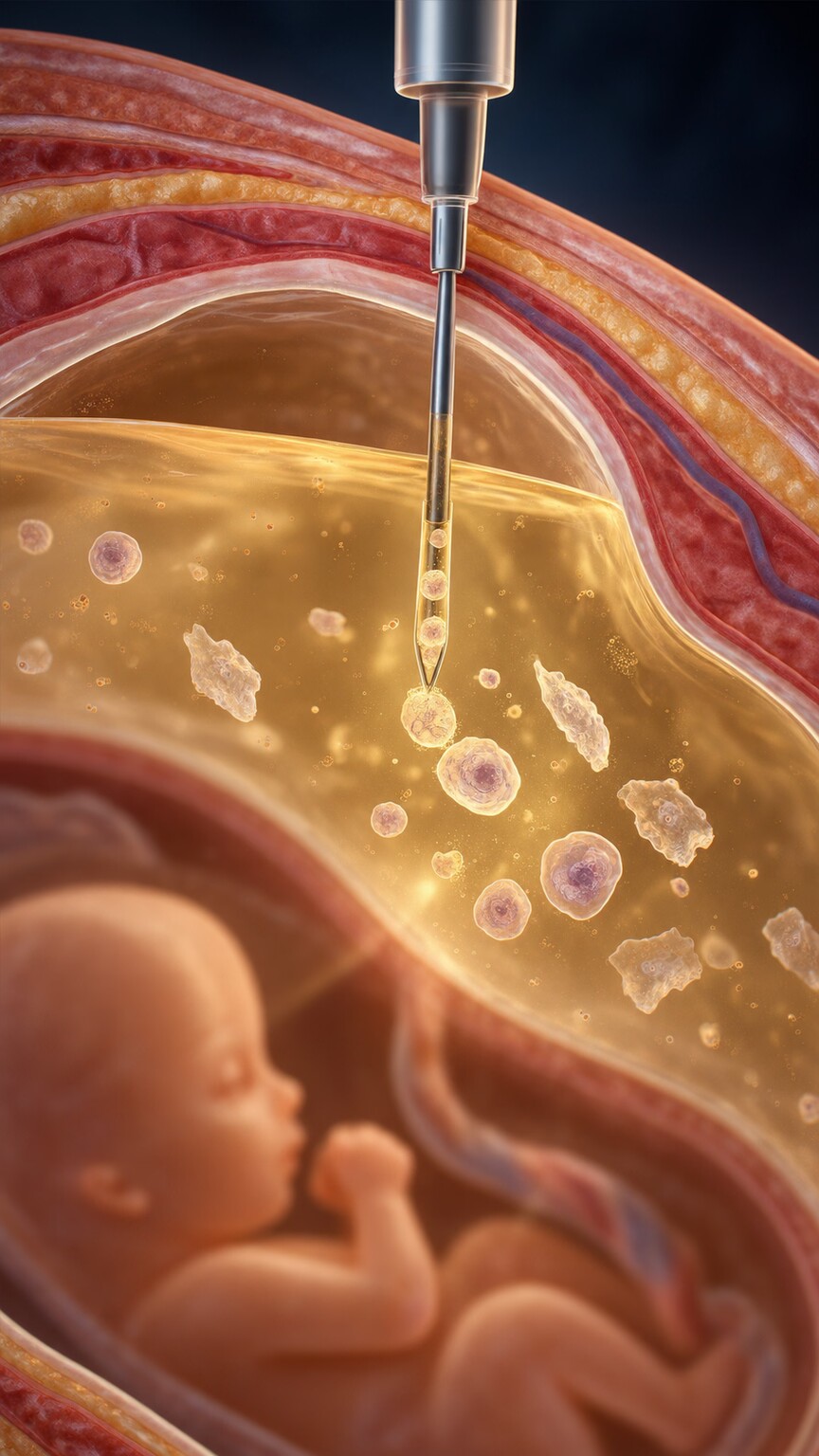
Figure 4. Amniocentesis withdraws a small sample of amniotic fluid, which contains skin and other cells the fetus has naturally shed, allowing direct examination of the fetus's own chromosomes.
Amniocentesis is the second diagnostic option, typically performed between weeks 15 and 20, later in pregnancy than CVS because it requires enough amniotic fluid to have accumulated for a safe, technically adequate sample. A thin needle, again guided continuously by real-time ultrasound, withdraws a small amount of the amniotic fluid that surrounds the baby — this fluid naturally contains skin cells and other cells the fetus has shed into it over time, and those cells are grown briefly in a lab culture before their chromosomes are examined directly.
Because amniocentesis samples fluid that has direct contact with the fetus itself rather than placental tissue, it avoids the rare but real complicating scenario of confined placental mosaicism — where a chromosomal difference is present only in placental cells and never actually reaches the fetus — that can occasionally muddy a CVS result. The procedure-related pregnancy loss risk for amniocentesis performed by an experienced specialist in current practice is generally cited as lower than the historical figures many people have heard, often quoted around 1 in 900 to 1 in 1,600 in more recent published data, though again, this exact figure should be discussed directly with the specialist performing the procedure rather than taken from a single source, since technique, gestational age, and individual clinical factors all influence it.
Beyond chromosomal analysis, the same amniotic fluid sample can be used to test for a handful of other conditions when clinically indicated, most notably neural tube defects like spina bifida, via a measurement of a protein called alpha-fetoprotein, and certain infections. This makes amniocentesis, in situations where multiple concerns exist at once, sometimes a more information-dense single procedure than choosing between separate, narrower tests. After the procedure, most specialists recommend a period of rest and avoiding strenuous activity for a day or two, and mild cramping, spotting, or fluid leakage in the following days should always be reported to the care team promptly, even though the overwhelming majority of amniocenteses proceed with no complications at all.
Screening Versus Diagnosis: The Distinction That Actually Matters Most
If there's one concept worth taking away from this entire article, it's this: NIPT and first-trimester combined screening are screening tests, while CVS and amniocentesis are diagnostic tests, and those two categories answer fundamentally different questions. A screening test estimates the statistical likelihood that a chromosomal difference is present — it can tell you that your personal risk went from a general population baseline to something much higher or much lower, but it cannot, on its own, confirm or rule anything out with certainty. A diagnostic test examines the fetus's actual chromosomes directly and gives a definitive answer.
This distinction has real emotional stakes, because a "high risk" or "positive" NIPT result is not the same thing as a diagnosis, even though it can feel exactly that alarming in the moment. Positive predictive value — the actual chance that a positive screening result reflects a true chromosomal difference rather than a false alarm — varies considerably depending on which condition is being screened for and the pregnant person's baseline age-related risk; for a younger patient with no other risk factors, a substantial share of positive NIPT results for rarer trisomies turn out, after diagnostic confirmation, not to reflect a true fetal chromosomal difference at all. This is precisely why a positive or high-risk screening result is always followed by an offer of diagnostic testing — CVS or amniocentesis — before any decisions are made, never treated as a final answer on its own.
Who Is Actually Offered Each Test, and Why the Recommendations Have Changed
Historically, invasive diagnostic testing was primarily offered to pregnant patients over age 35, reflecting the age-related rise in numerical chromosome differences, while younger patients were generally only offered combined screening. Current major professional guidelines have moved away from age as the primary deciding factor, instead recommending that every pregnant patient, regardless of age, be offered the choice of some form of chromosomal screening (most often NIPT) as part of routine prenatal care, with diagnostic testing offered directly to anyone who wants it, has a positive screening result, has a personal or family history of a chromosomal condition, or has an ultrasound finding suggestive of a possible chromosomal difference.
Certain situations shift a conversation more directly toward diagnostic testing from the start, bypassing screening altogether: a previous pregnancy or child affected by a chromosomal condition, a parent known to carry a balanced chromosomal rearrangement, or a detailed ultrasound finding — certain structural differences seen on a mid-pregnancy anatomy scan carry a meaningfully elevated association with an underlying chromosomal difference, prompting a direct recommendation for diagnostic testing rather than an intermediate screening step that would only delay a definitive answer.
It's worth noting explicitly that none of this testing is mandatory at any point in the decision tree, and declining every form of it is a completely legitimate choice some families make, for reasons ranging from personal values to a decision that the result, whatever it turned out to be, wouldn't change how the pregnancy would be managed. A prenatal care team's role is to make sure the option is offered and clearly explained, not to make the choice on a family's behalf, and declining testing carries no penalty in terms of the quality or thoroughness of the rest of a pregnancy's medical care.
What a Normal Result Actually Means (and Doesn't)
A normal, low-risk, or "negative" result on any of these tests is genuinely reassuring information, but it's worth understanding precisely what it covers and what it doesn't. NIPT and combined screening are validated specifically for the handful of common numerical chromosome differences they're designed to detect — a normal result substantially lowers the likelihood of those specific conditions but says nothing about the thousands of other genetic conditions caused by single-gene changes rather than whole-chromosome differences, which is a separate category of testing entirely. A normal CVS or amniocentesis result is more comprehensive, since it directly examines the full chromosome set (and, depending on the specific test ordered, sometimes smaller structural pieces via a chromosomal microarray), but even a completely normal chromosomal result doesn't rule out every possible health condition a baby could be born with, since plenty of conditions have no chromosomal basis at all.
Chromosomal Microarray: Seeing Smaller Than a Standard Karyotype Can

Figure 5. A chromosomal microarray compares thousands of small DNA segments against a reference pattern, detecting missing or duplicated pieces far smaller than a standard karyotype can resolve.
Once a CVS or amniocentesis sample is collected, the lab has more than one option for how to examine it, and the specific test ordered matters. A standard karyotype — the traditional approach of counting and visually examining chromosomes under a microscope — is excellent at catching whole extra or missing chromosomes and larger structural rearrangements, but it simply cannot see a piece of missing or duplicated genetic material below a certain size, roughly 5 to 10 million base pairs, since anything smaller doesn't change the chromosome's visible size or banding pattern enough to notice by eye.
A chromosomal microarray solves this by comparing the sample's DNA, piece by piece, against a reference pattern using a specialized chip containing thousands of probe points, each one checking a specific, tiny stretch of the genome for the correct amount of genetic material. This lets a microarray detect deletions and duplications far smaller than a karyotype can resolve — clinically significant findings that would have been entirely invisible under a microscope-based approach. Because of this added resolution, professional guidelines now generally recommend chromosomal microarray, rather than a standard karyotype alone, as the preferred first-line test on CVS or amniocentesis samples, particularly when an ultrasound has identified a structural finding, though it does come with one added layer of complexity: a microarray can occasionally detect a small change of uncertain significance, one whose real-world effect on development isn't yet well understood from published research, which genetic counseling specifically addresses when it comes up.
Risk, Cost, and Insurance: The Practical Side of the Decision
Beyond the biology, a handful of practical factors shape which test path a pregnant patient actually chooses, and it's worth addressing them directly rather than treating the decision as a purely clinical one. Cost and insurance coverage vary considerably: many insurance plans cover NIPT for patients considered higher risk (by age or other factors) without much friction, while coverage for patients at average risk who simply want the more accurate option can be inconsistent, sometimes requiring a documented clinical reason or resulting in an out-of-pocket cost that varies widely by lab and plan. It's reasonable, and genetic counselors and OB offices expect it, to ask directly about expected cost before any blood draw or procedure, rather than being surprised by a bill afterward.
Turnaround time also varies meaningfully across these options and can matter for pregnancies approaching a legal or personal decision-making timeline: NIPT results typically return in one to two weeks, a standard karyotype from CVS or amniocentesis usually takes one to two weeks as well since it requires growing living cells in culture first, while a rapid technique called FISH (fluorescence in situ hybridization) can give a preliminary answer for the most common trisomies within one to two days directly from a CVS or amniocentesis sample, well before the full karyotype or microarray result is ready, specifically to give families faster information when timing matters.
How to Actually Decide Which Path Fits Your Pregnancy

Figure 6. A genetic counseling conversation, weighing personal risk factors, timing, and how much certainty matters to a specific family, is typically what actually determines which testing path is the right fit.
There's no single right answer that applies to every pregnancy, but a few honest questions tend to clarify the decision quickly. How much does a definitive answer matter to you personally, versus a statistical estimate you'd be comfortable acting on either way? Some families want the added certainty a diagnostic test provides regardless of screening results, while others are fully comfortable with a low-risk screening result as their stopping point, feeling the added, if small, procedural risk isn't worth it given their own priorities. How would a confirmed diagnosis change what you'd do — some families want the information purely to prepare, medically and emotionally, for a baby with a known condition, while others are weighing it against other decisions; either use case is a legitimate reason to seek a definitive answer.
Cost, timing, and personal risk factors round out the practical side: a patient with a strong family history or a concerning ultrasound finding often has a clearer case for moving straight to diagnostic testing, while a patient with no particular risk factors who simply wants information often finds NIPT's combination of accuracy, safety, and simplicity fully sufficient. Whatever the starting point, a genetic counselor's job is specifically to walk through this exact decision without steering toward one outcome over another — the goal is a fully informed choice that fits the specific family in front of them, not a one-size-fits-all recommendation applied the same way to every pregnancy.
A Worked Example: How One Decision Point Actually Unfolds
Consider a 29-year-old in her eleventh week of pregnancy with no family history of any genetic condition, who is offered NIPT as part of routine first-trimester care. She proceeds with the blood draw, and two weeks later her result comes back low-risk across all the conditions it screens for. Given her age and this reassuring result, no further chromosomal testing is recommended, and her prenatal care continues on its standard schedule, with the anatomy ultrasound around week 20 serving as the next major structural checkpoint.
Now consider a 38-year-old in the same situation who receives a high-risk NIPT result specifically for trisomy 21. Her genetic counselor explains clearly that this is a screening result, not a diagnosis, and walks her through the option of amniocentesis to get a definitive answer, along with the procedure's approximate risk and timeline. She chooses to proceed, and the amniocentesis, performed at week 17, confirms a normal chromosome count — her NIPT result turns out to have been one of the false positives the screening test is statistically expected to occasionally produce, and she and her genetic counselor discuss why that happens (often related to a phenomenon called confined placental mosaicism, or simply the test's known false-positive rate) before she continues her pregnancy with that definitive reassurance in hand rather than lingering uncertainty from the screening result alone.
A third scenario illustrates how a structural ultrasound finding changes the calculus entirely. A 32-year-old with no prior risk factors has a mid-pregnancy anatomy scan that identifies a specific structural finding associated with an elevated chance of an underlying chromosomal difference. Rather than starting with NIPT, her maternal-fetal medicine specialist recommends proceeding directly to amniocentesis with chromosomal microarray, given that the ultrasound finding itself already represents concrete evidence worth investigating with the more comprehensive diagnostic test rather than an intermediate screening step. The microarray identifies a small deletion that a standard karyotype would not have detected, and genetic counseling then focuses on what published research says about that exact deletion's range of possible effects, connecting the family with a specialist experienced in that specific finding well before the baby is born, so a coordinated care plan is already in place at delivery.
Frequently Asked Questions
Is chromosomal testing mandatory during pregnancy?
No. It's offered as an option as part of routine prenatal care, and every pregnant patient has the right to decline any or all forms of this testing entirely, with no impact on the rest of their prenatal care.
Does NIPT tell you the baby's sex?
Yes, as a byproduct — since NIPT reads the relative amounts of chromosome material present, including the X and Y chromosomes, most labs report the likely fetal sex alongside the chromosomal screening results, generally with very high accuracy.
Can NIPT replace an ultrasound?
No. NIPT only analyzes chromosome material circulating in maternal blood and has no way to see physical or structural features of the developing baby, which is exactly what ultrasound is specifically designed to evaluate.
Is CVS or amniocentesis painful?
Most people describe the sensation as brief pressure or cramping rather than sharp pain, comparable to a strong menstrual cramp, and the procedure itself typically takes only a few minutes, though mild cramping can persist for a short time afterward.
What happens if a diagnostic test confirms a chromosomal condition?
Genetic counseling is offered to walk through exactly what the specific finding means for the pregnancy and the child's expected development, connect the family with condition-specific resources and specialists, and discuss the full range of options available going forward.
Can chromosomal testing be done for twins or other multiples?
Yes, though with added complexity. NIPT is generally less accurate for twin pregnancies since the placental DNA from each twin contributes separately to the overall fetal fraction, and CVS or amniocentesis for twins requires carefully sampling each sac individually, distinguishing which result belongs to which twin.
Does a normal chromosomal test guarantee a healthy baby?
No test can guarantee that. A normal chromosomal result substantially lowers the likelihood of the specific conditions it's designed to detect, but many health conditions have no chromosomal basis at all and aren't evaluated by this category of testing.
Conclusion
Prenatal chromosomal testing exists on a spectrum, from a zero-risk blood test that estimates probability to a diagnostic procedure that examines the fetus's actual chromosomes directly, and understanding which category a given test falls into is what makes a result — reassuring or concerning — genuinely interpretable rather than simply alarming. NIPT and combined screening tell you how your personal risk compares to the baseline; CVS and amniocentesis tell you, with direct certainty, what's actually there. Neither category is inherently the "right" choice for every pregnancy — the right combination depends on personal risk factors, how much certainty matters to that specific family, and how each option's timing and small procedural risks weigh against the value of an earlier or more definitive answer.
Whatever path a pregnancy takes through this decision tree, the conversation is meant to be collaborative rather than prescriptive: a genetic counselor or OB's role is to lay out the accurate tradeoffs of each option clearly, not to steer a family toward a predetermined choice. Bringing questions, even ones that feel basic, and asking directly about accuracy figures, cost, and timing for your specific situation, tends to produce a far more useful conversation than researching in isolation beforehand and arriving with conclusions already fixed. The science behind these tests continues to improve — NIPT's accuracy and the range of conditions it screens for have both expanded considerably since it first became available, and that trend is likely to continue, which is one more reason to have this conversation directly with your own care team rather than relying solely on information that may already be a few years out of date by the time you read it.
Still Not Sure What Your Results Mean?
Upload your labs and get a complete, visual, plain-language interpretation of every biomarker — delivered to your inbox in under 15 minutes.
Get My ReportThis article is for educational purposes only and does not constitute medical advice. Always consult your healthcare provider regarding your specific lab results.