
Why Would an Adult Be Referred for Chromosomal Testing?
Most people assume chromosomal testing is something that happens to babies — a needle in a pregnant belly, a worried conversation in a neonatal unit. So when a 34-year-old with three miscarriages, or a 41-year-old newly diagnosed with a certain type of leukemia, or a couple quietly struggling with infertility gets handed a requisition slip for a karyotype or a chromosomal microarray, the first reaction is often confusion: wait, isn't this a pregnancy test? It isn't. Adults are referred for chromosomal testing for five well-established clinical reasons, and none of them require you to be pregnant, a newborn, or visibly unwell. The most common is recurrent pregnancy loss or unexplained infertility, where a hidden chromosomal rearrangement in one partner can quietly derail conception or early development without ever causing a single day of illness. Others include a family history of a known chromosomal condition, developmental or intellectual differences that were never formally worked up in childhood, an adult cancer or blood disorder where the chromosomes inside the tumor cells themselves — not the person's own inherited chromosomes — carry diagnostic and treatment information, and atypical patterns of reproductive development or puberty. This article walks through each of those five paths in plain language, explains what the test actually involves, and clears up what your chromosomes even are and why counting and mapping them can answer questions that no other blood test can.
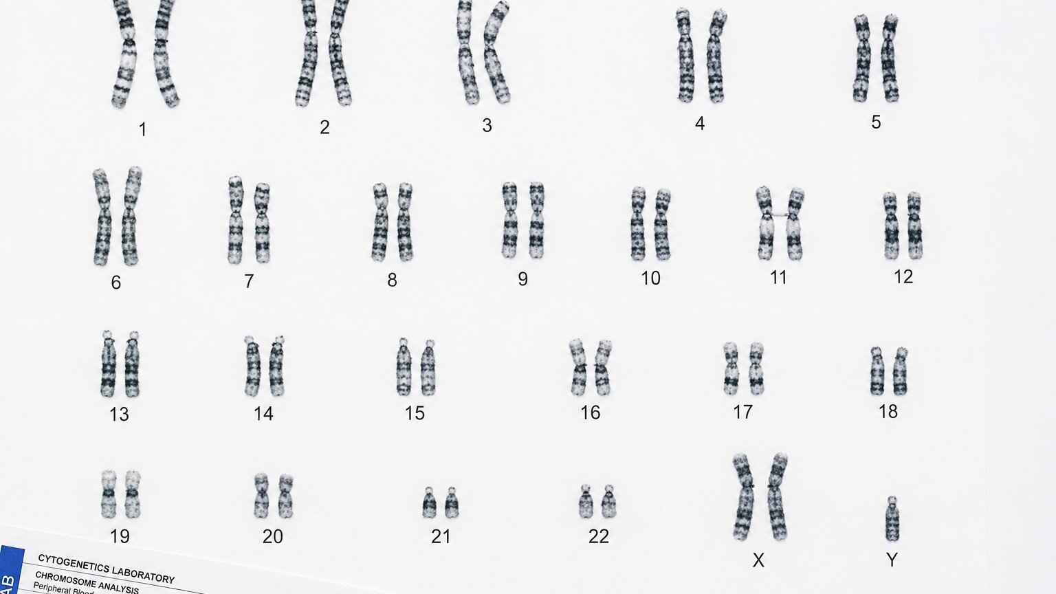
Figure 1. A karyotype arranges all 46 chromosomes from a single cell into 23 numbered pairs, allowing a cytogeneticist to compare their size, shape, and banding pattern.
What Chromosomal Testing Actually Looks For
Before getting into the reasons adults get referred, it helps to understand what the test is even counting. Every cell in your body (with the exception of eggs and sperm) carries 46 chromosomes — tightly coiled bundles of DNA — arranged in 23 pairs. One member of each pair came from your mother, one from your father. Chromosomes 1 through 22 are called "autosomes" and are the same in men and women; the 23rd pair is the sex chromosomes, XX or XY. Think of your chromosomes as 23 matched sets of encyclopedia volumes, each volume holding thousands of individual entries (genes). A gene test — the kind of DNA sequencing most people picture when they hear "genetic testing" — reads the sentences inside a single, specific volume, looking for a typo in one particular entry. Chromosomal testing does something completely different: it doesn't read the sentences at all. It checks whether all 46 volumes are present, whether any volume is missing a section, whether an extra copy of a volume got bound in by mistake, or whether two volumes accidentally got their chapters swapped. That distinction matters enormously for adults, because a person can have a completely normal set of "sentences" — no harmful gene mutations at all — and still carry a chromosomal rearrangement that only becomes visible once you zoom out and count and map the volumes themselves.
There are three main laboratory techniques used to do this, and which one gets ordered depends entirely on the clinical question. A standard karyotype is the oldest and most visual: a lab technician grows your white blood cells in a culture dish, chemically stops them mid-division (the only point in a cell's life when chromosomes condense enough to be photographed as distinct rod shapes), stains them with a dye that produces a striped banding pattern unique to each chromosome, and photographs the result under a microscope. A technician then physically arranges the 46 chromosomes into pairs by size and banding pattern, producing the classic image most people picture. A chromosomal microarray is a newer, higher-resolution technology that doesn't produce a picture at all — instead, it compares thousands of specific DNA markers scattered across your chromosomes to a reference set, flagging any spot where you have too much or too little genetic material, even in stretches far too small to see under a microscope. FISH (fluorescence in situ hybridization) sits in between: it uses glowing, color-coded molecular probes that stick to one specific, predetermined location on a chromosome, which makes it fast and targeted when a lab already suspects a very specific rearrangement rather than needing to scan the whole genome.
Curious what the rest of your lab results are actually telling you? Upload your results and get a complete, plain-language breakdown in under 15 minutes.
Analyze My ResultsReason 1: Recurrent Pregnancy Loss or Unexplained Infertility

Figure 2. A balanced translocation carried by one partner produces no symptoms in that person but can lead to repeated pregnancy loss when passed to an embryo in an unbalanced form.
This is, by a wide margin, the single most common reason a healthy adult with no symptoms at all ends up in a genetics clinic. When a couple experiences two or more pregnancy losses, or struggles with infertility that fertility specialists can't otherwise explain, standard practice is to test both partners' chromosomes — not the pregnancy tissue, the parents themselves. Here's the mechanism that makes this necessary: roughly 1 in 500 people in the general population carries what's called a "balanced translocation," meaning two chromosomes have swapped segments with each other, but no genetic material was actually lost or gained in the swap. Picture two of those encyclopedia volumes trading a chapter — volume 9 now has a chapter that used to belong to volume 22, and volume 22 has one that used to belong to volume 9. Every sentence, every piece of information, is still present somewhere in the set. Because nothing is missing or duplicated, a person carrying this exact rearrangement is almost always completely healthy, with no symptoms, no illness, and often no idea it exists.
The problem surfaces at the moment that person tries to make an egg or sperm cell. Egg and sperm formation requires the chromosome pairs to separate cleanly, one full set going into each reproductive cell. When one chromosome in the pair carries a translocated segment, that clean separation becomes a matter of chance, and a meaningful share of the resulting eggs or sperm end up with an unbalanced set — too much of one chapter, none of another. If one of those unbalanced cells is involved in conception, the resulting embryo is often missing genetic material it needs or carrying material it can't handle, and the pregnancy ends early, frequently before a woman even knows for certain she's pregnant, or in the first trimester. This can repeat pregnancy after pregnancy, entirely by chance, because the carrier parent is producing a mix of balanced and unbalanced reproductive cells every single cycle. A karyotype on both partners is what uncovers this — it's the only way to see the translocation, since it produces no symptoms, no abnormal hormone levels, and no signal on a typical fertility workup that would otherwise point a doctor toward it. Roughly 2 to 5% of couples with recurrent pregnancy loss are found to have exactly this kind of balanced chromosomal rearrangement in one partner when tested.
If a translocation is found, the news is rarely simple bad news — it reframes the situation. It explains a pattern that otherwise looked random and cruel, and it opens concrete paths forward: some couples go on to conceive without intervention, since a share of pregnancies from a balanced carrier are chromosomally normal; others pursue IVF with preimplantation genetic testing, screening embryos before transfer specifically for the unbalanced combinations that translocation can produce. Either way, the referral for chromosomal testing after pregnancy loss isn't a formality — it's often the single test most likely to actually explain what happened.
Two other structural quirks turn up through this same pathway and are worth knowing by name, since both can be mentioned on a lab report without much explanation attached. A "Robertsonian translocation" is a specific type where two of the five chromosomes that happen to have their genetic material clustered near one end (chromosomes 13, 14, 15, 21, and 22) fuse together at that end, effectively merging two chromosomes into one. Like a reciprocal translocation, a Robertsonian translocation carrier is usually healthy, but their reproductive cells can end up with an extra or missing copy of one of those chromosomes — this specific mechanism is behind a meaningful share of Down syndrome cases that run in families rather than occurring as an isolated, one-time event. The other is a "chromosomal inversion," where a segment of a single chromosome gets flipped end-to-end rather than swapped with another chromosome. Depending on exactly where the flipped segment sits, an inversion can interfere with the same clean pairing-and-separation process during egg or sperm formation, producing a similar pattern of pregnancy loss even though only one chromosome, not two, is technically involved. Clinical guidelines from reproductive medicine and genetics organizations generally recommend this kind of parental chromosome testing after two or more clinically recognized pregnancy losses, though some clinics begin the conversation after just one loss if other risk factors are present.
Reason 2: A Family History of a Known Chromosomal Condition
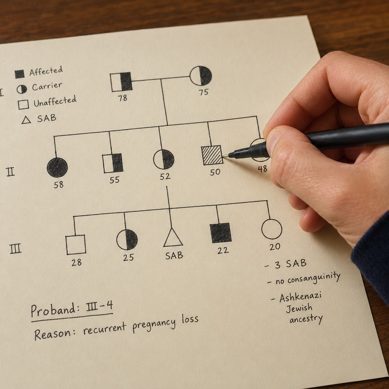
Figure 3. A three-generation pedigree chart is often the first tool a genetic counselor uses to identify who in a family may be silently carrying a balanced chromosomal rearrangement.
The second major path into adult chromosomal testing starts not with a symptom, but with a family conversation. If a couple has already had one child born with a chromosomal condition — Down syndrome caused by what's called a Robertsonian translocation, for instance, rather than the more common random error, or a child with a deletion or duplication syndrome — a genetic counselor will typically recommend testing both parents to find out whether one of them is a silent, balanced carrier of the same rearrangement, or whether it happened as a one-off event in that child alone. This distinction changes everything about future family planning: if a parent is a carrier, the recurrence risk for another pregnancy can be meaningfully elevated compared to the general population, and that same parent's siblings, adult children, and other relatives may also want to be tested, since the same silent rearrangement can be passed down through a family for generations without anyone knowing, each carrier living a completely ordinary, symptom-free life.
This is also where testing gets recommended for adults whose siblings or parents were diagnosed decades ago, sometimes back when genetic technology was far less precise than it is today. An adult in their 30s or 40s might be referred simply because an older sibling was diagnosed in childhood with a chromosomal condition and the family was never fully mapped, or because a distant relative's pattern of miscarriages or birth differences, once pieced together across a family tree, starts to look like a rearrangement running through the family line. A genetic counselor sitting down to build that three-generation pedigree — sketching out who had which pregnancies, which children, which diagnoses — is often the step that identifies which living adult relatives should actually be offered the blood test, turning a vague family story into a specific, testable hypothesis.
Reason 3: Developmental or Intellectual Differences Never Fully Worked Up in Childhood
Not everyone with a chromosomal difference gets identified as a child. Genetic testing technology has advanced enormously even within the last fifteen to twenty years — the chromosomal microarray that can now detect a tiny missing or duplicated segment invisible to older methods simply didn't exist, or wasn't standard practice, when many adults now in their 30s, 40s, or older were children. Someone who grew up with mild intellectual disability, learning differences, subtle physical features, or a cluster of minor health issues that were never tied together into a single diagnosis may reach adulthood having been told, at various points, that the cause was "just how they are," with no genetic explanation ever formally sought. As that person becomes an adult and starts to think about their own family planning, or as their own aging parents start asking questions about what caused their child's differences, a referral for chromosomal testing — usually a microarray, given its much higher resolution than an older-style karyotype — can retroactively supply an answer that was simply unavailable technology decades earlier.
This kind of adult referral also happens through genetic counseling clinics that specifically serve adults with intellectual or developmental disabilities who are aging out of pediatric care systems. A diagnosis found this way, even in adulthood, isn't just an academic label — it can open access to condition-specific medical monitoring (some chromosomal conditions carry known, manageable risks to the heart, thyroid, or other organs that benefit from routine screening once identified), connect a family to condition-specific support networks, and answer the recurrence-risk question for that adult's own future children or their siblings' children.
There's also a quieter version of this same referral path that has nothing to do with a formal disability diagnosis at all. Some adults carry a chromosomal variant so subtle — a small duplicated segment, a slightly rearranged stretch of one chromosome — that it produces no distinct syndrome, only a loosely defined pattern of traits: perhaps a lifelong difficulty with a specific type of learning, a cluster of minor physical features that never seemed connected to anything, or a family "look" that skipped some relatives and not others in a way nobody could explain. A microarray ordered in adulthood, often prompted by a completely unrelated reason like a child's own genetic workup turning up something that needs to be traced back to a parent, can surface this kind of variant for the first time in a person's 40s, 50s, or beyond, giving language to something that shaped their life quietly for decades without ever having a name.
Reason 4: Certain Cancers and Blood Disorders — Chromosomal Changes in Tumor Cells
This fourth reason works differently from the first three, and it's the one that surprises people most, because it has nothing to do with the chromosomes you were born with. Some cancers, particularly certain leukemias and lymphomas, are driven by a chromosomal rearrangement that develops later in life inside a single cell, purely by chance, and then gets copied into every descendant of that cell as the cancer grows. This is called an "acquired" chromosomal change, as opposed to the "constitutional" changes discussed above that are present in every cell of the body from conception onward and can be inherited. Because the rearrangement exists only inside the cancer cells — not in the rest of the body, and not passed down to children — testing for it uses a bone marrow or blood sample from the affected tissue itself, not a routine blood draw of the kind used for the other four reasons on this list.
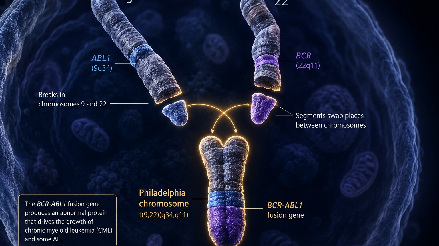
Figure 4. The Philadelphia chromosome, formed when chromosomes 9 and 22 exchange segments inside a bone marrow cell, is the defining acquired change behind chronic myeloid leukemia and is found only in the cancer cells, not the rest of the body.
The best-known example is the Philadelphia chromosome, the rearrangement behind chronic myeloid leukemia, formed when a piece of chromosome 9 and a piece of chromosome 22 trade places inside a single bone marrow cell. The resulting fusion creates a new, abnormal gene at the junction that drives uncontrolled cell growth — but critically, identifying this exact rearrangement doesn't just confirm the diagnosis, it directly determines treatment, since a class of drugs was designed specifically to block the protein this fusion produces. Similar diagnostic and treatment-guiding rearrangements exist across other leukemias and lymphomas, which is why an adult newly diagnosed with one of these blood cancers will typically have chromosomal or FISH testing done on the bone marrow sample as a routine, expected part of the diagnostic workup — not an unusual extra step, but a standard one that shapes which treatment path they're placed on and how their disease is expected to behave.
This same acquired-versus-constitutional distinction also explains why an adult can be told their cancer involves "a chromosomal abnormality" without that meaning anything about what their children could inherit, or what's happening in any other cell in their body — the change lives only inside the tumor, a genetic accident confined to one cell lineage rather than a trait carried since birth.
Reason 5: Atypical Reproductive Development or Puberty
The fifth reason centers on the sex chromosomes specifically — the 23rd pair, XX or XY — and how their number and structure can affect reproductive development in ways that sometimes aren't noticed until adolescence or adulthood. Conditions involving an atypical number of sex chromosomes, such as Klinefelter syndrome (an extra X chromosome in someone who is otherwise male, XXY instead of XY) or Turner syndrome (a missing or partial X chromosome in someone who is otherwise female, X instead of XX), don't always produce obvious signs in early childhood. Klinefelter syndrome in particular is thought to go undiagnosed in a majority of those who have it, often only coming to light when an adult male is evaluated for infertility, since the extra X chromosome commonly affects sperm production even when every other aspect of health and development seems unremarkable. Turner syndrome, similarly, can sometimes go unrecognized until a teenager doesn't begin puberty on the expected timeline, or an adult woman is being evaluated for irregular or absent periods and unexplained infertility.
Beyond the two most well-known examples, chromosomal testing can also be part of the workup for adults with differences of sex development more broadly — situations where reproductive anatomy, hormone patterns, or pubertal timing don't follow the typical pattern, and a clinician wants to understand whether the underlying cause involves the sex chromosomes themselves. In every one of these situations, the karyotype or microarray isn't ordered out of suspicion that something is "wrong" in a broad sense — it's ordered because knowing the precise chromosomal picture directly shapes what monitoring, hormone therapy, or fertility counseling actually makes sense for that specific person, replacing guesswork with a concrete answer.
How the Test Actually Works: From Blood Draw to Report
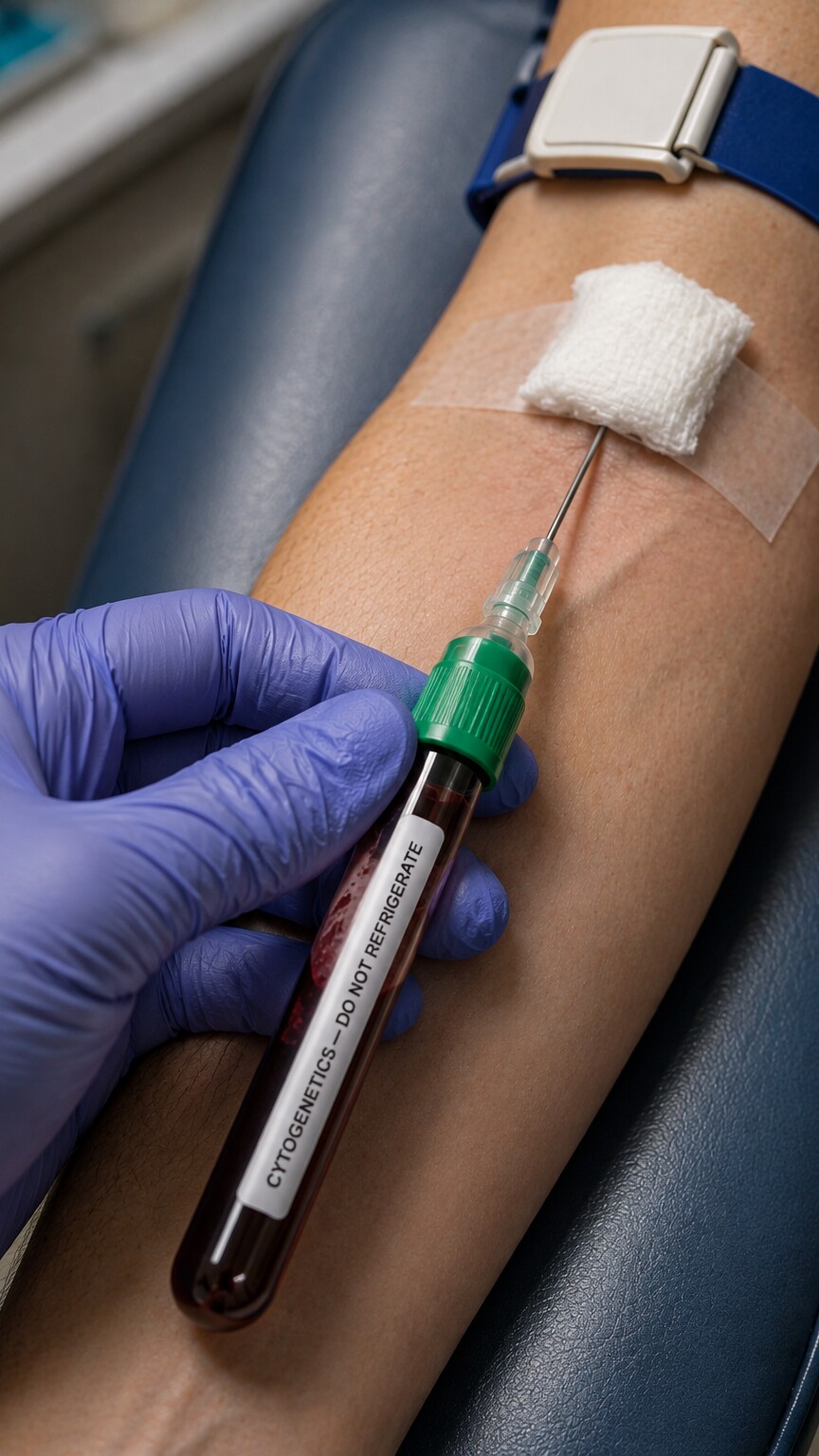
Figure 5. Chromosomal testing requires living white blood cells, which is why the sample is drawn into a specific tube type designed to keep the cells alive and dividing until they reach the cytogenetics lab.
For the constitutional reasons covered above — pregnancy loss, family history, developmental differences, and reproductive development — the test itself starts the same way any blood test does: a standard venous blood draw from the arm. What's different is the tube. Because a karyotype requires actual living, dividing cells rather than just DNA extracted from dead ones, the sample is collected into a specific tube (commonly a green-top sodium heparin tube) that keeps white blood cells alive in transit, rather than the tubes used for most routine chemistry panels. From there, the sample goes to a cytogenetics laboratory, a specialized lab distinct from the one that processes your everyday cholesterol or glucose panel.
In the lab, the white blood cells are placed in a culture medium and stimulated to divide over the course of several days. Once enough cells are actively dividing, a chemical is added that halts them at the exact stage of division — called metaphase — when chromosomes are condensed tightly enough to be visible and countable as distinct shapes under a microscope. A staining technique then produces a consistent, reproducible pattern of light and dark bands unique to each chromosome, the same way a barcode's black-and-white stripes let a scanner identify a specific product. A cytogeneticist, or increasingly an imaging system paired with expert review, photographs a cell in this state, digitally cuts out each of the 46 chromosomes, and arranges them into the 23 numbered pairs that make up the finished karyotype. A microarray, run on a different sample of extracted DNA rather than living cells, skips the cell culture and banding step entirely and instead runs the DNA against a chip containing thousands of reference markers, producing a computer-generated readout of gains and losses rather than a photograph. Turnaround time varies by method and lab, typically ranging from about one to three weeks for a karyotype, since it depends on cells actually growing and dividing in culture, and often somewhat faster for a microarray.
How Structural Rearrangements Are Actually Detected and Named
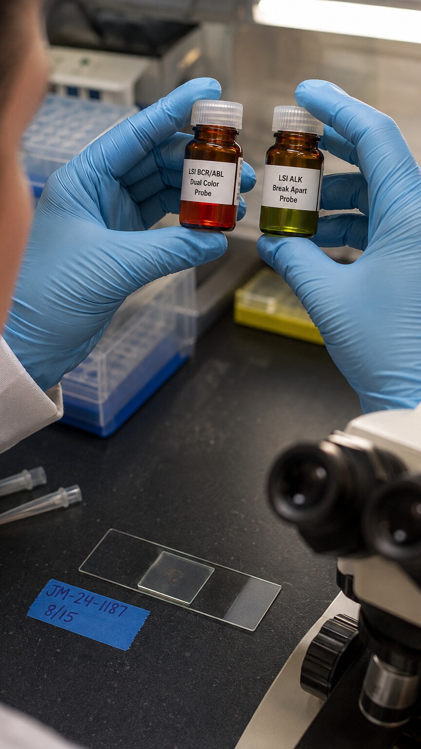
Figure 6. Fluorescent probes tagged in different colors bind to specific, predetermined locations on a chromosome, letting a lab confirm or rule out a rearrangement in hours rather than the days a full karyotype requires.
When a karyotype or microarray does turn up a rearrangement, the lab report describes it using a standardized shorthand called ISCN (International System for Human Cytogenomic Nomenclature) notation — dense strings like "46,XX,t(9;22)(q34;q11)" that look intimidating but decode into plain information once you know the pattern. The first number is the total chromosome count (46 is typical); the letters that follow indicate the sex chromosome pattern (XX or XY); anything after that describes what's unusual — "t" for translocation, followed by which two chromosomes are involved and the specific bands where the exchange occurred. A deletion is marked with "del," a duplication with "dup," an extra whole chromosome with a plus sign, a missing one with a minus sign.
This is also where FISH earns its place in the toolkit. If a clinician already has a strong, specific suspicion — say, confirming the Philadelphia chromosome in a suspected leukemia, or checking a family member for one exact translocation already identified in a relative — a fluorescent probe designed to bind only to that specific chromosomal region can confirm or rule it out within hours, using a small, targeted test rather than the broader, slower process of building and analyzing a full karyotype from scratch. It won't catch anything unexpected elsewhere in the genome, since it's only looking where it's told to look, but for a known, specific question, it's fast, precise, and often the most efficient tool for the job.
What the Results Actually Mean: Three Common Outcomes
A chromosomal test report generally lands in one of three places, and knowing the shape of each possibility before the results arrive tends to make the waiting far less nerve-wracking. The first and most common outcome, especially for adults tested after pregnancy loss or infertility, is a normal result: 46 chromosomes, correctly arranged, nothing unusual detected. A normal result doesn't mean nothing is going on biologically — plenty of pregnancy loss and infertility has causes that have nothing to do with chromosome structure at all, from uterine anatomy to clotting disorders to causes that remain unidentified even after a thorough workup — but it does rule out this particular explanation, which is valuable information in its own right and often shifts the next steps of a workup in a different direction.
The second outcome is a clearly abnormal result: a named translocation, a deletion, an extra or missing chromosome, something with a well-established clinical meaning that a genetic counselor can explain in detail, attach statistics to, and use to guide concrete next steps, whether that's a specific approach to future pregnancies, a referral for condition-specific monitoring, or connecting the person to others who carry the same finding. This is usually the most actionable of the three outcomes, even when the news itself is hard to hear, precisely because so much is already known about it from other patients and published research.
The third outcome is the one that tends to cause the most confusion: a "variant of uncertain significance," often shortened to VUS. This means the lab did find something — a small duplicated or deleted segment, typically — but current scientific knowledge isn't yet sufficient to say with confidence whether that particular change causes problems or is simply a harmless quirk of normal human variation, the genetic equivalent of a birthmark. This isn't a failure of the test; it reflects an honest limit of current medical knowledge, since human chromosomes vary from person to person more than most people realize, and databases of "normal" variation are still being built out region by region. When a VUS comes back, genetic counselors often recommend testing other family members — a finding that turns up in a completely healthy parent or grandparent, carried without any apparent effect for decades, is much more reassuring than one that appears for the first time in the person being tested, and can sometimes reclassify a finding from uncertain to likely benign. A related outcome specific to a small subset of results is mosaicism, where only some of the body's cells carry the chromosomal change and others don't, a distinction covered in more depth in a related article linked at the bottom of this page.
Preparing for a Chromosomal Testing Referral
Because chromosomal testing sits under the umbrella of clinical genetics rather than routine bloodwork, the path to actually getting tested usually runs through a genetic counselor, either as a direct referral from a primary care doctor, OB-GYN, fertility specialist, or oncologist, or as a self-scheduled appointment at a genetics clinic in regions where that's available without a referral. The first appointment is rarely just a blood draw — it typically starts with an in-depth conversation covering personal medical history, reproductive history, and a detailed family history spanning at least three generations, since that family picture often shapes which specific test gets ordered and how the results, once they arrive, should be interpreted. Bringing whatever family medical information is available — ages and causes of relevant diagnoses, any prior genetic testing results in the family, a rough sense of how many pregnancies and losses have occurred across relatives — makes this conversation far more productive, even if the information feels incomplete or secondhand.
Insurance coverage for chromosomal testing varies by the reason it's being ordered and by individual plan, but testing ordered for a recognized medical indication — recurrent pregnancy loss, infertility, a relevant family history, or a cancer diagnosis — is frequently covered, particularly when a genetic counselor's clinical documentation supports the medical necessity of the test. It's reasonable, and genetic counselors expect it, to ask directly about expected turnaround time, out-of-pocket cost, and what happens next depending on each possible result, before the blood draw itself. Most genetics clinics also schedule a dedicated results appointment rather than delivering findings by phone or portal message alone, specifically because a chromosomal result — whether reassuringly normal, clearly abnormal, or an uncertain variant — usually benefits from being walked through by someone trained to explain exactly what it does and doesn't mean for that person and their family.
Frequently Asked Questions
Does a chromosomal referral mean a doctor suspects cancer or a serious genetic disease?
Not by default. The single most common reason adults are referred is recurrent pregnancy loss or infertility, where the person being tested is almost always completely healthy — the test is looking for a silent, balanced rearrangement that affects reproduction without causing any illness in the carrier. Chromosomal testing after a cancer diagnosis is a different, separate use of the same underlying technology, applied to the tumor cells rather than the person's own inherited chromosomes.
If I'm found to be a balanced translocation carrier, does that mean something is wrong with me?
No. A balanced translocation carrier has all the same genetic material as anyone else — it's just arranged differently between two chromosomes. This typically causes no symptoms and no health problems in the carrier themselves. The clinical significance shows up specifically in reproduction, when chromosomes need to separate cleanly to form eggs or sperm.
Is chromosomal testing the same as the genetic sequencing panels sold directly to consumers?
No, and the difference matters. Consumer DNA panels typically look at specific, well-studied single-letter variations across your genome, not the overall structure, number, or arrangement of your chromosomes. A karyotype or microarray answers a different question entirely — whether the chromosomes themselves are the right number and correctly arranged — and can only be ordered through a healthcare provider and processed by a certified cytogenetics laboratory.
How long does it take to get chromosomal test results back?
A standard karyotype typically takes one to three weeks, since it depends on white blood cells actually growing and dividing in a culture before they can be photographed and analyzed. A chromosomal microarray, which works directly from extracted DNA rather than living cultured cells, is often somewhat faster. A targeted FISH test looking for one specific, already-suspected rearrangement can sometimes return results within a day or two.
Do I need a genetic counselor to order this test, or can my regular doctor do it?
A primary care doctor, OB-GYN, fertility specialist, or oncologist can technically order a chromosomal test, but most will refer to a genetic counselor or clinical geneticist first, since choosing the right test (karyotype versus microarray versus targeted FISH) depends on the specific clinical question, and interpreting a complex result usually benefits from someone trained specifically in that interpretation. In many places this referral is a standard, expected part of a recurrent pregnancy loss, infertility, or relevant family-history workup rather than an extra or unusual step.
Will a chromosomal test affect my health or life insurance?
In the United States, the Genetic Information Nondiscrimination Act (GINA) prohibits health insurers and most employers from using genetic test results, including chromosomal testing, to make coverage or employment decisions. GINA's protections do not extend to life, disability, or long-term care insurance, however, so it's worth discussing this distinction with a genetic counselor before testing if it's a concern, particularly if you're in the process of applying for one of those policy types.
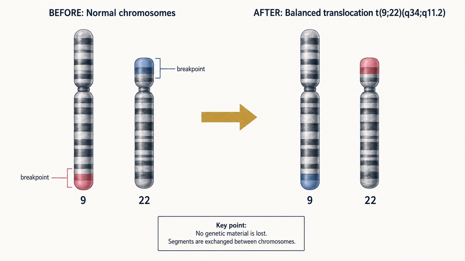
Figure 7. In a balanced translocation, segments trade places between two chromosomes with no genetic material lost overall — which is why the carrier is typically healthy even though the arrangement can disrupt reproduction.
Conclusion
Chromosomal testing in adults isn't a leftover pediatric test that occasionally gets misapplied — it's a purpose-built tool for five specific, well-defined clinical situations, each with its own logic. It explains pregnancy losses that otherwise look random. It clarifies risk for families who already know a chromosomal condition runs somewhere in their line. It can retroactively answer questions about developmental differences that older technology simply couldn't resolve. It guides treatment for certain cancers by reading the chromosomes inside the tumor itself. And it explains patterns of reproductive development that don't follow the expected timeline. In every one of these cases, the referral exists because counting and mapping all 46 chromosomes answers a question that no gene panel, no hormone test, and no imaging study can answer on its own.
What ties all five reasons together is that a chromosomal referral is rarely a vague, catch-all gesture — it's ordered because a clinician has a specific, answerable question in mind, and this particular test, out of everything else available, is the one built to answer it. That's worth holding onto if you're the one holding the requisition slip: the test exists to replace an unanswered question with a concrete one, whether the eventual answer is reassuring, actionable, or somewhere in between. If you've been referred for this kind of testing, the fact that it was ordered says far more about the specific question your clinician is trying to answer than it does about your overall health.
Still Not Sure What Your Results Mean?
Upload your labs and get a complete, visual, plain-language interpretation of every biomarker — delivered to your inbox in under 15 minutes.
Get My ReportThis article is for educational purposes only and does not constitute medical advice. Always consult your healthcare provider regarding your specific lab results.