
Target Cells on a Blood Smear Aren't Always a Cause for Alarm
If your blood smear report mentions "target cells," the name alone can sound like something went wrong — a bullseye pattern showing up where a normal red blood cell should be. But target cells, also called codocytes, are one of the more mechanically well-understood findings in hematology, and a small number of them scattered across an otherwise normal smear is often a benign, incidental observation rather than a sign of disease. What actually matters isn't whether target cells are present at all, but how many there are and what other findings accompany them. This article explains exactly why red blood cells take on this distinctive shape in the first place, walks through the handful of genuinely different conditions that can cause it, and covers how a doctor decides whether a target cell finding needs any further workup at all.
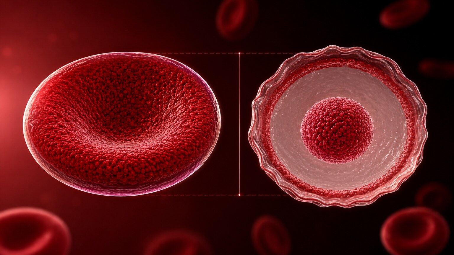
Figure 1. A target cell forms when a red blood cell has more membrane surface area than its hemoglobin content can properly fill, causing the excess membrane to redistribute and create a dense central spot surrounded by a pale ring — the classic bullseye pattern.
Why a Blood Smear Looks at Shape at All
It's worth understanding why a blood smear even bothers examining the shape of individual cells in the first place, since most routine blood testing today is done through automated analyzers that count and measure cells without a person ever looking at them directly under a microscope. These automated machines are excellent at counting cells and measuring their average size, but they're far more limited when it comes to recognizing specific, distinctive shape abnormalities, especially subtler ones or shapes that only affect a portion of the total cell population. A blood smear, where an actual person examines a thin layer of blood spread across a glass slide under a microscope, catches exactly this kind of detail that automated counting alone would miss entirely.
This is precisely the role target cells play in this bigger picture: they're a shape-based clue, invisible to routine automated counting, that becomes visible only through this direct microscopic examination. A blood smear is typically ordered specifically when an automated blood count shows an abnormality that needs closer visual interpretation, or when a doctor has a specific clinical suspicion that benefits from directly seeing the cells themselves rather than relying on the automated summary numbers alone.
Some automated analyzers do include flags or alerts suggesting a possible shape abnormality worth a closer look, based on subtler statistical patterns in how the machine's own measurements are distributed across all the cells it counted, but these flags function more like a prompt than a diagnosis — they tell the laboratory that a manual review is warranted, without themselves identifying the specific shape involved. The actual identification of a shape like a target cell, and the judgment about how many are present and what that likely means, still comes from a trained person examining the slide directly.
Why the Bullseye Shape Forms in the First Place
A healthy red blood cell is shaped like a biconcave disc — think of a donut where the hole didn't quite punch all the way through, thinner in the center and thicker around the rim. This shape isn't accidental; it's the natural result of a normal, balanced ratio between the amount of cell membrane surrounding the cell and the amount of hemoglobin (the oxygen-carrying protein) packed inside it. When that ratio gets thrown off — specifically, when a cell ends up with more membrane surface area than its hemoglobin content can properly support and fill out — the cell can no longer hold its normal biconcave shape.
Instead, the excess membrane redistributes itself, folding the cell's outer edges inward and creating a small pool of hemoglobin-rich material in the very center, surrounded by a pale, hemoglobin-poor ring, with a second darker ring around the outer rim. Viewed under a microscope, this pattern looks remarkably like an archery target or a bullseye, which is exactly where the name comes from. Every single cause of target cells covered in this article, despite being biologically very different conditions, ultimately produces this same underlying mechanical imbalance between membrane and hemoglobin.
This ratio between a cell's surface area and its internal volume even has a formal name in hematology: the surface-area-to-volume ratio. A normal biconcave red blood cell has a relatively high surface-area-to-volume ratio for its size, which is actually part of what makes it so efficient at exchanging oxygen and carbon dioxide across its membrane, and part of what gives it the flexibility to squeeze through the body's narrowest capillaries. A target cell, by definition, has pushed this ratio even higher than normal, since it's carrying more membrane than its internal hemoglobin volume calls for — which is a useful way to think about every one of the specific causes covered in the rest of this article, since each one, through a completely different biological pathway, ultimately pushes this same single ratio in the same direction.
It's worth noting that target cells aren't unique in being explained by this membrane-to-volume principle — several other red blood cell shape abnormalities seen on a smear are explained by the opposite imbalance, where a cell has too little membrane relative to its volume, producing a different, rounder shape called a spherocyte instead of the flattened, bullseye-producing excess seen in target cells. Understanding this shared underlying framework is part of what makes red blood cell shape analysis such a genuinely informative diagnostic tool, rather than a collection of disconnected, memorized shape names.
Curious what the rest of your CBC or blood smear findings are telling you? Upload your results and get a complete, plain-language breakdown in under 15 minutes.
Analyze My ResultsCause One: Liver Disease and Altered Cholesterol Metabolism
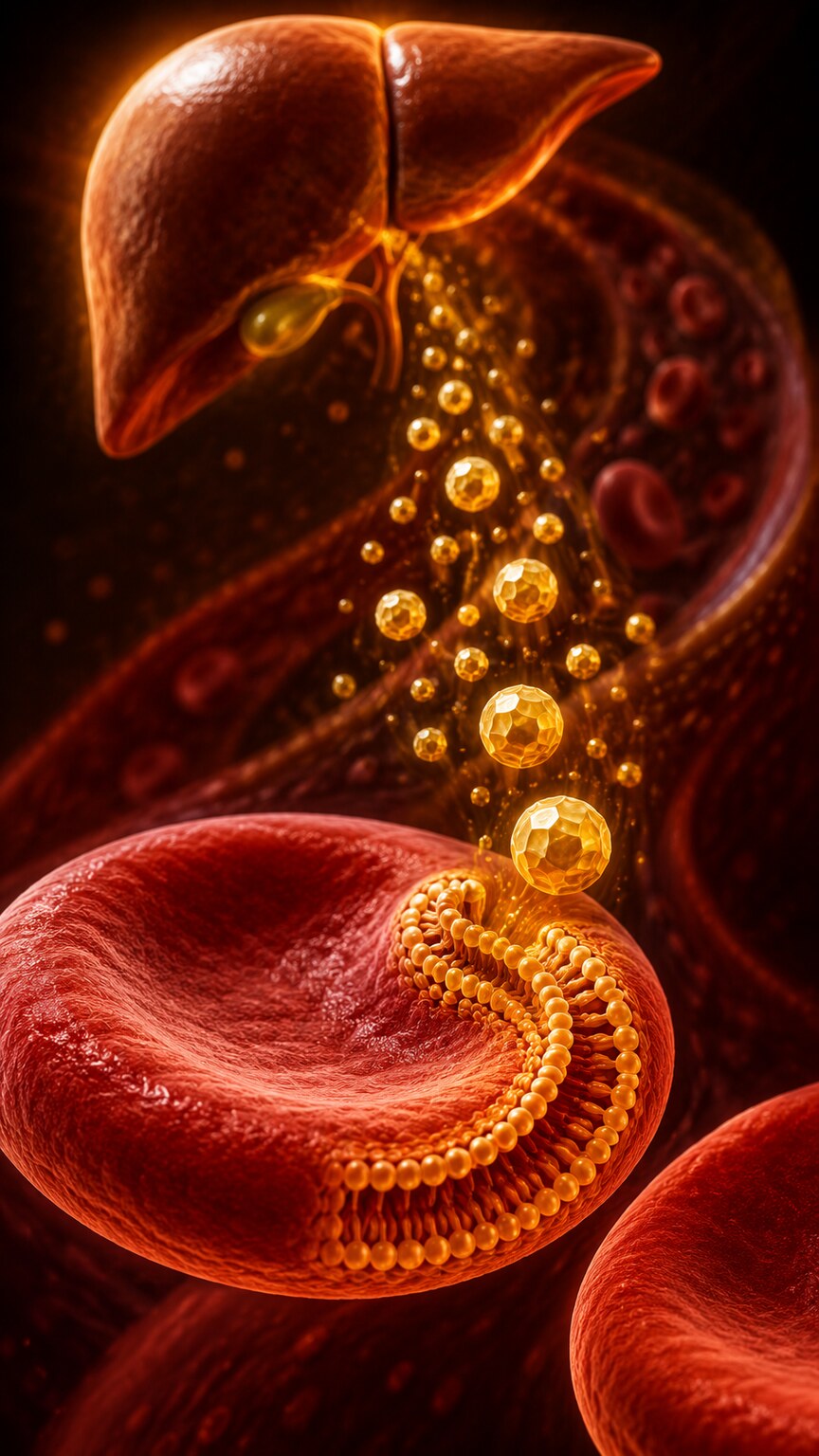
Figure 2. In liver disease, disrupted bile flow and lipid metabolism raise circulating free cholesterol, some of which gets absorbed directly into red blood cell membranes, expanding their surface area beyond what their hemoglobin content proportionally fills.
One of the most common causes of target cells is chronic liver disease, particularly conditions involving obstructed bile flow, such as cirrhosis or cholestatic liver disease. The liver plays a central role in regulating cholesterol and other fat-based molecules called lipids circulating in your blood. When liver function is impaired, this regulation breaks down, and levels of free cholesterol in the bloodstream can rise. Red blood cell membranes are themselves built largely from cholesterol and other lipids, and they can absorb some of this excess circulating cholesterol directly into their own membrane structure.
As a red blood cell's membrane absorbs more cholesterol than usual, its total surface area expands, while its internal hemoglobin content stays the same. This creates exactly the membrane-to-hemoglobin mismatch described above, and target cells begin appearing on the smear. In advanced liver disease, this can affect a large enough proportion of circulating red blood cells that target cells become one of the more prominent and clinically useful findings on the smear, sometimes prompting further liver-specific testing even before other liver symptoms become obvious.
This liver-driven mechanism tends to progress alongside how advanced the underlying liver condition is, meaning the number of target cells present can loosely track with disease severity in some patients, though it's far from a precise or reliable severity measurement on its own. In more advanced, longstanding cholestatic liver disease, cell membrane changes can become severe enough to produce a related, more extreme shape sometimes described separately, where the membrane abnormality is pronounced enough to create spiny, irregular projections rather than the cleaner bullseye pattern of a typical target cell — a distinction a pathologist will note specifically, since it can suggest a more advanced stage of the same underlying liver-driven lipid disturbance.
Cause Two: Thalassemia and Reduced Hemoglobin Production
Thalassemia is a group of inherited conditions in which the body produces less of one of the protein chains that make up hemoglobin than it normally would. Unlike liver disease, where the membrane side of the ratio is the problem, thalassemia approaches the same imbalance from the opposite direction: the cell's membrane forms normally, but there simply isn't enough hemoglobin being produced to properly fill it. The practical result is the same mismatch, and the same bullseye shape, even though the underlying cause is essentially the reverse mechanism.
Target cells are a particularly well-known and expected finding in thalassemia, often appearing in meaningful numbers even in mild, minimally symptomatic forms of the condition sometimes called thalassemia trait, where a person carries the genetic change but has few or no symptoms in daily life. This is actually one of the more useful diagnostic clues a hematologist relies on: someone with mildly low hemoglobin, normal or only slightly abnormal iron studies, and a smear showing multiple target cells is a classic pattern that specifically points toward thalassemia rather than the far more common iron deficiency anemia, which can otherwise look superficially similar on a basic blood count alone.
There are two broad categories of thalassemia, based on which specific protein chain of hemoglobin is underproduced, called alpha thalassemia and beta thalassemia, and both can produce target cells, though the severity and typical target cell burden can differ between them and between their various subtypes. More severe forms of thalassemia, where a larger share of normal hemoglobin production is affected, tend to produce a correspondingly larger number of target cells, along with more pronounced anemia and other accompanying smear findings, while milder trait forms may show only a modest number of target cells alongside blood counts that are only subtly abnormal or occasionally even within the normal reference range.
Because thalassemia is genetic and typically present from birth, a smear showing a thalassemia-consistent target cell pattern in a young child, particularly alongside a family history of similar findings, carries different diagnostic weight than the same finding appearing for the first time in an adult with no prior blood count abnormalities — the latter scenario makes an acquired cause, like liver disease, meaningfully more likely to be at least part of the explanation, since thalassemia itself doesn't newly develop later in life the way an acquired condition can.
This age-of-onset reasoning is a genuinely useful diagnostic shortcut worth remembering: a finding present since childhood, especially one shared by close relatives, points toward an inherited cause almost by definition, while the same finding appearing for the first time later in life, with no prior history of it, points the workup much more firmly toward one of the acquired causes discussed elsewhere in this article.
Cause Three: Iron Deficiency Anemia
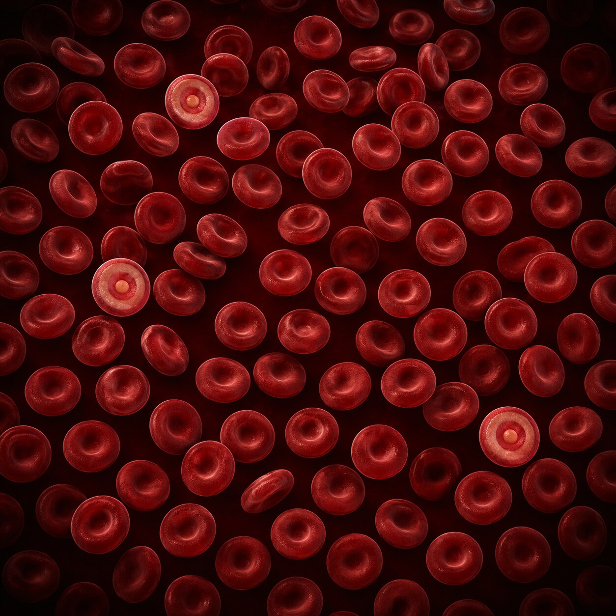
Figure 3. A small handful of scattered target cells, surrounded predominantly by normally shaped red blood cells, is frequently reported as an incidental finding and generally carries far less clinical weight than a smear where target cells make up a large proportion of all cells present.
Iron deficiency anemia can also produce target cells, through a mechanism related to, but distinct from, thalassemia's. Without enough iron, the body can't produce sufficient hemoglobin, again leaving the cell's normally formed membrane without enough internal hemoglobin content to properly fill it out. Because iron deficiency anemia is so much more common than thalassemia, especially in certain populations, it's often the more likely explanation for a smaller number of target cells accompanying a low hemoglobin result, though the two conditions require different follow-up (iron studies for suspected iron deficiency, hemoglobin electrophoresis for suspected thalassemia) and can sometimes coexist, complicating the picture further.
This is exactly why the quantity of target cells matters so much in interpretation. A smear showing only a small scattering of target cells, surrounded predominantly by normally shaped cells, is generally treated very differently from a smear where target cells make up a substantial proportion of all red blood cells present — the former is frequently reported as an incidental, low-significance finding, while the latter usually prompts a more focused workup for one of the specific underlying causes covered in this article.
Iron deficiency anemia's relationship to target cells is also worth understanding through the lens of severity and progression, since the underlying mechanism intensifies as iron stores become more depleted over time. In the earliest stages of iron deficiency, before hemoglobin production is meaningfully affected, target cells are unlikely to appear at all, since the cell's membrane-to-hemoglobin ratio hasn't yet been pushed out of its normal range. As iron deficiency progresses and hemoglobin production genuinely falls short, the membrane-to-hemoglobin imbalance becomes more pronounced, and target cells become correspondingly more likely to appear and more numerous when they do. This progression is part of why target cells are more typically associated with moderate-to-severe iron deficiency anemia rather than the earliest, mildest stages of iron depletion.
It's also worth mentioning that iron deficiency anemia and thalassemia aren't mutually exclusive explanations for the same smear — someone can genuinely have both conditions simultaneously, particularly since iron deficiency is common enough in the general population that it can coexist with an underlying thalassemia trait a person may not have previously known they carried. In cases where the clinical picture doesn't cleanly point to one single explanation, iron studies and hemoglobin electrophoresis are sometimes both ordered together specifically to sort out whether one, the other, or both conditions are contributing to the findings on the smear.
Cause Four: Splenectomy and Reduced Spleen Function
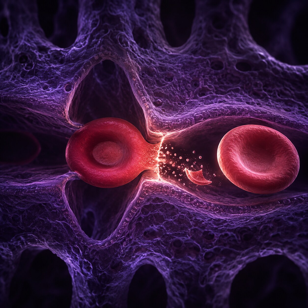
Figure 4. The spleen normally acts as a quality-control filter, trimming small amounts of excess membrane from red blood cells as they pass through its tissue — after splenectomy, this remodeling no longer happens, and cells with mildly excess membrane persist as target cells rather than being corrected.
The spleen plays a role in this story that's easy to overlook: as red blood cells circulate through the spleen's tissue over their roughly 120-day lifespan, the spleen acts as a kind of ongoing quality-control filter, gradually trimming away small amounts of excess or damaged membrane material and helping maintain each cell's proper membrane-to-hemoglobin ratio. When the spleen is surgically removed, a procedure called splenectomy, or when spleen function is otherwise reduced, this ongoing remodeling process stops.
Without this correction happening, red blood cells with even a mild degree of membrane excess, which might have been trimmed back to normal by a functioning spleen, are left uncorrected and persist as target cells in circulation. This is why target cells are such a commonly expected, and generally unremarkable, finding on the blood smear of anyone who has had a splenectomy for any reason, and their presence in this specific context isn't interpreted as a sign of a separate underlying problem the way it might be in someone with an intact spleen.
Beyond surgical splenectomy, several other conditions can produce a similar reduction in effective spleen function without the organ actually being removed, a state hematologists refer to as functional hyposplenism or, when severe, functional asplenia. Certain inherited blood disorders, some autoimmune conditions, and conditions that cause the spleen to become infiltrated or damaged over time can all reduce its filtering capacity to a degree that mimics what happens after surgical removal, producing target cells and other related smear findings through essentially the same loss-of-remodeling mechanism. This is one reason a smear pattern consistent with reduced spleen function, in someone who hasn't actually had a splenectomy, can itself be a useful clue prompting further evaluation of spleen size and function through imaging or other testing.
The spleen's filtering role also connects to a related, easily overlooked clinical point: because the spleen is one of the body's key defenses against certain types of bacterial infection, reduced spleen function, whether from surgical removal or a functional cause, carries its own separate set of health implications beyond the blood smear finding itself, including specific vaccination recommendations that doctors typically discuss with anyone who has had a splenectomy or is found to have significantly reduced spleen function.
Cause Five: Certain Hemoglobin Variants
Beyond thalassemia, several other inherited hemoglobin variants can also produce target cells, most notably hemoglobin C disease, in which the body produces a structurally altered form of hemoglobin that doesn't fold into its normal shape as efficiently. This altered hemoglobin can crystallize inside the cell in a way that changes how the cell's membrane behaves, producing target cells that are often quite prominent and numerous on the smear, sometimes serving as one of the more visually distinctive clues suggesting this specific hemoglobin variant before confirmatory hemoglobin electrophoresis testing is performed.
Some people carry more than one of these hemoglobin variants simultaneously, or carry a hemoglobin variant alongside thalassemia, and these combined conditions can produce an even more pronounced target cell pattern than either condition alone would. This is part of why a hematologist reviewing a smear with numerous target cells will often specifically ask about family history and ancestry, since several of the hemoglobin variants associated with this finding are more common in certain populations, and that context can meaningfully narrow down which specific confirmatory test makes the most sense to order next.
Hemoglobin C is far from the only variant in this category. Combined states, such as hemoglobin SC disease, where a person carries one copy each of the hemoglobin S variant (associated with sickle cell disease) and the hemoglobin C variant, are known to produce a notably prominent target cell pattern, often alongside other distinctive shape changes on the same smear. This particular combination illustrates a broader point worth remembering throughout this whole topic: target cells frequently appear alongside other shape abnormalities rather than in isolation, and the complete combination of shapes present on a single slide, not any one shape considered alone, is often what allows a hematologist to arrive at a specific, confident interpretation.
How Target Cells Are Distinguished From Other Similar-Looking Shapes
Under the microscope, a few other red blood cell abnormalities can superficially resemble a target cell to an untrained eye, and part of a pathologist's expertise involves reliably telling them apart. A cell that's simply been cut through at an unusual angle during the smear preparation process, called an artifactual "pseudo-target" appearance, can occasionally mimic a true target cell without actually reflecting any real membrane-to-hemoglobin imbalance at all. Experienced smear reviewers account for this possibility by looking at the overall pattern and consistency across many cells on the slide, rather than drawing conclusions from a single cell's appearance in isolation, since a true target cell finding should show up consistently across a meaningful number of cells if it reflects a genuine underlying process rather than a preparation artifact.
Another related but distinct shape, called a folded cell, can also superficially resemble a target cell in certain orientations, though it forms through simple mechanical folding during smear preparation rather than reflecting any true membrane-to-hemoglobin ratio abnormality. Distinguishing a genuine target cell from these look-alike possibilities is part of why blood smear interpretation, despite modern automation elsewhere in laboratory medicine, still relies on trained human expertise rather than being something a machine can reliably substitute for.
How a Pathologist Actually Decides How Much a Target Cell Finding Matters

Figure 5. A pathologist reviewing a smear weighs the quantity of target cells present alongside every other finding on the same slide and the accompanying complete blood count, since no single cell shape is interpreted in isolation from the rest of the picture.
When a laboratory professional reviews a blood smear under the microscope, target cells are never assessed in isolation — they're interpreted alongside everything else visible on the same slide and the results of the complete blood count that typically accompanies it. A pathologist looks at roughly what percentage of red blood cells show the target pattern, whether other abnormal shapes are present at the same time (since several of the causes above can produce more than one kind of shape change simultaneously), and how the hemoglobin, red blood cell size, and other CBC values line up with each of the specific causes described earlier in this article.
This combined pattern-matching is what actually drives the decision about whether target cells warrant further workup at all. A small number of target cells alongside an entirely normal CBC and no other smear abnormalities is frequently dismissed as a non-specific, low-significance finding not worth pursuing further. A larger number of target cells alongside abnormal hemoglobin, abnormal red blood cell size, or other accompanying shape changes is a different story, and typically prompts specific follow-up testing tailored to whichever of the causes above seems most likely given the full picture.
Some laboratories formalize this by using a semi-quantitative grading system on the report itself, describing the target cell finding as occasional, mild, moderate, or marked, roughly corresponding to increasing proportions of affected cells, rather than reporting an exact percentage for every single sample. This kind of grading gives the ordering doctor a quick, standardized sense of severity without requiring an exhaustive cell-by-cell count, and it's this graded language, alongside the accompanying CBC values, that a doctor is actually reading when deciding whether the finding needs any further attention.
What Kind of Follow-Up Testing Usually Comes Next

Figure 6. When a target cell finding is significant enough to warrant follow-up, the specific tests ordered next — liver function tests, hemoglobin electrophoresis, or iron studies — depend on which underlying cause the rest of the clinical picture points toward.
If target cells are numerous enough, or the surrounding clinical picture suggests they're clinically significant rather than incidental, the specific follow-up testing ordered depends heavily on which underlying cause seems most plausible. Suspected liver disease is typically followed up with liver function tests, measuring enzymes and other markers that reflect how well the liver is working. Suspected thalassemia or a hemoglobin variant is typically followed up with hemoglobin electrophoresis, a test that separates and identifies the different types of hemoglobin present in the blood. Suspected iron deficiency is typically followed up with iron studies, including ferritin, which reflects the body's stored iron reserves.
None of this follow-up testing is triggered automatically just because target cells appear on a report — it's a clinical judgment call made by whoever is reviewing the full smear and blood count together, weighing the quantity of target cells against every other piece of available context, exactly as described in the previous section.
What to Expect if You're Asked to Come Back for Repeat Testing
If your doctor does want to pursue further testing after a target cell finding, it's worth knowing roughly what that process looks like, since the tests involved differ meaningfully from a standard blood draw in a few respects. Hemoglobin electrophoresis and related tests that specifically identify hemoglobin variants are typically sent to a specialized reference laboratory rather than processed on-site at every clinic, meaning results can take longer to come back than a routine complete blood count, sometimes a week or more depending on the lab. Iron studies and liver function tests, by contrast, are usually available much faster, often within a day or two, since these are more routine tests run at most standard clinical laboratories.
It's also worth knowing that a family history conversation is a genuinely useful thing to prepare for before this kind of follow-up appointment, particularly if thalassemia or a hemoglobin variant is suspected. Because these conditions are inherited, knowing whether parents, siblings, or other close relatives have ever been told they have "thalassemia trait," anemia of unclear cause, or any known hemoglobin abnormality can meaningfully speed up how quickly a doctor arrives at a confident interpretation, sometimes even before the confirmatory test results themselves come back.
If a hemoglobin variant or thalassemia is ultimately confirmed, genetic counseling is sometimes offered as an additional next step, particularly for people of reproductive age, since these conditions can be passed on to children and some combinations of inherited hemoglobin variants carry more significant health implications than either variant alone. This isn't a step that applies to everyone with target cells on a smear — it's specifically relevant once a genetic cause has actually been confirmed through the appropriate testing described above, not something triggered by the initial smear finding by itself.
None of this needs to happen quickly or urgently in the vast majority of cases. Because target cells reflect a chronic, stable underlying process in almost every scenario described in this article, rather than a rapidly evolving emergency, there's typically no rush involved in scheduling this kind of follow-up testing, and it's entirely reasonable to take the time to gather relevant family history and ask questions before moving forward with any specific next step. Patience here is not a missed opportunity — it's simply matching the pace of the workup to the pace of the underlying biology itself.
Why This Finding Illustrates a Broader Principle in Lab Interpretation
Target cells are a genuinely useful example of a broader principle worth keeping in mind whenever you're looking at any single finding on a lab report: almost nothing in laboratory medicine is inherently good or bad in total isolation from everything else. The exact same microscopic finding — a red blood cell with a bullseye shape — can be a complete non-issue in one clinical context (a person with a normal CBC and no other findings, or someone with a known prior splenectomy) and a meaningful diagnostic clue in another (a person with unexplained anemia and abnormal liver enzymes, or a family history of thalassemia). The finding itself doesn't change; what changes is the surrounding context that gives it meaning.
This is exactly why a single line on a lab report, viewed without the rest of the picture around it, can be genuinely difficult to interpret correctly even for people with some medical knowledge, and why the full pattern across an entire report, alongside relevant history and other testing, is what actually drives a confident, accurate interpretation rather than any one number or finding reviewed by itself. Keeping this principle in mind is genuinely useful well beyond target cells specifically — it applies just as much to an unfamiliar biomarker name, an unusual enzyme level, or any other isolated flag that might otherwise seem alarming without its surrounding context.
Frequently Asked Questions
Do a few target cells on my report mean something is wrong with my liver?
Not necessarily. A small number of target cells is a common, often incidental finding with several possible explanations, only one of which is liver disease. It's typically interpreted alongside your full CBC and other smear findings rather than as a standalone signal of liver problems.
Is it normal to have target cells after having my spleen removed?
Yes. The spleen normally trims excess membrane from red blood cells, and without it, cells with even mild membrane excess persist as target cells. This is an expected, generally unremarkable finding after splenectomy rather than a sign of a separate problem.
How can target cells help tell thalassemia apart from iron deficiency?
Both can cause target cells through a similar membrane-to-hemoglobin mismatch, but the combination of mildly low hemoglobin, largely normal iron studies, and multiple target cells is a classic pattern pointing toward thalassemia, whereas abnormal iron studies point more toward iron deficiency.
How many target cells is considered a lot?
There's no single universal cutoff, since interpretation depends on the full clinical picture, but a small handful scattered among mostly normal cells is generally treated very differently from a smear where target cells make up a substantial proportion of all red blood cells present.
Can target cells appear for more than one reason at the same time?
Yes. It's possible to have more than one contributing cause simultaneously, such as iron deficiency alongside an unrecognized thalassemia trait, or liver disease alongside a prior splenectomy. When the clinical picture doesn't cleanly point to a single explanation, more than one type of follow-up testing may be ordered together.
Will target cells go away if the underlying cause is treated?
It depends on the cause. Target cells related to reversible conditions, like iron deficiency, can decrease as the underlying deficiency is corrected. Target cells related to inherited conditions like thalassemia, or to a permanent change like splenectomy, are more likely to remain a stable, ongoing feature of future blood smears.
Conclusion
Target cells form through one consistent mechanical principle — a red blood cell with more membrane than its hemoglobin content can properly fill — even though the specific reason that imbalance occurs varies widely, from liver disease and altered cholesterol metabolism, to thalassemia and iron deficiency's reduced hemoglobin production, to the loss of the spleen's normal membrane-trimming function after splenectomy. A small number of target cells on an otherwise unremarkable smear is frequently a benign, incidental finding, while a larger, more prominent pattern, especially alongside other abnormal results, is what actually prompts further investigation. If your own report mentions target cells, the quantity and the company they keep on the rest of your results matter far more than the finding by itself, and that's exactly the context worth discussing with your doctor rather than something to research and worry over on your own.
Understanding the mechanism behind this one specific finding also offers a useful template for approaching any unfamiliar term on a lab report going forward: rather than treating an unusual word as inherently alarming, it's worth asking what physical process actually produces that finding, and what else on the same report either supports or argues against each of the possible explanations. That approach turns an intimidating vocabulary word into a genuinely understandable piece of your own biology, which is exactly the goal behind explaining a finding like this one in the first place, rather than leaving you to sit with an unfamiliar term and no real sense of what it actually means for your own health.
Still Not Sure What Your Results Mean?
Upload your labs and get a complete, visual, plain-language interpretation of every biomarker — delivered to your inbox in under 15 minutes.
Get My ReportThis article is for educational purposes only and does not constitute medical advice. Always consult your healthcare provider regarding your specific lab results.