
Teardrop-Shaped Red Blood Cells Can Point to Bone Marrow Issues
A red blood cell shaped like a teardrop, medically called a dacrocyte, isn't a random cosmetic quirk — it's the physical fingerprint of a cell that had to squeeze and stretch its way out of crowded or scarred territory to reach your bloodstream at all. Unlike many other cell shape changes that reflect something happening inside the cell itself, teardrop cells reflect something happening to the cell from the outside, specifically inside the bone marrow where red blood cells are made. A small number of these cells can be an unremarkable, nonspecific finding. A significant number, especially alongside a few other specific clues, is one of the more reliable visual signals in hematology that the bone marrow itself deserves a closer look. This article explains exactly how the teardrop shape actually forms, walks through the handful of genuinely different bone marrow conditions that can produce it, and covers the specific accompanying pattern that turns this finding from a curiosity into a real diagnostic lead.
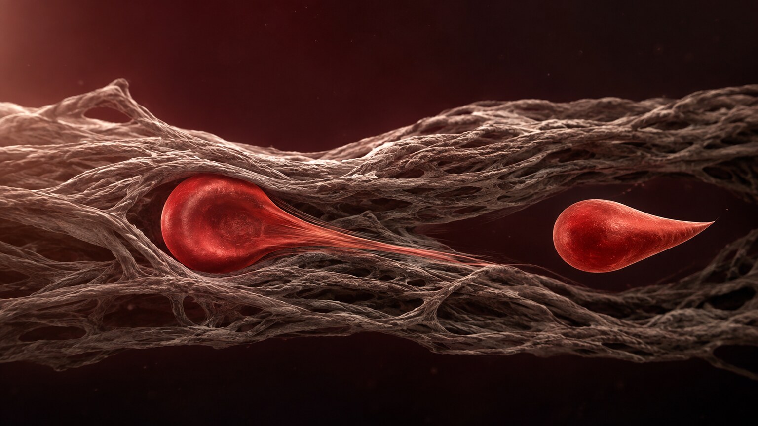
Figure 1. A teardrop cell forms mechanically, not chemically — as a developing red blood cell squeezes through dense fibrous scar tissue on its way out of the bone marrow, its trailing edge stretches into a tail before finally pinching free.
Where Red Blood Cells Actually Come From
To understand why teardrop cells form, it helps to understand where red blood cells come from in the first place, and just how much work is packed into that journey. Every red blood cell in your body starts life deep inside your bone marrow, the soft, spongy tissue found within the hollow centers of certain bones, where a specialized stem cell divides and matures through several distinct stages over roughly a week before finally becoming a fully functional red blood cell ready to enter your bloodstream. At the very last stage of this process, the maturing cell has to physically expel its own nucleus and then navigate its way out of the marrow tissue itself, squeezing between the specialized cells and blood vessel walls that make up the marrow's internal architecture before finally reaching the general circulation.
This exit process, under normal, healthy conditions, happens smoothly enough that it leaves no visible mark on the cell's final shape — a healthy marrow's open, well-organized structure simply doesn't put enough mechanical stress on an exiting cell to distort it. Teardrop cells only become a meaningful finding when something about that exit pathway has changed significantly enough to leave a visible, physical imprint on the cells passing through it, which is exactly the scenario the rest of this article explores.
This entire production and release process runs continuously, around the clock, generating an enormous number of new red blood cells every single day just to keep pace with the ordinary, ongoing turnover of a person's existing blood supply. That sheer, continuous throughput is part of why even a moderate disruption to the marrow's normal exit architecture can show up relatively quickly and consistently on a blood smear — it isn't a rare, occasional event being caught by chance, but a steady, high-volume process being visibly altered in a way that repeat sampling reliably picks up.
Why the Teardrop Shape Forms: A Mechanical Injury, Not a Chemical One
Most of the red blood cell shape abnormalities covered elsewhere in hematology come from something wrong inside the cell — an imbalance between its membrane and its hemoglobin content, a genetic defect in one of its structural proteins, or damage from an outside chemical process. Teardrop cells are different: they form primarily through direct physical force applied to an otherwise normal cell as it tries to leave the bone marrow and enter circulation.
Inside healthy bone marrow, developing red blood cells pass relatively freely through open, spongy marrow tissue on their way into the bloodstream. When that marrow tissue becomes replaced by dense, fibrous scar tissue, or becomes overcrowded with abnormal cells that shouldn't be there, developing red blood cells trying to exit have to squeeze through a much tighter, less forgiving space than usual. As a cell forces its way through this narrowed passage, its trailing edge can get physically stretched out into an elongated tail before the cell finally pulls free — and that stretched, pinched shape is exactly what a teardrop cell looks like under the microscope.
This mechanical explanation has been directly supported by microscopy studies examining bone marrow tissue itself, which have captured red blood cells caught mid-transit, visibly deformed and elongated precisely at the narrow points where they're squeezing past dense fibrous strands or crowding cells, essentially freezing the exact moment of deformation this article is describing. This kind of direct visual confirmation is part of why the mechanical explanation for teardrop cell formation is so well accepted in hematology, rather than being an inferred, indirect theory — it's been observed happening in real time.
It's also worth noting that a cell doesn't need to be permanently damaged by this process to end up with a teardrop shape — the deformation happens to the cell's membrane specifically, and depending on how much the membrane recoils afterward, some teardrop cells retain a fairly pronounced, elongated tail while others show only a subtler, more modest point at one end. This range of severity is part of why an experienced pathologist evaluates not just whether teardrop cells are present, but how pronounced and how numerous they are, since both details carry additional information about how significant the underlying marrow disruption actually is.
Curious what the rest of your CBC or blood smear findings are telling you? Upload your results and get a complete, plain-language breakdown in under 15 minutes.
Analyze My ResultsCause One: Primary Myelofibrosis, the Classic Cause
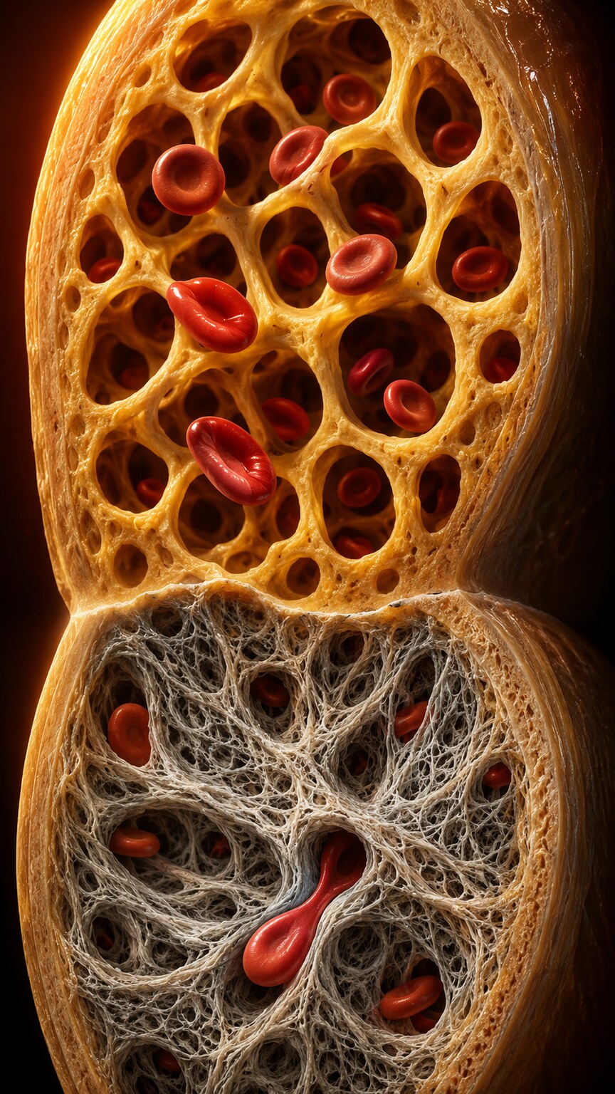
Figure 2. In primary myelofibrosis, the bone marrow's normal open architecture is gradually replaced by dense collagen scar tissue, narrowing the pathways developing blood cells must squeeze through to reach circulation.
Primary myelofibrosis is the condition most classically associated with teardrop cells, and it's the textbook example of exactly the mechanism described above. It's a disorder in which the bone marrow progressively develops scar tissue, called fibrosis, gradually replacing the normal, open marrow architecture that blood cells need in order to develop and exit efficiently. As this scarring worsens, the physical squeeze every developing red blood cell has to endure on its way out becomes more severe, and teardrop cells become correspondingly more numerous and more visually pronounced on the blood smear.
Primary myelofibrosis belongs to a broader category of blood disorders called myeloproliferative neoplasms, conditions in which the bone marrow overproduces one or more types of blood cells due to an acquired genetic change in the marrow's own stem cells. In primary myelofibrosis specifically, that overproduction is accompanied by, and eventually overtaken by, this progressive scarring process, which is what ultimately drives the teardrop cell pattern along with the anemia that commonly develops as the disease advances and the marrow's actual blood-producing capacity declines.
The disease typically follows a recognizable trajectory, though the pace varies considerably from person to person. In its earliest, so-called prefibrotic phase, the marrow may show only minimal scarring, with relatively few teardrop cells and blood counts that can even appear mildly elevated rather than low, since the marrow is still hyperactive at this stage rather than failing. As fibrosis progresses into its more overt, later phase, scarring becomes more extensive, teardrop cells become considerably more numerous and pronounced, and the marrow's actual capacity to produce healthy blood cells begins to decline, producing the anemia and other blood count abnormalities more classically associated with advanced disease. This staged progression is part of why the same underlying diagnosis, primary myelofibrosis, can look quite different on a blood smear depending on exactly when in this trajectory a given patient's sample happens to be examined.
Symptoms tend to track loosely with this same progression. Early-stage disease is often entirely asymptomatic, discovered incidentally during routine bloodwork ordered for an unrelated reason, with teardrop cells sometimes being one of the very first visible clues before any symptoms develop at all. More advanced disease commonly produces fatigue related to worsening anemia, abdominal discomfort or a sense of fullness related to spleen enlargement (itself partly driven by the extramedullary hematopoiesis described later in this article), unintentional weight loss, night sweats, and a generally reduced sense of well-being — a cluster of symptoms collectively reflecting both the marrow's declining function and the body's compensatory efforts elsewhere.
Cause Two: Secondary Myelofibrosis From Other Blood Disorders
Myelofibrosis doesn't always start out as primary myelofibrosis from the beginning — it can also develop as a later stage of two other, related myeloproliferative neoplasms: polycythemia vera, a condition involving overproduction of red blood cells, and essential thrombocythemia, a condition involving overproduction of platelets. Both of these conditions can, over years, progress into a fibrotic, scarred marrow state that looks and behaves essentially like primary myelofibrosis, complete with the same teardrop cell pattern, even though the disease didn't begin as myelofibrosis originally.
This progression, sometimes called post-polycythemia vera myelofibrosis or post-essential thrombocythemia myelofibrosis depending on which condition it evolved from, is part of why patients already diagnosed with either of these two conditions are typically monitored over time with periodic blood counts and smears, watching specifically for early signs of this kind of transition, including the gradual appearance of teardrop cells, well before the disease has progressed far enough to cause dramatic symptoms.
All three of these related conditions — primary myelofibrosis, polycythemia vera, and essential thrombocythemia — share a common underlying biological thread despite their different starting presentations: each is driven by an acquired mutation in a small number of genes controlling how aggressively bone marrow stem cells signal themselves to divide and produce blood cells. This shared genetic root is part of why one condition can transform into another over time, and it's also why testing for these specific mutations, described in more detail later in this article, plays a role across all three related conditions rather than being useful only for diagnosing myelofibrosis specifically.
The rate at which polycythemia vera or essential thrombocythemia progresses into secondary myelofibrosis varies considerably between patients, and not every person with either underlying condition will eventually experience this transition at all — many live for decades with a stable, well-managed version of their original diagnosis and never develop significant marrow fibrosis. This is precisely why ongoing monitoring matters rather than assuming transformation is inevitable: catching the earliest signs, teardrop cells among them, allows a hematologist to confirm and address a genuine transition promptly if and when it actually begins, rather than either missing it or over-treating a stable condition that was never going to progress in the first place.
Cause Three: Bone Marrow Infiltration by Cancer
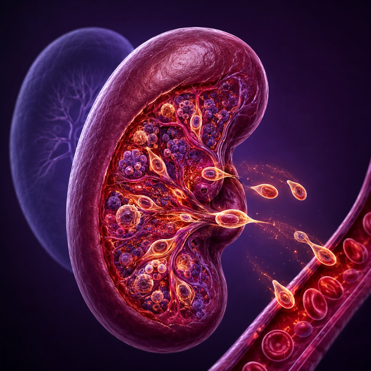
Figure 3. When bone marrow function is significantly compromised, the spleen and liver can partially resume the blood-cell-producing role they perform before birth, a process called extramedullary hematopoiesis, which itself contributes additional teardrop cells to circulation.
A separate cause entirely, called myelophthisic process, involves the bone marrow's normal architecture being physically crowded out and disrupted by something that doesn't belong there at all — most often cancer cells, whether from leukemia originating in the marrow itself, lymphoma involving the marrow, or cancer that has spread to the bone marrow from somewhere else in the body, such as breast, prostate, or lung cancer. In every one of these scenarios, the invading abnormal cells physically distort and crowd the marrow's normal structure, creating the same kind of tight, deforming passage that produces teardrop cells, through essentially the same underlying mechanical principle as fibrosis, just with a different invading material doing the crowding.
When the marrow's blood-producing capacity is significantly compromised, whether by fibrosis or by this kind of infiltration, the body has a remarkable backup system: the spleen and liver, which actually produced blood cells for the developing fetus before birth, can partially reactivate this same function in adulthood, a process called extramedullary hematopoiesis, literally meaning blood production happening outside the marrow. This compensatory production, however, happens in tissue that was never really built for it, and cells produced this way are themselves prone to being released with irregular shapes, including teardrop cells, adding a second, separate source of the same finding on top of whatever's happening in the marrow directly.
This compensatory process is also a major reason significant spleen enlargement, called splenomegaly, is such a common accompanying finding in advanced myelofibrosis and other conditions producing severe marrow dysfunction. As the spleen ramps up its blood-producing role to compensate for the failing marrow, its own tissue structure expands considerably, sometimes dramatically, to accommodate this unfamiliar new function. A markedly enlarged spleen, identified either through physical examination or imaging, alongside teardrop cells and a leukoerythroblastic pattern on the blood smear, further reinforces the likelihood that significant marrow pathology is genuinely present, since these findings tend to travel together as different visible expressions of the same underlying process.
The specific types of cancer capable of infiltrating bone marrow this way deserve a bit more detail, since the term covers a genuinely broad category. Leukemias, cancers originating from blood-forming cells themselves, are perhaps the most direct example, since the marrow is quite literally where these cancer cells originate and multiply. Lymphomas, cancers of the lymphatic system, can also spread into and infiltrate bone marrow tissue as part of their disease progression, even though they don't originate there. And solid tumors — cancers that begin in an organ like the breast, prostate, lung, or several others — can, in more advanced stages, metastasize specifically to bone, sometimes infiltrating the marrow space within that bone as part of that spread. Each of these represents a genuinely different underlying cancer with a different treatment approach, even though all three can produce essentially the same teardrop cell pattern through the same shared mechanical crowding principle.
Cause Four: Severe Thalassemia and Other Causes of Marrow Stress
Beyond fibrosis and infiltration, teardrop cells can also appear, generally in smaller numbers, in conditions that place significant ongoing stress on the bone marrow without physically scarring or crowding it. Severe forms of thalassemia, the inherited condition involving reduced hemoglobin chain production covered in more depth elsewhere on this site, can produce enough disorganized, ineffective red blood cell development within the marrow that some developing cells emerge misshapen, including some with a teardrop appearance, even without any true fibrosis being present. Severe megaloblastic anemia, caused by significant vitamin B12 or folate deficiency, can produce a similar effect through its own disruption of normal cell development within the marrow.
In these conditions, teardrop cells tend to appear as one abnormal shape among several others rather than as the single dominant, defining finding they become in myelofibrosis, which is part of why the overall pattern of accompanying findings, covered in the next section, matters so much for correctly distinguishing between these different possible explanations.
It's worth understanding why severe thalassemia produces this effect even without true fibrosis, since the mechanism differs somewhat from the physical crowding described in the previous sections. In severe thalassemia, the marrow attempts to compensate for the body's chronic shortage of properly functioning hemoglobin by dramatically ramping up red blood cell production, sometimes to many times the normal rate. This intense overproduction floods the marrow with an unusually large number of developing cells simultaneously, creating a kind of internal crowding through sheer volume rather than through scar tissue or invading cancer cells, and that crowding alone can be enough to physically deform some cells on their way out, producing a smaller-scale version of the same teardrop effect.
Megaloblastic anemia works through a related but distinct pathway: severe vitamin B12 or folate deficiency disrupts the normal process of DNA synthesis inside developing blood cells, causing many of them to grow abnormally large and structurally disorganized before they're ready to be released. This disorganized development can itself produce misshapen cells, including some teardrop forms, though megaloblastic anemia's more classic and defining smear finding is actually a different one — unusually large red blood cells called macrocytes — with teardrop cells appearing as a secondary, less prominent feature alongside that primary finding rather than as the dominant abnormality.
Teardrop Cells as a Nonspecific Finding: When a Few Don't Mean Much
Before moving on to the pattern that actually raises genuine concern, it's worth being explicit about the opposite side of this topic: a small number of teardrop cells, on their own, isn't automatically evidence of any of the significant conditions described above. Teardrop cells can appear in modest numbers in a range of milder, more common situations — including some cases of ordinary iron deficiency anemia, certain other mild anemias, and occasionally even in blood samples from people with no identifiable underlying blood disorder at all, sometimes attributed to minor, transient stress on the marrow or even artifacts introduced during the smear preparation process itself.
This is precisely why an experienced pathologist doesn't treat every single teardrop cell as an automatic red flag requiring urgent workup. Context, quantity, and the accompanying pattern on the same smear all factor into how seriously a given teardrop cell finding is taken, exactly the same interpretive principle that applies to nearly every red blood cell shape finding covered across hematology — the shape itself is a clue, not a verdict, and its true significance only emerges once it's considered alongside everything else visible on the same slide.
This same nuance also applies when a smear is read from a sample that wasn't handled quite ideally before reaching the lab — a delayed transport time, an improperly prepared slide, or a sample that sat too long before being smeared can all occasionally introduce artifactual cell distortions that superficially resemble true teardrop cells without reflecting any real marrow pathology at all. An experienced reviewer accounts for this possibility as well, generally by looking for consistency of the finding and by correlating it against the rest of the clinical picture rather than reacting to an isolated, unexplained shape abnormality in a sample with no other supporting evidence behind it.
The Pattern That Actually Matters: A Leukoerythroblastic Smear
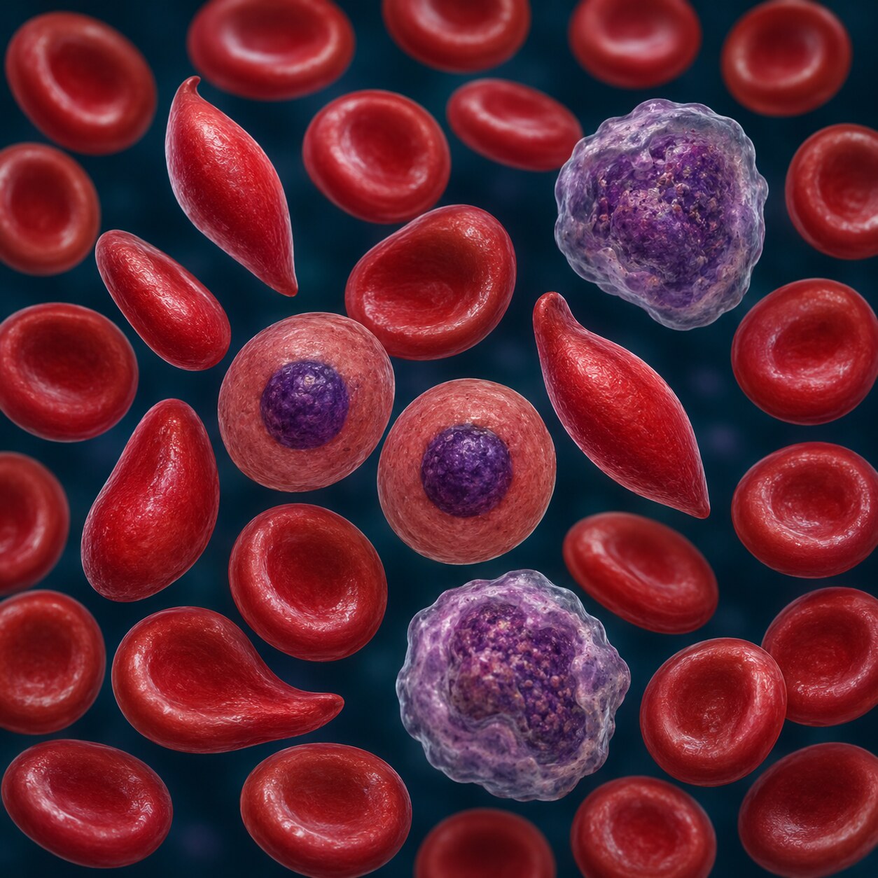
Figure 4. A leukoerythroblastic smear — teardrop cells appearing together with immature, nucleated red blood cells and immature white blood cells that shouldn't normally be in circulation at all — is the specific combined pattern that most strongly points toward bone marrow scarring or infiltration.
A handful of scattered teardrop cells, on their own, without anything else unusual accompanying them, is a relatively nonspecific finding that a pathologist may note without particular alarm, since a small number can appear incidentally in various mild or transient conditions. What genuinely raises concern for significant bone marrow pathology is a specific combined pattern called a leukoerythroblastic smear, where numerous teardrop cells appear together with two other things that shouldn't normally be present in circulating blood at all: nucleated red blood cells (immature red blood cells that still contain their nucleus, normally kept within the marrow until that nucleus is expelled) and immature white blood cells at various early stages of development that should also, under normal circumstances, remain within the marrow until fully mature.
This specific three-part combination — teardrop cells, nucleated red blood cells, and immature white blood cells, all appearing together — is highly suggestive that the bone marrow's normal architecture has been significantly disrupted, either by fibrosis or by infiltration, severely enough that cells at every stage of development are being pushed out into circulation before they're actually ready, essentially because the marrow itself no longer has the normal structural framework to properly regulate and time their release. Seeing teardrop cells in isolation prompts a note and possible follow-up; seeing a full leukoerythroblastic pattern prompts a considerably more urgent, focused workup.
It's worth understanding why nucleated red blood cells and immature white blood cells specifically, rather than any other kind of abnormal finding, are the ones that pair so meaningfully with teardrop cells in this pattern. Under normal circumstances, the bone marrow acts as a kind of quality-control checkpoint, holding developing cells back until they've reached full maturity and are genuinely ready to function in circulation. A severely disrupted marrow architecture, whether scarred by fibrosis or crowded by infiltrating cells, loses much of this normal gatekeeping ability, essentially because the structural framework that normally regulates an orderly, staged release has itself been damaged or displaced. Immature cells that would ordinarily be retained and allowed to finish developing get pushed out prematurely instead, alongside the mechanically deformed teardrop cells experiencing the same disrupted exit pathway. All three findings are, in this sense, different visible symptoms of the exact same underlying structural breakdown.
The term "leukoerythroblastic" itself reflects this combination directly in its own name — "leuko" referring to the immature white blood cells, "erythro" referring to the immature (nucleated) red blood cells, and "blastic" referring to the early developmental stage both cell types are caught at when this pattern is present. Recognizing this specific combined terminology on a lab report, rather than seeing "teardrop cells" reported in isolation, is itself a signal that the finding has already been flagged by the reviewing pathologist as clinically significant, since the leukoerythroblastic label is specifically reserved for this recognized combined pattern rather than being applied loosely to any single abnormal cell shape on its own.
It's also worth noting what a leukoerythroblastic pattern does not, by itself, tell a doctor: it strongly suggests significant marrow disruption is present, but it doesn't specify which of the several possible underlying causes — fibrosis, cancer infiltration, or another less common process entirely — is actually responsible. That distinction is precisely what the follow-up testing described in the next section exists to resolve, since the blood smear alone, however informative, can only narrow the possibilities down rather than pinpoint the exact diagnosis on its own.
What Follow-Up Testing Typically Looks Like

Figure 5. Once a leukoerythroblastic pattern is identified, the specific next step is typically a bone marrow biopsy, which directly examines the marrow tissue itself rather than relying solely on the indirect clues visible in circulating blood.
When a pathologist identifies a significant number of teardrop cells, particularly alongside the leukoerythroblastic pattern described above, the next step is generally a bone marrow biopsy — a procedure that directly samples marrow tissue, usually from the back of the pelvic bone, allowing a pathologist to examine the marrow's actual architecture under the microscope rather than only inferring what's happening there indirectly through circulating blood cell shapes. This direct examination can confirm the presence and degree of fibrosis, or identify infiltrating cancer cells directly, in a way that circulating blood alone can only strongly suggest.
If myelofibrosis or a related myeloproliferative neoplasm is suspected specifically, genetic testing looking for one of three specific acquired mutations — in genes called JAK2, CALR, or MPL — is typically performed as well, since the large majority of cases of primary myelofibrosis and its related conditions carry one of these three mutations. Identifying which specific mutation is present, if any, doesn't just help confirm the diagnosis; it also carries real prognostic and treatment-planning value, since these different mutations are associated with somewhat different expected disease courses and can influence which treatment approach a hematologist recommends.
These three genes all converge on a shared underlying biological pathway, one that normally helps regulate how strongly bone marrow stem cells respond to the hormonal signals that tell them to divide and produce more blood cells. A mutation in any one of the three genes tends to lock this pathway into an overactive state, causing the marrow to behave as though it's constantly receiving a strong "produce more blood cells" signal, regardless of the body's actual needs — this shared mechanism is why three genetically distinct mutations can all produce such similar clinical pictures, since they're all ultimately disrupting the same regulatory system, just at slightly different points along it.
Testing for these mutations is typically done using a blood sample rather than requiring the bone marrow biopsy sample specifically, since the mutation, having arisen in the marrow's stem cells, is also present in the mature blood cells those stem cells eventually produce and release into circulation. This means genetic testing and the bone marrow biopsy are often pursued together but through separate sample types, each contributing a different, complementary piece of the overall diagnostic picture — the biopsy showing the marrow's actual physical architecture, and the genetic test identifying the specific molecular driver behind it.
When a Bone Marrow Biopsy Confirms the Diagnosis

Figure 6. A bone marrow biopsy sample is examined for the actual degree of fibrosis or cellular infiltration present, information that directly shapes ongoing management decisions and helps distinguish among the several possible underlying causes covered in this article.
Once a bone marrow biopsy and any relevant genetic testing are complete, the specific findings determine the path forward, which varies considerably depending on the underlying cause identified. Confirmed primary or secondary myelofibrosis is managed with its own specific set of treatment approaches, ranging from careful monitoring in milder, early cases to targeted medications or, in eligible patients, stem cell transplantation for more advanced disease. Marrow infiltration by cancer is managed according to the specific cancer identified, following an entirely different treatment pathway tailored to that cancer type rather than to the blood cell shape finding that originally prompted the workup.
In every case, the biopsy result is what ultimately transforms an indirect clue — a distinctive cell shape visible on a blood smear — into a specific, actionable diagnosis, which is exactly the role this kind of direct tissue examination is meant to play whenever indirect findings in circulating blood raise a strong enough suspicion to warrant it.
For patients diagnosed with myelofibrosis specifically, ongoing management typically involves periodic reassessment, since the disease's pace and severity can shift meaningfully over time. Repeat blood counts and smears, tracking whether teardrop cells and the broader leukoerythroblastic pattern are becoming more or less pronounced, serve as one relatively simple, noninvasive way to monitor disease activity between the more involved bone marrow biopsies, which aren't typically repeated as frequently given their more invasive nature. In this way, the same shape finding that often first raises the initial suspicion also becomes a practical tool for tracking the disease over the months and years that follow a confirmed diagnosis.
Frequently Asked Questions
Do a few teardrop cells on my report automatically mean I have myelofibrosis?
No. A small number of scattered teardrop cells is a relatively nonspecific finding that can appear incidentally in various conditions. Significant concern arises specifically when teardrop cells appear in large numbers alongside a leukoerythroblastic pattern, not from a few isolated cells alone.
What is a leukoerythroblastic smear, exactly?
It's a specific combined pattern where teardrop cells appear together with immature, nucleated red blood cells and immature white blood cells that shouldn't normally be circulating. This full pattern together is a much stronger signal of significant bone marrow disruption than teardrop cells alone.
Why does a bone marrow biopsy get ordered instead of just repeating the blood test?
Because a blood smear only shows indirect evidence, in the form of cell shapes, of what's happening inside the marrow. A biopsy directly examines the marrow tissue itself, confirming the actual degree of fibrosis or the presence of infiltrating cells in a way circulating blood alone cannot.
Are JAK2, CALR, and MPL mutations the actual cause of myelofibrosis?
They're acquired genetic changes found in the large majority of primary myelofibrosis cases, and identifying which one is present helps confirm the diagnosis and provides prognostic information, though testing for them is done specifically because myelofibrosis is already suspected, not as a general screening test.
Can polycythemia vera or essential thrombocythemia turn into myelofibrosis?
Yes, in some patients. Both conditions can progress over time into a fibrotic marrow state that behaves like primary myelofibrosis, including the same teardrop cell pattern. This is why patients with either diagnosis are typically monitored periodically for early signs of this transition.
Does an enlarged spleen relate to teardrop cells?
Often, yes. When bone marrow function declines significantly, the spleen can partially take over blood cell production, a process called extramedullary hematopoiesis, which itself contributes additional teardrop cells and commonly causes the spleen to enlarge as it adapts to this unfamiliar role.
Conclusion
Teardrop-shaped red blood cells form through a distinctly mechanical process — a cell physically squeezed and stretched as it forces its way out of scarred or overcrowded bone marrow tissue — which is why this particular shape points so specifically toward the marrow itself rather than toward a chemical or membrane-level problem inside the cell. Primary and secondary myelofibrosis are the classic causes, bone marrow infiltration by cancer produces the same shape through a related crowding mechanism, and severe thalassemia or megaloblastic anemia can contribute smaller numbers through general marrow stress. What ultimately separates a benign, incidental finding from a genuine red flag is the surrounding pattern: a full leukoerythroblastic picture, not teardrop cells in isolation, is what typically prompts the bone marrow biopsy that can actually confirm what's happening and guide what comes next.
Understanding this single principle — that a cell's shape can encode real, physical information about the environment it just came from, not only about its own internal chemistry — is a useful lens for thinking about blood smear findings more broadly, and teardrop cells remain one of the clearest, most direct examples of that principle in all of hematology, a shape that quite literally carries the physical memory of the difficult journey it took just to reach circulation at all.
Still Not Sure What Your Results Mean?
Upload your labs and get a complete, visual, plain-language interpretation of every biomarker — delivered to your inbox in under 15 minutes.
Get My ReportThis article is for educational purposes only and does not constitute medical advice. Always consult your healthcare provider regarding your specific lab results.