
What Are Schistocytes and Why Do They Appear on a Smear?
If a technologist looking at your blood under a microscope reports seeing schistocytes, they're describing something very specific: red blood cells that have been physically torn apart while moving through your bloodstream, rather than cells that simply look unusual because of a size or color problem. A schistocyte is a fragment — a broken-off piece of what used to be a normal, round red blood cell — and finding even a small number of them on a smear is one of the few results in laboratory medicine that can shift a routine blood draw into an urgent same-day phone call from your doctor's office. This article explains exactly what a schistocyte is, the mechanical process that creates one, the specific conditions capable of causing this kind of damage, and why clinicians treat this particular finding with a level of urgency that most abnormal lab results don't get.
What a Schistocyte Actually Looks Like Under the Microscope
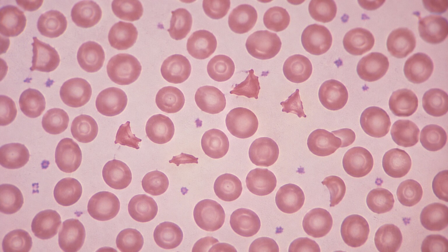
Figure 1. Schistocytes appear on a stained smear as small, jagged, irregularly shaped fragments — helmet cells, triangles, and crescents — mixed in with normal round red blood cells.
To a trained eye, a schistocyte doesn't look like a red blood cell that's slightly off — it looks like a piece of one. A healthy red blood cell, when stained and viewed under a microscope, is a smooth, round disc with a lighter, dimpled center, similar in outline to a doughnut viewed from above. A schistocyte breaks that pattern entirely: it's smaller, it has hard angles instead of a smooth curve, and it often takes on one of a few recognizable shapes that hematologists learn to spot by name. "Helmet cells" are among the most classic — fragments with two sharp points and a flattened edge, resembling a tiny military helmet. Others look like simple triangles, crescents, or commas, and some are so small and irregular that they're best described just as "fragments" without a more specific shape name. What unites all of them is a jagged, cut edge — the visual signature of a cell that was sliced rather than one that gradually changed shape over its lifespan, which is exactly what separates a schistocyte from other oddly shaped red blood cells a smear might reveal.
This distinction matters diagnostically, because plenty of other red blood cell shapes look unusual without meaning the same thing. A spherocyte, for example, is a red blood cell that has lost surface area but kept a smooth, round outline, usually because its outer membrane was damaged from the inside rather than physically cut. A sickle cell has a smooth, curved, elongated shape caused by abnormal hemoglobin bending the entire cell, not slicing off a piece of it. A schistocyte, by contrast, is defined by that broken, angular edge — direct physical evidence that something in the bloodstream cut a piece off a cell that started out whole. Laboratories typically only flag the finding as clinically significant once schistocytes make up more than about 1% of the red blood cells counted on the slide, since a rare, isolated fragment can occasionally appear on an otherwise unremarkable smear without indicating disease — it's the presence of multiple fragments, seen consistently across the slide, that turns this from a curiosity into a genuine red flag.
How a Healthy Red Blood Cell Turns Into Broken Fragments
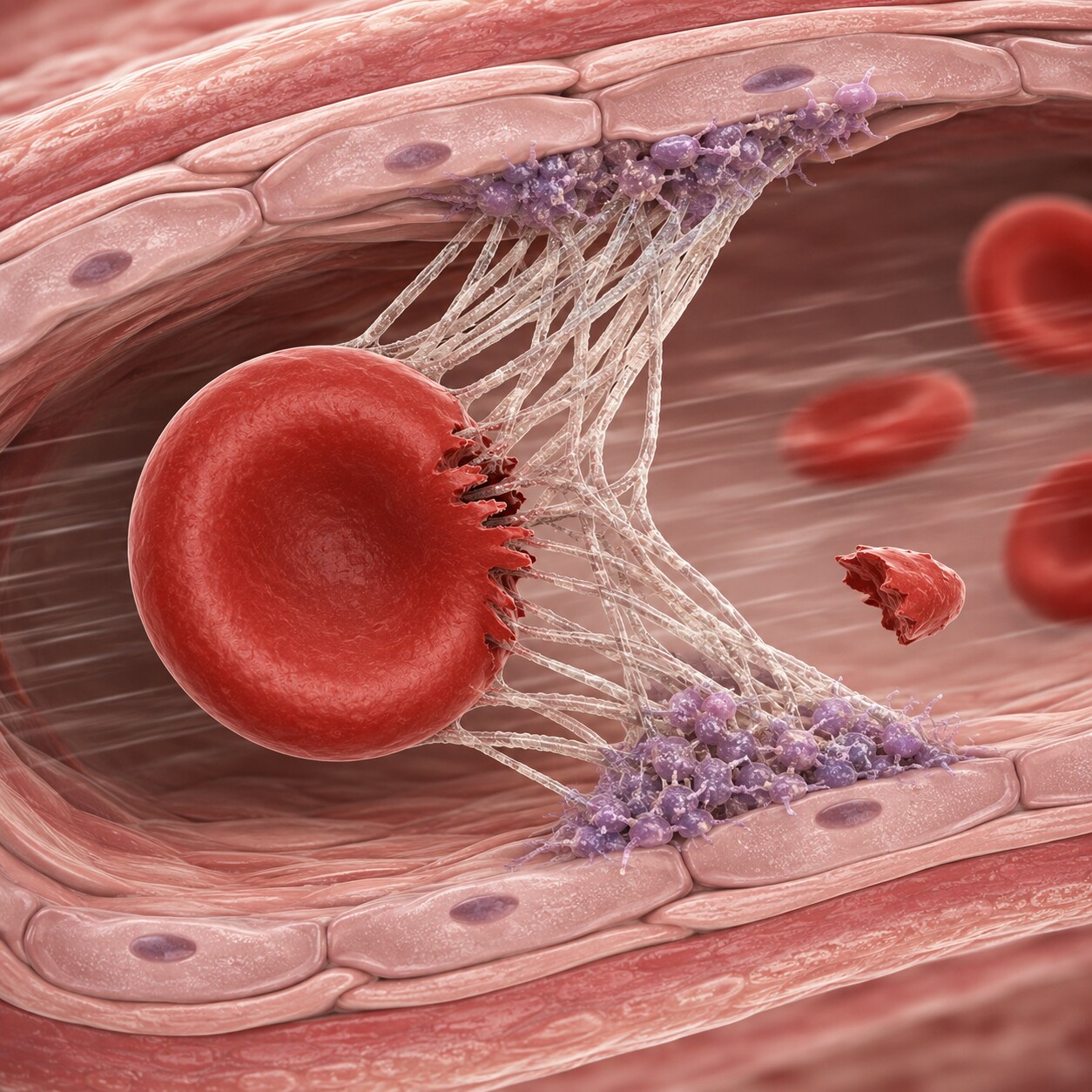
Figure 2. Strands of fibrin — a clotting protein — mesh across a narrowed vessel, and the force of blood flow drags red blood cells across the mesh, physically shearing pieces off.
Every red blood cell is built to be remarkably flexible. It has to squeeze through capillaries narrower than its own resting width, fold nearly in half, and spring back to shape thousands of times over its roughly 120-day lifespan, all without breaking. That flexibility depends on a healthy, elastic outer membrane and an open, unobstructed path to travel through. Schistocytes form when that path stops being open — when something inside a small blood vessel creates a physical obstacle the cell can't simply bend around, and the force of ongoing blood flow drags the cell across that obstacle hard enough to shear a piece off, the way a piece of fruit gets sliced if you drag it across a cheese grater rather than cutting it cleanly with a knife.
In the great majority of cases, that obstacle is fibrin — a stringy, mesh-like protein that's normally produced only at the site of an injury to seal a cut and stop bleeding. In the conditions that cause schistocytes, fibrin strands and small platelet clumps form inside intact small blood vessels — arterioles and capillaries — where they have no business being, based on tiny, inappropriate clots called microthrombi. As blood keeps flowing past these strands under normal circulatory pressure, red blood cells get pushed against and through the mesh over and over, and each pass shears off a little more of the cell until what's left is a jagged fragment instead of a full, round disc. This general process has a name in medicine — microangiopathic hemolytic anemia, often abbreviated MAHA — which breaks down plainly as "small-vessel-related destruction of red blood cells causing anemia." It isn't a single disease; it's a description of what's happening mechanically, and a wide range of very different underlying conditions can all produce that same mechanical result.
It's worth being clear about what schistocyte formation is not. It isn't caused by an infection directly attacking red blood cells, and it isn't caused by a nutritional deficiency the way some other forms of anemia are. It's purely mechanical — a matter of physical force acting on a cell that has nowhere safe to go. That's part of why the finding is taken so seriously: it's direct, visible proof that something is actively and repeatedly damaging the smallest vessels in the body in real time, right now, not evidence of a slow-building deficiency that can be addressed on a leisurely timeline.
Thrombotic Thrombocytopenic Purpura (TTP): The Cause That Demands Same-Day Action
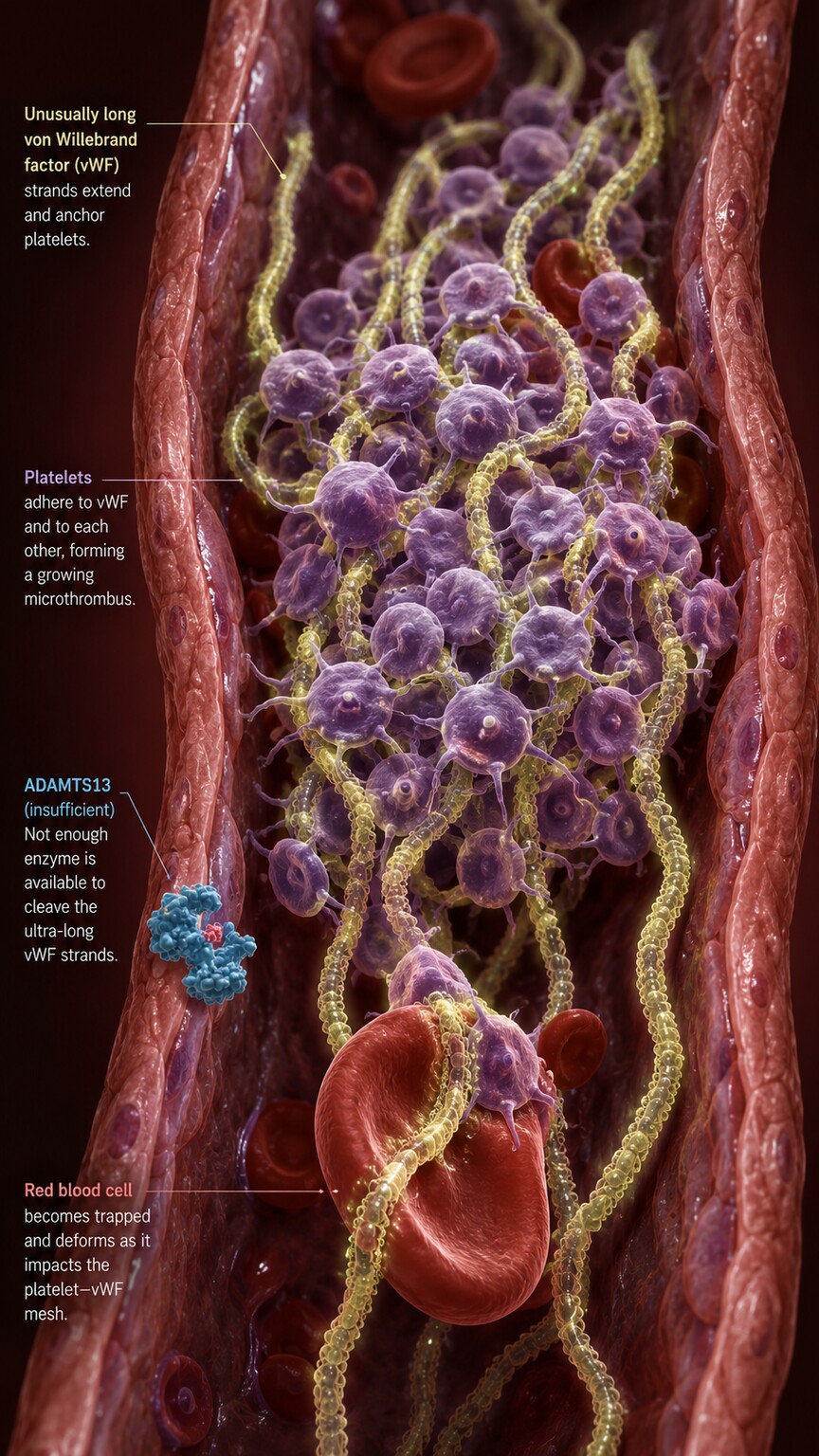
Figure 3. Without enough working ADAMTS13 enzyme, unusually large strands of von Willebrand factor stay intact and trap platelets into microscopic clots throughout the small vessels.
Of all the conditions that cause schistocytes, thrombotic thrombocytopenic purpura, or TTP, is the one clinicians are most anxious to rule out first, because untreated TTP has historically carried a mortality rate above 90%, while prompt treatment brings survival above 80–90%. That gap, driven almost entirely by how quickly treatment starts, is the single biggest reason a smear showing schistocytes gets escalated immediately rather than filed away for a routine follow-up appointment. TTP's root cause is a shortage of a specific enzyme called ADAMTS13, whose normal job is to trim down an unusually long, sticky clotting protein called von Willebrand factor into shorter, more manageable pieces. When ADAMTS13 is severely deficient — most often because the immune system has mistakenly produced antibodies against it, and less commonly due to an inherited genetic mutation — von Willebrand factor stays abnormally long and sticky, and those oversized strands snag passing platelets, forming small clots scattered throughout the body's smallest vessels. Red blood cells forced through this platelet-and-protein mesh are what get shredded into schistocytes.
Because TTP consumes platelets to build these tiny clots, it produces a very specific, recognizable trio of findings on routine labs: schistocytes on the smear, a low platelet count, and signs of active red blood cell destruction such as a high LDH (an enzyme released when cells break down) and a low haptoglobin (a protein that gets used up mopping up the debris from broken red blood cells). Some people with TTP also develop neurological symptoms — confusion, headache, or even seizures — because these same tiny clots can form in vessels supplying the brain, along with kidney involvement if the clots affect the kidneys. Because waiting for a specialized ADAMTS13 lab test to come back — which can take days — would cost precious time, the standard approach when TTP is strongly suspected is to begin plasma exchange therapy right away, often the same day the smear is reviewed, swapping out the patient's plasma (which contains the antibodies and abnormal von Willebrand factor) for donor plasma containing normal, functioning ADAMTS13.
Curious what the rest of your complete blood count is telling you? Upload your results and get a complete, plain-language breakdown in under 15 minutes.
Analyze My ResultsHemolytic Uremic Syndrome (HUS): The Cause Most Often Seen in Children
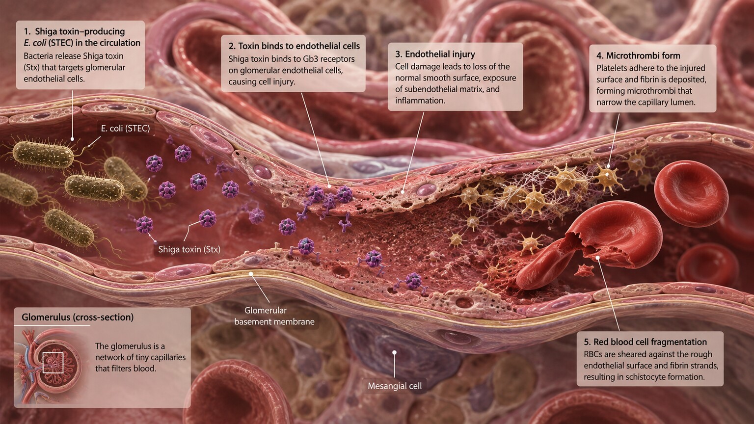
Figure 4. Shiga toxin, produced by certain strains of E. coli, injures the delicate lining of small kidney vessels, triggering the same clot-and-shear process that produces schistocytes.
Hemolytic uremic syndrome, or HUS, produces the identical schistocyte-and-platelet pattern seen in TTP, but through a completely different starting point and in a different typical patient. The most common form, sometimes called "typical" HUS, follows an infection with a specific strain of bacteria — most notoriously E. coli O157:H7, usually picked up from undercooked ground beef, unpasteurized dairy or juice, or contaminated produce and water. This strain produces a toxin, called Shiga toxin, that directly damages the delicate cells lining small blood vessels, particularly in the kidneys. That injury triggers the same kind of inappropriate clot formation seen in TTP, and red blood cells forced through those clots get sheared into schistocytes in the same mechanical way.
HUS is disproportionately a disease of young children, and it typically follows a recognizable pattern: a child develops what looks like ordinary gastroenteritis, often with bloody diarrhea, and then, five to ten days later, develops pale skin, unusual bruising, decreased urination, and swelling — signs that the kidneys are struggling and that red blood cells and platelets are being consumed. Because the kidneys bear the brunt of the vessel damage in typical HUS, kidney function often declines more prominently here than in TTP, and some children require temporary dialysis while the acute injury resolves. A rarer form, called atypical HUS, isn't triggered by an infection at all but by a problem in part of the immune system called the complement system, which becomes chronically overactive and damages small vessels on an ongoing basis; it tends to run a more chronic, relapsing course and is managed with different, complement-targeted medications rather than antibiotics or plasma exchange alone.
Disseminated Intravascular Coagulation (DIC): When Clotting Runs Out of Control Everywhere
Disseminated intravascular coagulation, or DIC, is a third major route to schistocyte formation, and it's less a disease in its own right than a dangerous complication that other severe illnesses can trigger. DIC happens when something — severe infection (sepsis), major trauma, certain cancers, or complications of childbirth are among the most common triggers — causes the body's clotting system to activate everywhere at once, all through the bloodstream, rather than staying contained at a single site of injury the way clotting is supposed to work. This widespread, inappropriate clotting uses up clotting factors and platelets far faster than the body can replace them, and it produces exactly the kind of fibrin mesh throughout small vessels that shears red blood cells into schistocytes.
What makes DIC distinct from TTP and HUS on lab testing is that it consumes clotting factors broadly, not just platelets, so it typically shows abnormal results across a wider set of coagulation tests — prolonged clotting times, low fibrinogen (a core clotting protein), and a markedly elevated D-dimer (a fragment produced when clots break down) — in addition to schistocytes and a falling platelet count. Because DIC often accompanies dangerously ill patients who are already being closely monitored in a hospital setting for the underlying trigger, it's frequently caught in that context rather than through a first, unexpected lab finding — but the underlying mechanical damage to red blood cells is identical to what happens in TTP and HUS, just triggered by a different starting problem.
HELLP Syndrome and Severe Hypertension in Pregnancy
Pregnancy introduces its own specific route to schistocyte formation: HELLP syndrome, whose name is itself a summary of its key lab findings — Hemolysis, Elevated Liver enzymes, and Low Platelets. HELLP is considered a severe variant of preeclampsia, the pregnancy complication involving high blood pressure and organ strain, and it typically develops in the third trimester, though it can occasionally appear shortly after delivery. The extremely high blood pressure and vessel dysfunction seen in severe preeclampsia and HELLP damage small vessels throughout the body, including the placenta and liver, triggering the same kind of localized clotting and red blood cell shearing seen in the other conditions on this list. A pregnant person found to have schistocytes alongside a rising blood pressure, upper abdominal pain, and abnormal liver tests is being evaluated for an obstetric emergency, and delivery of the baby — even if premature — is often the definitive treatment once HELLP is confirmed, since the underlying process typically doesn't fully resolve until the pregnancy ends.
Malignant hypertension — blood pressure so severely elevated that it causes acute organ damage on its own, entirely separate from pregnancy — can produce this same picture in anyone, at any age. Extremely high pressure damages the lining of small vessels directly, provoking the identical clot-and-shear cycle, and schistocytes found alongside a blood pressure reading in the range of 180/120 mmHg or higher, particularly with signs of kidney, brain, or eye involvement, point toward this cause specifically.
Mechanical Causes: Heart Valves and Circulatory Support Devices
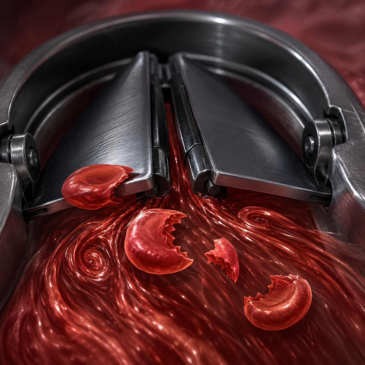
Figure 5. The rigid metal or carbon leaflets of a mechanical heart valve create small zones of turbulent, high-shear blood flow that can physically fragment red blood cells over time.
Not every cause of schistocytes involves inappropriate clotting at all — a meaningful share come from purely mechanical trauma imposed by hardware inside the circulatory system itself. Mechanical prosthetic heart valves, made of rigid metal or carbon rather than the soft tissue used in biological valve replacements, close and open with a snapping motion that creates small pockets of highly turbulent, high-velocity blood flow around their leaflets. Red blood cells caught in that turbulence can be physically torn as they pass through, producing a low-grade, chronic stream of schistocytes that shows up on smears from many people with these valves, even when the valve itself is functioning exactly as designed. This is such a well-recognized pattern that it has its own name — mechanical hemolytic anemia — and it's generally mild and well tolerated, monitored periodically rather than treated aggressively, unless the fragmentation becomes severe enough to cause a meaningful anemia or unless it worsens suddenly, which can be an early clue that the valve itself has developed a problem, such as a leak around its edge.
A similar mechanism explains why people with ventricular assist devices — mechanical pumps implanted to support a failing heart — and those on certain forms of extracorporeal life support, such as ECMO, are also routinely monitored for this same low-grade fragmentation. Severe narrowing of a native heart valve, most notably severe aortic stenosis, can occasionally produce a milder version of this same picture even without any prosthetic hardware involved, since a narrowed valve opening forces blood through at unusually high velocity and turbulence in a similar way.
Other, Less Common Contributors
Beyond the major categories above, a handful of other situations can produce schistocytes, usually in smaller numbers or in more specific clinical contexts. Severe burns covering a large portion of the body can directly damage circulating red blood cells from heat exposure and the resulting changes in blood vessel walls. Certain autoimmune conditions that inflame and damage small blood vessels, broadly called vasculitis, can create the same kind of vessel-lining injury that triggers clot-and-shear damage seen in TTP and HUS. Some cancers, particularly certain aggressive tumors and specific blood cancers, can trigger a cancer-associated form of microangiopathic hemolytic anemia through mechanisms that overlap with DIC. Organ transplant recipients occasionally develop a condition called transplant-associated thrombotic microangiopathy, sometimes linked to certain anti-rejection medications, that produces this same pattern in the newly transplanted organ's blood supply. Severe vitamin B12 deficiency, though it doesn't cause true mechanical fragmentation, can occasionally produce a look-alike picture on smear review, which is one of several reasons a full clinical and lab picture — not a single finding in isolation — is what actually establishes the cause.
Cells That Get Mistaken for Schistocytes
Part of what makes smear interpretation a genuine skill rather than a simple checklist is that a handful of other abnormal red blood cell shapes can superficially resemble a schistocyte to an inexperienced eye, even though they point toward entirely different problems. Bite cells, for instance, have a semicircular chunk missing from one edge, as though something had literally taken a bite out of the cell — but rather than mechanical shearing from fibrin strands, bite cells form when the spleen's resident immune cells physically remove clumps of damaged, oxidized hemoglobin from an otherwise intact cell, most classically seen in G6PD deficiency during an oxidative stress event triggered by certain medications, infections, or foods like fava beans. Blister cells, sometimes seen alongside bite cells in the same condition, have a thin, cleared-out area near the membrane's edge that looks almost like a bubble, reflecting hemoglobin that has denatured and pulled away from the cell's outer wall.
Keratocytes, sometimes called "horn cells," have one or two pointed, horn-like projections rather than the multiple sharp, irregular angles typical of a schistocyte, and they can appear in some of the same conditions that cause true schistocytes, which occasionally makes the distinction more a matter of degree than a hard line. Acanthocytes, spiky cells covered in irregular, blunt projections across their whole surface rather than one or two sharp edges, point toward a different set of causes entirely, including certain liver diseases and rare inherited lipid disorders, and are read as a separate finding from schistocytes despite a passing visual resemblance at first glance. This is precisely why blood smear review is performed by trained laboratory professionals and pathologists rather than left to automated pattern-matching alone — the clinical implications of correctly telling these shapes apart are significant, and a bite cell mistakenly logged as a schistocyte could send a workup down the wrong path entirely, delaying the correct diagnosis while an unrelated, incorrect one is pursued.
What Other Lab Findings Usually Accompany Schistocytes
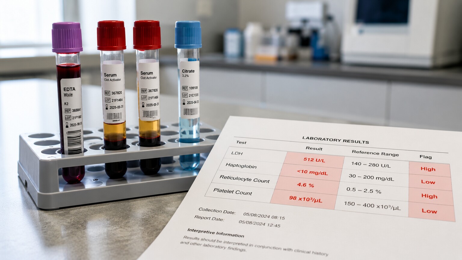
Figure 6. Schistocytes rarely appear alone on a lab report — a rising LDH, a falling haptoglobin, a falling platelet count, and a rising reticulocyte count typically accompany the finding.
Because schistocyte formation is really just the visible tail end of active red blood cell destruction happening throughout the body, a handful of other lab values almost always move in a predictable direction alongside it, and a clinician reviewing your results reads these together as one connected story rather than as separate, unrelated numbers. LDH (lactate dehydrogenase) is an enzyme found inside virtually all cells, including red blood cells, and it spills into the bloodstream in large quantities whenever cells rupture — so a high LDH is one of the most sensitive general markers that active hemolysis is happening somewhere. Haptoglobin moves in the opposite direction: this protein's job is to bind up free hemoglobin released by broken red blood cells and help clear it from circulation, and it gets used up and depleted so quickly during active hemolysis that a low or unmeasurable haptoglobin is one of the more specific clues pointing toward this process.
The platelet count is another critical piece of the puzzle, and in most of the major causes discussed above — TTP, HUS, DIC, HELLP — it runs low, because platelets are being consumed to build the same small clots that are shredding red blood cells in the first place. The reticulocyte count, which measures how many immature red blood cells the bone marrow is releasing, typically runs high, reflecting the bone marrow working overtime to replace red blood cells that are being destroyed faster than normal. Indirect bilirubin, a breakdown product of hemoglobin, is often mildly to moderately elevated for the same underlying reason, and it's part of why some people with active hemolysis develop a faint yellowish tint to their skin or eyes. Seeing this entire pattern together — schistocytes, high LDH, low haptoglobin, low platelets, high reticulocytes — is what allows a lab result to be read confidently as microangiopathic hemolytic anemia rather than being dismissed as an isolated, possibly meaningless smear observation.
How Many Schistocytes Is "Too Many"? Reading the Percentage
Schistocytes are typically reported as a rough percentage of the total red blood cells counted while a technologist reviews the slide, and that percentage carries real diagnostic weight. A very small number — well under 1% — can sometimes be seen in people with no significant underlying disease, since a handful of red blood cells can be mechanically damaged during ordinary blood draws or specimen handling, or from minor, clinically unimportant vessel irregularities. Once schistocytes reliably make up more than roughly 1% of red blood cells on a well-prepared slide, though, the finding is generally considered clinically significant and worth pursuing, and the higher that percentage climbs, the more strongly it points toward one of the serious, actively destructive processes covered above rather than an incidental artifact. Some laboratories use a semi-quantitative scale — rare, few, moderate, or many — rather than a precise percentage, but the underlying clinical logic is the same: a handful of scattered fragments is a different situation than fields of the microscope crowded with them.
It's worth understanding that automated blood cell analyzers, the machines that generate most routine complete blood count results, are notoriously unreliable at detecting schistocytes on their own. These machines are excellent at counting cells and estimating their average size, but they weren't designed to reliably distinguish a jagged fragment from a normally shaped small red blood cell using the flags and indices they generate. This is precisely why a manual review of the smear by a trained eye — a human physically looking through a microscope — remains the gold standard for this specific finding, and it's one of the clearest examples of why an automated report reading "normal" doesn't always tell the full story, particularly if a clinician has a specific reason to suspect one of the conditions discussed above and requests a manual smear review directly.
What Happens After Schistocytes Are Found: The Diagnostic Workup
Once a smear comes back showing a clinically significant number of schistocytes, the workup that follows moves quickly and in a fairly predictable sequence, because the goal is to identify or rule out TTP within hours, not days. A complete blood count with platelet count, LDH, haptoglobin, reticulocyte count, and a coagulation panel (including fibrinogen and D-dimer) are typically drawn immediately, since together they distinguish TTP and HUS, which usually spare the broader clotting system, from DIC, which disrupts it broadly. A blood sample is often sent for an ADAMTS13 activity level, though because this specialized test can take one to several days to return from a reference laboratory, treatment decisions for suspected TTP are rarely delayed while waiting on the result. Kidney function tests (creatinine and blood urea nitrogen) help gauge how much kidney involvement is present, which is particularly relevant for HUS. In a child with a preceding diarrheal illness, a stool culture and specific testing for Shiga toxin help confirm typical HUS. In anyone pregnant or recently postpartum, blood pressure, liver enzymes, and a careful review of pregnancy-related symptoms screen for HELLP syndrome and severe preeclampsia. A recent medical history — including any prosthetic heart valve, recent chemotherapy, autoimmune disease diagnosis, or severe infection — helps direct the workup toward the mechanism most likely to be at play in that specific person.
Why This Finding Is Often Treated as an Emergency Before the Cause Is Even Confirmed
One detail that surprises a lot of people is that treatment for the most dangerous possibility — TTP — is frequently started before the diagnosis is fully confirmed, rather than after. This "treat first, confirm later" approach exists specifically because of the stark difference in outcomes between prompt and delayed treatment described earlier: since plasma exchange is relatively safe and ADAMTS13 results can take days, most hematology teams consider the risk of treating a case that turns out not to be TTP to be far smaller than the risk of withholding treatment from a case that is. This is a genuine exception to the more cautious, wait-for-a-confirmed-diagnosis approach used for most other conditions in medicine, and it's a direct reflection of just how narrow the window for effective intervention in TTP actually is. For HUS and DIC, treatment is more centered on supportive care and addressing the underlying trigger — antibiotics are specifically avoided in suspected Shiga toxin-producing E. coli infection, since some evidence suggests they may worsen toxin release, while DIC management focuses on treating whatever severe illness is driving it, alongside careful blood product support.
Recovery and Long-Term Monitoring After the Acute Episode
What happens after the immediate crisis passes depends heavily on which underlying condition caused the schistocytes in the first place, and for several of these conditions, "recovered" doesn't mean the story is over. People who survive an episode of TTP face a genuine risk of relapse — roughly one in three will experience at least one recurrence, sometimes years later, which is why survivors are typically followed with periodic ADAMTS13 activity testing even after their platelet count and smear have fully normalized. A persistently very low ADAMTS13 level between episodes, even without active symptoms, can flag someone as being at meaningfully higher risk for a future flare, and some patients are managed with ongoing immune-suppressing medication specifically to keep antibody production against ADAMTS13 in check over the long term.
Children who recover from typical, Shiga toxin-related HUS generally do well overall, but a meaningful minority are left with some degree of lasting kidney impairment, high blood pressure, or protein in the urine that only becomes apparent on follow-up testing months or years after the acute illness resolved, which is why pediatric nephrologists typically recommend periodic kidney function and blood pressure checks for a period of time after discharge rather than considering the child fully cleared the moment the acute illness passes. Atypical HUS, driven by the complement system rather than an infection, tends to behave differently again — it can relapse without warning, and many people with a confirmed genetic predisposition remain on complement-targeted medication indefinitely to prevent recurrence rather than stopping treatment once one episode resolves.
DIC and HELLP syndrome generally follow a more reassuring pattern once the underlying trigger is addressed: DIC typically resolves as the triggering illness — sepsis, trauma, or an underlying cancer — is brought under control, and HELLP syndrome characteristically improves within days of delivery, since removing the placenta removes the driver of the vessel damage in the first place. For anyone whose schistocytes trace back to a mechanical heart valve or a circulatory support device, monitoring is simply ongoing by nature, since the mechanical stress that causes the fragmentation doesn't go away as long as the hardware remains in place — periodic blood counts simply become part of routine follow-up for as long as the device is in use.
Frequently Asked Questions
Can schistocytes appear on a smear without any serious underlying condition?
A very small number, generally well under 1% of red blood cells, can occasionally appear without significant disease, sometimes from minor mechanical stress during the blood draw itself. Once the count rises above that threshold and appears consistently across the slide, it's considered clinically significant and worth investigating rather than dismissing as incidental.
Is a schistocyte the same thing as a sickle cell or a spherocyte?
No. All three are abnormal red blood cell shapes, but they form through completely different processes. A schistocyte is a physically sheared fragment with jagged edges. A sickle cell is bent into a crescent shape by abnormal hemoglobin fibers. A spherocyte is a rounded cell that has lost membrane surface area from an internal defect, not from external cutting.
Why does finding schistocytes sometimes lead to treatment before all the test results are back?
Because thrombotic thrombocytopenic purpura (TTP), one of the most dangerous causes, is fatal in the large majority of untreated cases but highly treatable when caught early. Since the specialized ADAMTS13 test used to confirm TTP can take days, treatment is often started as soon as the condition is strongly suspected, rather than waiting for full confirmation.
Do mechanical heart valves always cause schistocytes?
Many people with mechanical prosthetic heart valves have a small, chronic, low-level number of schistocytes on their smear simply from normal turbulent flow around the valve leaflets, and this is usually mild and well tolerated. A sudden increase or a new, more severe pattern can be a clue that the valve itself has developed a problem and warrants further evaluation.
What other tests are usually ordered alongside a smear showing schistocytes?
A platelet count, LDH, haptoglobin, reticulocyte count, and coagulation panel (including fibrinogen and D-dimer) are typically drawn together, since the specific pattern across these tests helps distinguish TTP and HUS from DIC. Kidney function tests and, when relevant, an ADAMTS13 activity level or a stool test for Shiga toxin may follow depending on the clinical picture.
Conclusion
A schistocyte is direct, visible evidence that red blood cells are being physically torn apart somewhere inside your smallest blood vessels — not a subtle shift in shape from a slow-developing deficiency, but the mechanical result of an active, ongoing process. That's exactly why the finding carries the weight it does in a lab report: it points toward a relatively short list of specific, serious conditions — TTP, HUS, DIC, HELLP syndrome, severe hypertension, or mechanical damage from a heart valve or circulatory device — several of which are true medical emergencies where speed of treatment directly determines outcome. If you or a family member has been told a smear showed schistocytes, the appropriate response isn't to search for reassurance that it's probably nothing — it's to make sure the fuller workup described in this article, including a platelet count, LDH, haptoglobin, and a clear conversation with your medical team about which of these underlying causes is being considered, happens promptly.
Still Not Sure What Your Results Mean?
Upload your labs and get a complete, visual, plain-language interpretation of every biomarker — delivered to your inbox in under 15 minutes.
Get My ReportThis article is for educational purposes only and does not constitute medical advice. Always consult your healthcare provider regarding your specific lab results.