
What Does Finding Nucleated Red Blood Cells on a Smear Mean?
A red blood cell isn't supposed to have a nucleus. By the time it's ready to leave the bone marrow and start its 120-day career carrying oxygen through your bloodstream, the cell has already ejected its nucleus entirely, trading that storage space for more room to pack in hemoglobin. So when a lab report notes nucleated red blood cells — sometimes abbreviated NRBCs, and occasionally called normoblasts on an older report — on a peripheral blood smear from anyone past the first week or two of life, it's flagging something specific: cells that left the marrow before they were finished, or under pressure severe enough to override the normal release checkpoint entirely. This article walks through what that release checkpoint actually is, the handful of distinct stress pathways that can force it open, why an automated cell counter mistakes these cells for white blood cells and has to mathematically correct for the error, and why this particular finding carries unusually serious weight when it turns up in a hospitalized adult.
What a Nucleated Red Blood Cell Actually Is
Every red blood cell starts life inside the bone marrow as a nucleated precursor, going through a defined sequence of maturation stages with increasingly technical names — proerythroblast, basophilic erythroblast, polychromatic erythroblast, and finally orthochromatic erythroblast — as it shrinks, loses its internal machinery, and packs itself with hemoglobin. At the final stage before release, the cell physically expels its nucleus in a process called enucleation: the nucleus condenses into a small, dense sphere and is pinched off and engulfed by a specialized marrow macrophage, leaving behind a slightly larger, still-immature cell called a reticulocyte, which finishes its maturation in the bloodstream over about a day before becoming a fully mature red blood cell. That expelled nucleus is not a byproduct the body discards carelessly — the whole sequence is tightly choreographed, timed, and normally kept entirely contained within the marrow until it's complete.
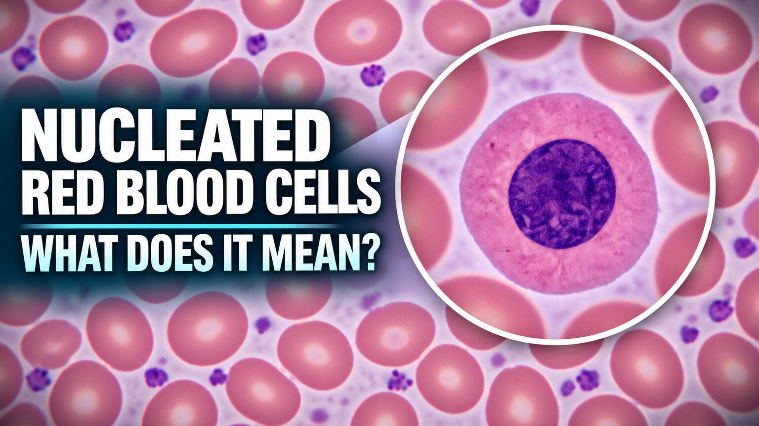
A nucleated red blood cell on a peripheral smear, then, is a cell that either escaped before finishing that sequence — meaning the marrow released a precursor stage rather than a properly enucleated one — or, far less commonly, briefly retained its nucleus after being pushed out under extreme pressure before finally shedding it in the bloodstream itself instead of inside the marrow. Under a microscope, it appears as a red blood cell containing a small, round, densely stained purple-blue nucleus, usually smaller and rounder than a comparable white blood cell's nucleus, and often accompanied by the more basophilic, bluish-tinted cytoplasm typical of an immature cell rather than the salmon-pink of a fully mature one. Recognizing that specific combination — red cell cytoplasm plus a compact round nucleus — is what separates a nucleated red blood cell from a lymphocyte or another white cell sitting nearby on the same field.
The One Major Exception: Newborns

Figure 1. Newborns normally carry a measurable number of nucleated red blood cells in their first hours of life — the same finding that signals serious marrow stress in an adult is expected, self-resolving physiology here.
Before going further, one exception has to be stated plainly, because it changes the entire interpretation of this finding: nucleated red blood cells are a completely normal part of a newborn's blood in the first hours and days of life. A fetus develops in a relatively low-oxygen environment compared to life outside the womb, and to compensate, fetal bone marrow (along with the liver and spleen, which still produce blood cells before birth) runs at a naturally higher output, releasing a small, expected number of nucleated red blood cells into circulation as a routine byproduct of that accelerated production. In a healthy full-term newborn, this count typically clears from the bloodstream within the first three to seven days as the marrow's release checkpoint matures and oxygen delivery stabilizes outside the womb. A pediatric lab report showing nucleated red blood cells in a two-day-old infant is, on its own, rarely a cause for concern; the same finding persisting well beyond the first week, or appearing at an unusually high count even in the first days, is what prompts a closer look for something like intrauterine hypoxia, a difficult delivery, or a maternal-fetal blood group incompatibility. Everything described in the rest of this article, by contrast, concerns what this finding means outside of that narrow, expected newborn window — in older infants, children, and especially adults, where the release checkpoint should already be fully intact.
Why the Automated Analyzer Gets Confused First
Before a human even looks at a slide, a nucleated red blood cell has already caused a specific, well-documented problem upstream, at the automated analyzer. Most modern hematology analyzers count white blood cells using a method that identifies and lyses (bursts open) the red blood cells in a sample, then counts whatever intact nucleated structures remain, on the assumption that anything with a nucleus left standing after that step must be a white blood cell. A nucleated red blood cell defeats that assumption directly: its nucleus survives the lysing step just as a genuine white blood cell's would, so the analyzer counts it as one, silently inflating the reported white blood cell count by exactly the number of nucleated red blood cells actually present. This isn't a rare edge case in laboratory medicine — it's common enough that it has a standard, named correction built into routine practice.
The fix is a straightforward piece of arithmetic once the smear has been reviewed and the nucleated red blood cells counted per 100 white blood cells: Corrected WBC = (Uncorrected WBC × 100) ÷ (100 + NRBC count per 100 WBC). A patient whose analyzer reports a white blood cell count of 20,000 per microliter, but whose smear shows 25 nucleated red blood cells for every 100 white cells counted, actually has a true white blood cell count closer to 16,000 once the correction is applied — a meaningful difference that could otherwise be misread as a more severe degree of infection or leukocytosis than is truly present. This is precisely why finding nucleated red blood cells on a smear isn't just a standalone data point sitting next to the rest of the report; it actively changes how at least one other number on that same report, the white blood cell count, needs to be interpreted.
Curious what the rest of your complete blood count is telling you? Upload your results and get a complete, plain-language breakdown in under 15 minutes.
Analyze My ResultsThe Core Mechanism: A Release Checkpoint Under Pressure
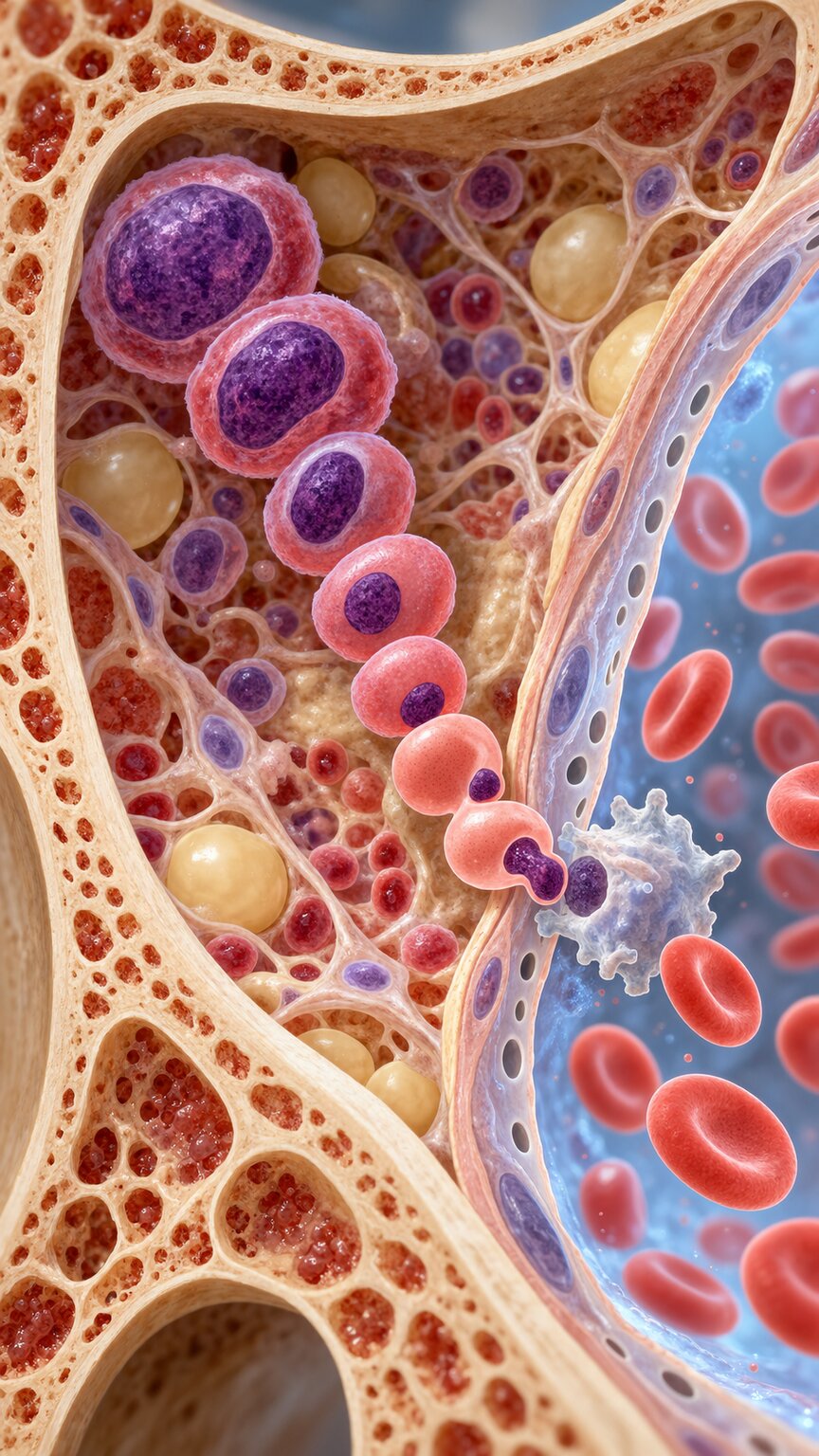
Figure 2. Under normal conditions, the marrow's sinusoidal barrier holds every red cell precursor back until it has fully matured and shed its nucleus.
Every distinct cause of nucleated red blood cells described in the following sections ultimately routes through the same physical checkpoint, so it's worth understanding that checkpoint before looking at what breaks it. Bone marrow is organized around a network of thin-walled blood vessels called sinusoids, and the cells lining those sinusoids act as a physical and functional barrier, allowing only genuinely mature cells to squeeze through small pores in that lining and enter the bloodstream. A nucleated precursor is simply too large and structurally rigid, nucleus intact, to fit through those pores under ordinary circumstances — it has to finish enucleating first, shrinking and remodeling its own cytoskeleton, before it's physically capable of passing through.
Three broad categories of pressure can override this barrier: a marrow driven to produce red blood cells so urgently and at such volume that precursors get pushed toward the sinusoids before they've finished maturing; a marrow whose own architecture has been physically damaged or crowded, so the sinusoidal barrier itself no longer holds cells back reliably; and a body that has lost its normal downstream filter — the spleen — which would otherwise catch and remove any nucleated red blood cell that did slip through before it could recirculate freely. Nearly every specific medical cause discussed below is really just a different route into one of these three categories, which is why nucleated red blood cells function less like a single diagnosis and more like a warning light that several different dashboards can trigger.
Severe Hemolysis: When Demand Overwhelms the Marrow's Normal Pace
The first and most direct route is simple overproduction under extreme time pressure. When red blood cells are being destroyed faster than the marrow can normally replace them — a state called hemolysis — the marrow responds to the resulting drop in oxygen-carrying capacity by ramping up erythropoietin-driven production dramatically, sometimes to six to eight times its baseline output. At that pace, the marrow's own quality-control step, holding each precursor back until enucleation is fully complete, starts to slip; precursors are shuttled toward the sinusoids on a compressed timeline, and a measurable fraction slip through the barrier with their nucleus still attached. This is why nucleated red blood cells are a well-recognized companion finding in severe hemolytic anemias, including advanced sickle cell disease, and it's a useful example of how findings across different articles on this same topic connect: a smear already showing sickle-shaped cells and Howell-Jolly bodies commonly shows nucleated red blood cells riding alongside them, all three reflecting the same underlying story of a marrow working at maximum, urgent capacity.
Severe hemolytic anemia from other causes — autoimmune destruction of red blood cells, certain inherited enzyme deficiencies, or a mismatched blood transfusion — produces the same pattern through the same basic logic, regardless of what's actually destroying the red blood cells. The common thread across every hemolytic cause is a very high reticulocyte count appearing together with nucleated red blood cells, since both findings are direct evidence of the same accelerated, urgently paced production process; seeing one without the other in a severely anemic patient is actually the more unusual finding and often prompts a second look at whether something is also suppressing the marrow's ability to respond.
Severe iron deficiency anemia and vitamin B12 or folate deficiency can occasionally produce a small number of nucleated red blood cells as well, though through a somewhat different route than outright hemolysis. In these deficiency states, red blood cell production is frequently ineffective — the marrow is working hard and producing precursors in large numbers, but a meaningful fraction of those precursors are structurally abnormal enough that they never survive long enough to leave the marrow as healthy, mature cells, dying instead within the marrow itself in a process called ineffective erythropoiesis. That same abnormal maturation process can occasionally let a poorly formed precursor slip past the sinusoidal barrier before properly enucleating, though this route typically produces a far smaller nucleated red blood cell count than the dramatic surges seen in severe hemolytic anemia or advanced sickle cell disease, and it usually resolves once the underlying nutritional deficiency is corrected with appropriate supplementation.
Hypoxia: When the Whole Body Is Starved for Oxygen

Figure 3. Chronic, severe oxygen deprivation — from advanced lung disease, congenital heart disease, or critical illness — drives the same urgent erythropoietin surge as hemolysis, through a different starting problem.
A second, mechanistically distinct route arrives at the same destination: sustained, severe hypoxia. The kidneys constantly monitor how much oxygen is reaching their own tissue as an indirect proxy for how much oxygen is reaching the body as a whole, and when that oxygen delivery drops significantly and stays down, they respond by releasing more erythropoietin, the hormone that signals the marrow to accelerate red blood cell production. Chronic, severe lung disease, certain congenital heart defects that mix oxygen-poor and oxygen-rich blood, and prolonged critical illness with unstable oxygenation can all sustain this signal strongly enough, for long enough, to push the marrow into the same accelerated, checkpoint-slipping mode of production described in the hemolysis section above — the difference is that here, the trigger is a genuine shortage of oxygen delivery rather than red blood cells being destroyed prematurely.
This route matters clinically because it means nucleated red blood cells shouldn't automatically be read as a sign of a blood disorder specifically — in a patient with known severe chronic obstructive pulmonary disease or an uncorrected congenital heart defect, this finding may simply reflect a marrow doing exactly what it's supposed to do in response to a real, ongoing oxygen deficit elsewhere in the body. Distinguishing this route from a primary blood or marrow problem usually relies on the broader clinical picture — oxygen saturation readings, known lung or heart disease history, and whether the red blood cell count itself is elevated (as expected from a compensatory hypoxic response) rather than low, as it typically is in the hemolytic and marrow-infiltration causes described elsewhere in this article.
People living at high altitude for extended periods offer a useful, non-disease illustration of this same hypoxia-driven mechanism at a much milder intensity. Residents of mountain communities several thousand meters above sea level naturally run a higher red blood cell count and hemoglobin than people at sea level, as a permanent physiological adaptation to the thinner air, and a small number of nucleated red blood cells can occasionally appear as part of that same adaptive process during periods of particularly demanding physical exertion at altitude. This isn't considered a disease state at all — it's simply the same erythropoietin-driven pathway operating at a lower, sustainable intensity in a body that has had time to adjust, which is a useful contrast to the much more urgent, higher-volume version of the same signal seen in acute severe lung failure or unstable congenital heart disease.
Functional Asplenia and Splenectomy: Losing the Downstream Filter
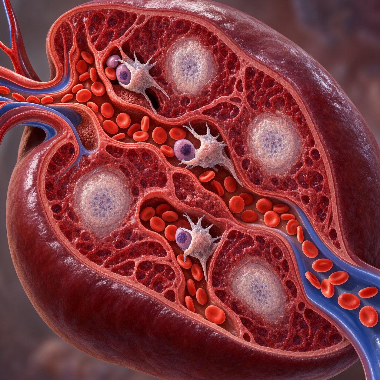
Figure 4. A functioning spleen acts as a second checkpoint, catching and removing any nucleated red blood cell that slips past the marrow's own barrier.
Even when a small number of nucleated red blood cells do slip past the marrow's sinusoidal barrier under normal, non-crisis conditions, there's typically a second line of defense: the spleen. As blood passes through the spleen's tight, winding network of vessels, it acts as a quality-control filter, physically trapping and removing abnormal or immature cells, nucleated red blood cells included, before they can recirculate freely through the rest of the body. This is a different role from the spleen's more commonly discussed job of removing worn-out old red blood cells at the end of their normal lifespan — here, it's specifically screening for cells that shouldn't be in general circulation in the first place, regardless of age.
Remove that filter — through a surgical splenectomy, or through functional asplenia, where the spleen is technically present but has been scarred and shrunk into uselessness, as happens progressively in sickle cell disease — and even a modest, otherwise unremarkable number of nucleated red blood cells slipping out of the marrow starts showing up reliably on a peripheral smear, simply because nothing downstream is catching them anymore. This is an important distinction from the hemolysis and hypoxia routes described above: in someone without a functioning spleen, nucleated red blood cells can appear even without a dramatic surge in marrow production, purely because the removal side of the equation has been eliminated rather than the production side being accelerated. A lab report on someone with a known splenectomy showing a small, stable number of nucleated red blood cells alongside other splenectomy-associated findings — Howell-Jolly bodies and target cells among them — is often simply confirming an expected, chronic baseline rather than signaling a new, acute problem.
Bone Marrow Infiltration: When the Factory Floor Itself Is Damaged
The most clinically serious route is direct physical disruption of the marrow's own architecture — a process doctors call myelophthisis, from Greek roots meaning "marrow wasting." Cancer that has spread to the bone (metastatic breast, prostate, or lung cancer being common sources), primary blood cancers like leukemia, and a scarring, fibrotic process called myelofibrosis can each replace or crowd out the marrow's normal structural framework, including the very sinusoidal barrier described earlier that's supposed to hold immature cells back. Rather than a checkpoint slipping under time pressure, this is a checkpoint that has been physically damaged and can no longer function as a gatekeeper at all — cells at every stage of development, mature and immature alike, get pushed out into circulation more or less indiscriminately.
This particular route rarely produces nucleated red blood cells in isolation. It's most often accompanied by teardrop-shaped red blood cells and immature white blood cells appearing on the same slide, together forming a specific, named pattern called a leukoerythroblastic smear — a pattern covered in detail in a related article on this site about teardrop cells, since the two findings share this exact same root cause. The practical significance of recognizing nucleated red blood cells as part of that fuller leukoerythroblastic pattern, rather than reading them as an isolated finding, is that they point toward a considerably more urgent underlying process — one that typically warrants an expedited bone marrow biopsy — than nucleated red blood cells appearing on their own alongside evidence of ordinary hemolysis or hypoxia.
Not sure whether your own results point to something urgent or something explainable? Upload your labs and get a clear, plain-language breakdown in under 15 minutes.
Analyze My ResultsHow Many Is Too Many? Quantifying the Finding

Figure 5. Nucleated red blood cells are reported per 100 white blood cells counted — the specific ratio, not just their presence, shapes how the finding is weighed clinically.
Unlike some smear findings that are reported simply as present or absent, nucleated red blood cells are quantified with a specific ratio: the number seen per 100 white blood cells counted during the manual differential, written on a report as, for example, "NRBC: 5/100 WBC." A very small number — one or two per 100 white blood cells — is common enough in significant but non-catastrophic hemolysis or hypoxia that many labs don't flag it as urgent on its own. Counts climbing into the double digits, and particularly counts exceeding 10 per 100 white blood cells in a hospitalized adult, are treated with considerably more concern, both because they suggest more severe underlying marrow stress and because, independent of the underlying cause, higher absolute counts of circulating nucleated red blood cells have themselves been associated with worse clinical outcomes in critically ill patients, a connection explored further in the next section.
Some laboratories report an additional, related figure called the absolute nucleated red blood cell count, calculated by applying that same per-100-white-blood-cell ratio to the total white blood cell count rather than leaving it as a bare ratio — for example, a ratio of 5 per 100 white blood cells in a patient with 8,000 white blood cells per microliter works out to an absolute count of roughly 400 nucleated red blood cells per microliter of blood. This absolute figure is increasingly favored in research settings and in some hospital protocols because it accounts for the fact that a given ratio means something different depending on how many total white blood cells that ratio is being calculated against, letting clinicians and researchers compare nucleated red blood cell burden more consistently across patients with very different total white counts.
The ratio format also matters for a subtler reason: it's precisely this same per-100-white-blood-cell count that feeds directly into the corrected white blood cell count formula described earlier. A report that lists nucleated red blood cells without also correcting the white blood cell count alongside them is, in a strict technical sense, an incomplete report — the two numbers are meant to be read and adjusted together, not in isolation from each other.
Why This Finding Carries Extra Weight in Critically Ill Adults
Outside of the newborn period, nucleated red blood cells found in a hospitalized, critically ill adult — particularly someone in an intensive care unit — have accumulated a substantial body of clinical research specifically because of how strongly they track with severity of illness and outcome. Multiple ICU-focused studies have found that patients with nucleated red blood cells appearing in their blood have meaningfully higher mortality rates than similarly ill patients without them, and that the association holds up even after adjusting for other known severity scores. The leading explanation isn't that the nucleated red blood cells themselves cause harm — rather, their presence is thought to reflect just how severely and broadly disrupted a critically ill body's regulatory systems have become, touching oxygen delivery, inflammatory signaling, and bone marrow function all at once, so their appearance functions as a kind of composite marker of overall physiological strain rather than a marker tied to any single organ system.
This is a meaningfully different clinical context than the sickle cell disease, chronic lung disease, or post-splenectomy scenarios described earlier in this article, where nucleated red blood cells often represent an expected, chronic, and relatively stable baseline finding in someone whose underlying condition is already well understood. A brand-new appearance of nucleated red blood cells in someone who is acutely, severely unwell and didn't have this finding before is treated with more urgency precisely because it's occurring against a backdrop where the underlying cause hasn't yet been identified or stabilized, rather than confirming something already known and accounted for.
What Typically Happens Next

Figure 6. The follow-up workup is driven entirely by which of the underlying mechanisms — hemolysis, hypoxia, splenic loss, or marrow infiltration — the rest of the clinical picture points toward.
Because nucleated red blood cells can arise from such genuinely different underlying processes, there's no single standard follow-up test that applies universally the way there might be for a more specific finding. Instead, a doctor typically works backward from the rest of the available information already at hand: a reticulocyte count and markers of hemolysis (such as bilirubin and LDH) if hemolytic anemia seems likely from the rest of the smear and history; a pulse oximetry reading and pulmonary function history if hypoxia from lung or heart disease is the more plausible explanation; confirmation of splenectomy status or an abdominal ultrasound checking spleen size and structure if functional asplenia hasn't already been established; and, if the accompanying findings suggest a leukoerythroblastic pattern with teardrop cells and immature white blood cells, an expedited referral for bone marrow biopsy to directly evaluate the marrow's own architecture.
Age and known medical history do a great deal of the initial narrowing before any new tests are even ordered. Nucleated red blood cells found in a person with well-documented sickle cell disease point overwhelmingly toward the same hemolytic and post-splenectomy mechanisms already established for that diagnosis, while the same finding appearing unexpectedly, for the first time, in someone without any of these known conditions understandably prompts a more open, thorough evaluation to establish which of the underlying pathways is actually responsible before management can be planned.
How quickly this workup moves also depends heavily on setting. In an outpatient clinic, a small, incidental nucleated red blood cell count found on a routine annual blood panel in an otherwise well person is often addressed with a relatively unhurried pace — a repeat smear in a few weeks, alongside basic iron studies and kidney function testing, is a common first step, since the most likely explanations in that setting tend to be mild, treatable, and not urgent. In a hospital or emergency department, the same finding in someone who is acutely unwell is folded immediately into the broader emergency workup already underway, rather than treated as a separate problem requiring its own dedicated visit — this is one of the clearest examples of how identical laboratory findings can trigger very different paces of response purely based on the clinical context surrounding them.
Common Misconceptions About This Finding
A few misunderstandings come up often enough with this particular finding that they're worth addressing directly. The first is assuming that any nucleated red blood cell automatically means leukemia or another blood cancer — in reality, the far more statistically common causes across all patients are severe hemolysis, chronic hypoxia, and post-splenectomy states, with marrow infiltration from cancer representing a minority of cases overall, even though it's the cause that understandably generates the most concern when people research the finding online. The accompanying findings on the same smear, not the nucleated red blood cells alone, are what actually separate a benign chronic pattern from one that warrants urgent cancer-focused evaluation.
A second misconception is treating the corrected white blood cell count as a minor technical footnote rather than something that meaningfully changes clinical decision-making. A falsely elevated, uncorrected white blood cell count in someone with a significant nucleated red blood cell burden can make an infection look more severe than it actually is, or make a borderline white count look unambiguously high when the corrected number would actually fall within a normal range — which is exactly why a lab report listing nucleated red blood cells should, in a technically complete report, always list the corrected white blood cell figure alongside the raw one rather than leaving that correction for someone else to calculate later.
A third misconception, more common among patients researching their own results than among clinicians, is assuming that finding a very small number — one or two per 100 white blood cells — carries the same weight as finding a large number in the double digits. The dose matters enormously here: a trace finding in someone with a known, explained cause is often noted and left alone entirely, while a large, unexplained burden is what actually drives urgent further testing. Reading a lab report's raw presence-or-absence language without noticing the actual ratio reported alongside it can make a reassuring finding sound far more alarming than it is.
How This Finding Fits Into the Rest of the Complete Blood Count
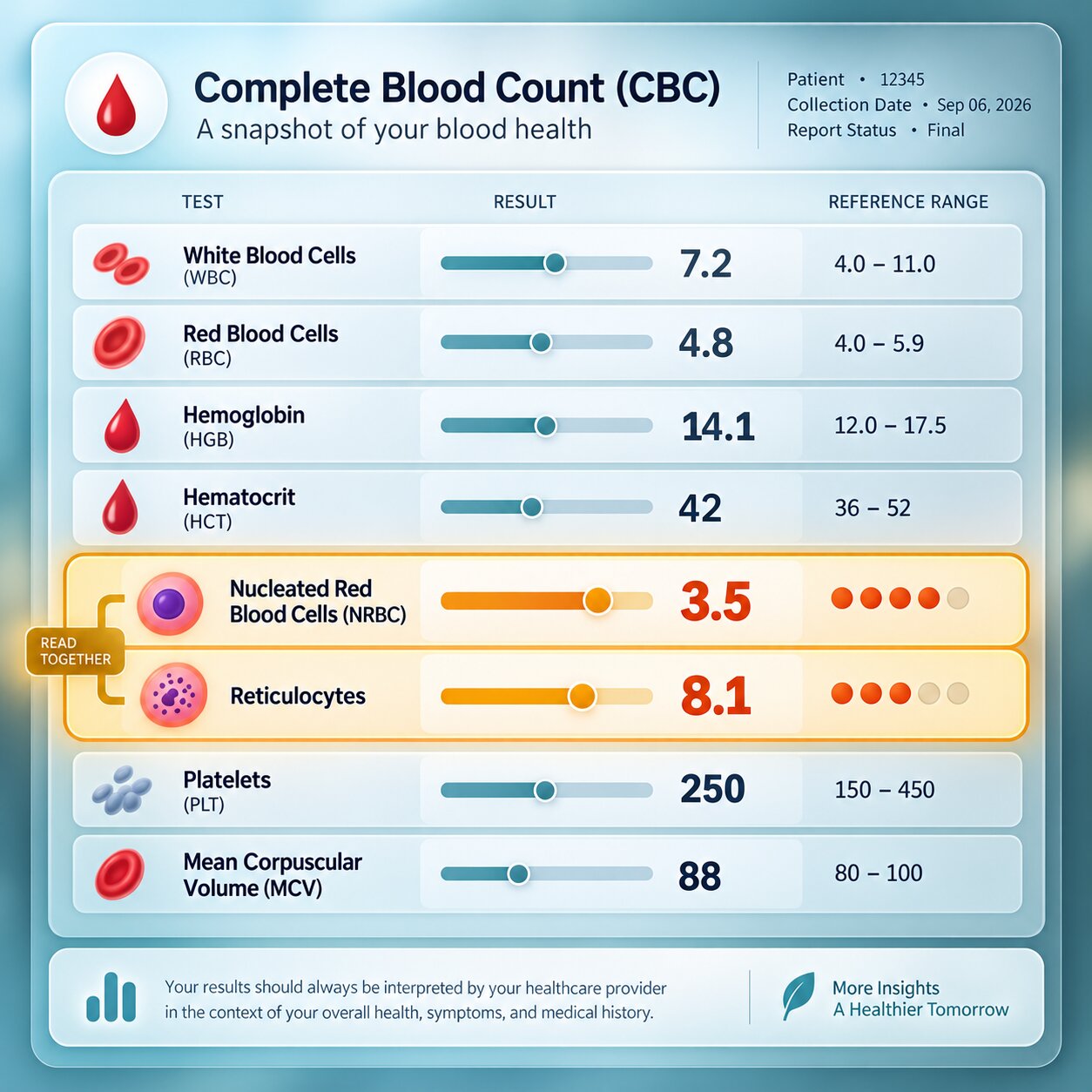
Figure 7. Nucleated red blood cells are almost never read alone — the reticulocyte count, hemoglobin trend, and white cell differential together tell a fuller, more specific story.
As with most smear findings discussed elsewhere on this site, nucleated red blood cells are rarely meaningful in complete isolation from the rest of a complete blood count. A falling or already-low hemoglobin alongside a high reticulocyte count and nucleated red blood cells builds a consistent picture of active hemolysis or significant blood loss being urgently compensated for. A normal or elevated hemoglobin alongside nucleated red blood cells, particularly in someone with known chronic lung or heart disease, is more consistent with a hypoxia-driven compensatory response rather than blood loss or destruction. And nucleated red blood cells appearing together with teardrop cells, immature white blood cells at multiple stages, and an otherwise unexplained drop across several blood cell lines at once (red cells, white cells, and platelets together) is the pattern most concerning for marrow infiltration, since it suggests the marrow's overall production and gatekeeping capacity is being compromised across the board rather than in just one cell line. Reading the nucleated red blood cell count as one thread within this larger pattern, rather than as a number that stands entirely on its own, is what actually allows it to point toward a specific, actionable cause.
Frequently Asked Questions
Is finding nucleated red blood cells on a smear always serious?
Not always — it depends heavily on age and context. In a newborn's first few days, it's expected physiology. In an adult with a known, stable condition like a prior splenectomy or chronic sickle cell disease, it can simply confirm an already-understood baseline. A new, unexplained appearance in an acutely ill adult, on the other hand, is taken more seriously and typically prompts further evaluation.
Why does finding nucleated red blood cells change my white blood cell count?
Automated analyzers count anything with an intact nucleus left after the red cells are lysed as a white blood cell, so a nucleated red blood cell gets miscounted as one. Labs correct for this using the number of nucleated red blood cells seen per 100 white blood cells, which lowers the reported white count to reflect the true number.
Can nucleated red blood cells appear without any red blood cell disorder at all?
Yes. Severe, sustained hypoxia from chronic lung or heart disease can drive the same accelerated marrow production seen in hemolytic anemia, without any red blood cells actually being destroyed prematurely. A missing or non-functioning spleen can also allow a small, otherwise unremarkable number to circulate simply because nothing is filtering them out anymore.
Why do nucleated red blood cells matter so much in critically ill patients?
Multiple studies of ICU patients have found that their presence is associated with meaningfully higher mortality, likely because it reflects how broadly disrupted a critically ill body's oxygen delivery, inflammatory, and marrow systems have become, rather than pointing to any single treatable cause on its own.
What other findings usually accompany nucleated red blood cells on the same smear?
It depends on the cause. Hemolysis usually pairs them with a high reticulocyte count; hypoxia pairs them with a normal or high hemoglobin; and marrow infiltration pairs them with teardrop cells and immature white blood cells, forming a pattern called a leukoerythroblastic smear.
Conclusion
A nucleated red blood cell showing up outside the bone marrow, past the newborn period, is the visible result of one of three overridden safeguards: a production line moving too urgently to finish its own quality check, a structural gatekeeper physically damaged and no longer holding cells back, or a downstream filter that's no longer there to catch what slips through. Which of those three is actually responsible determines everything about how seriously the finding is treated and what gets tested next — from a straightforward hemolytic workup, to confirming a well-known chronic condition, to an urgent bone marrow biopsy. It also quietly reshapes at least one other number on the same report, since an accurate white blood cell count depends on correcting for exactly how many of these immature cells the analyzer mistakenly counted as something else. Taken together with the rest of the complete blood count and smear, rather than read in isolation, this finding is one of the more mechanistically specific windows laboratory medicine offers into what the bone marrow is actually experiencing at the moment the blood was drawn.
Still Not Sure What Your Results Mean?
Upload your labs and get a complete, visual, plain-language interpretation of every biomarker — delivered to your inbox in under 15 minutes.
Get My ReportThis article is for educational purposes only and does not constitute medical advice. Always consult your healthcare provider regarding your specific lab results.