
What Does It Mean If Your Blood Smear Shows Spherocytes?
Seeing the word "spherocytes" on your blood smear report is understandably unsettling when nobody has explained what it means yet. In plain terms, a spherocyte is a red blood cell that has lost its normal shape and turned into a small, dense ball — and finding them under the microscope tells your doctor that somewhere in your body, red blood cells are being broken down and cleared out faster than they should be. That's the short version. The longer, more useful version is that spherocytes point toward a fairly specific and fairly short list of causes, ranging from a condition you were born with, to one your immune system developed on its own, to a handful of rarer triggers tied to infection, burns, or a recent transfusion. This guide walks through what a pathologist is actually looking at under the microscope when they flag spherocytes, how many is considered clinically significant versus incidental, and what your doctor will realistically do next to figure out which of these several explanations actually fits your specific situation.
Curious what the rest of your bloodwork is telling you? Upload your results and get a complete, plain-language breakdown in under 15 minutes.
Analyze My ResultsWhat a Blood Smear Actually Is (and How It's Different From a Routine CBC)
Most blood tests, including a standard complete blood count, are read entirely by a machine — an automated analyzer pushes your blood through narrow channels, counts and sizes thousands of cells per second, and spits out numbers like your red blood cell count, hemoglobin, and the various CBC indices. A blood smear is something different and, in a sense, more old-fashioned: a small drop of your blood is spread into a thin film across a glass slide, stained with a dye, and examined directly by a person looking through a microscope. That human step matters, because an automated analyzer is very good at counting and measuring, but it can't always recognize an unusual cell shape the way a trained eye can. A cell that gets counted correctly as "one red blood cell" by the machine might, under the microscope, turn out to be an abnormally shaped cell carrying an important diagnostic clue the machine has no way of flagging. Spherocytes are a perfect example of exactly that kind of clue — a shape the analyzer counts without comment, but a human reader immediately recognizes as significant.
Why a Doctor Would Order a Blood Smear in the First Place
A blood smear isn't usually anyone's very first test — it tends to get ordered as a deliberate follow-up once something else has already raised a question. The most common trigger is an abnormal or unexplained finding on a routine CBC: unexpected anemia, red blood cell indices that don't fit a typical pattern, or, often overlooked by patients, an automated "flag" the analyzer itself generates when it detects something about the cell population it isn't confident interpreting on its own. Symptoms can prompt one directly too — unexplained fatigue, pale skin, yellowing of the eyes or skin, dark urine, or a family history of anemia or gallstones at a young age are all reasons a doctor might request a manual smear alongside or instead of routine bloodwork. It's also a standard part of the workup for a newly enlarged spleen, since an overactive spleen filtering out abnormal cells is exactly the mechanism behind several of the conditions covered below. In hospitalized or critically ill patients, a smear can be ordered urgently, within hours, when a doctor needs to understand quickly why someone's red blood cell count is dropping.
How a Blood Smear Is Actually Prepared and Read
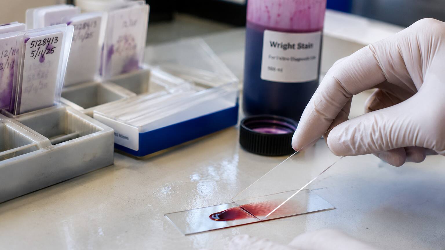
Making a usable blood smear is a more precise process than it might sound. A technologist places a small drop of blood near one end of a glass slide, then uses a second slide held at an angle to push the drop across the surface in a single smooth motion, spreading it into a thin, feathering film — this is often called a wedge or push smear. Once dried, the slide is stained, most commonly with a dye called Wright's stain, which colors different cell structures in shades of pink, purple, and blue so their internal features become visible under magnification. The technologist or pathologist doesn't examine the whole slide equally; they focus on a specific "monolayer" region, roughly in the middle of that feathered spread, where red blood cells sit in a single layer, separated from one another, and each one shows its normal central lighter area clearly. Areas where the smear is too thick distort cell shape through overlap, and areas too far out on the feathered edge can make normal cells look artificially flattened or spread out — so reading the correct zone of the slide, under high oil-immersion magnification, is what makes the difference between an accurate read and a misleading one.
This manual step is also why a blood smear is often ordered as a follow-up rather than a first-line test: it takes real time and real expertise, so it's typically reserved for situations where an automated CBC has already come back with something unexplained — an unusual index, a flag from the analyzer itself, or symptoms that don't add up. When a smear is read, the pathologist is doing three things at once: assessing red blood cell shape and size, examining white blood cells for abnormal forms, and checking that platelets look adequate and appropriately sized. Spherocytes fall into that first category, and their presence is noted directly on the report, often alongside a rough estimate of how many were seen.
What Spherocytes Actually Look Like Under the Microscope
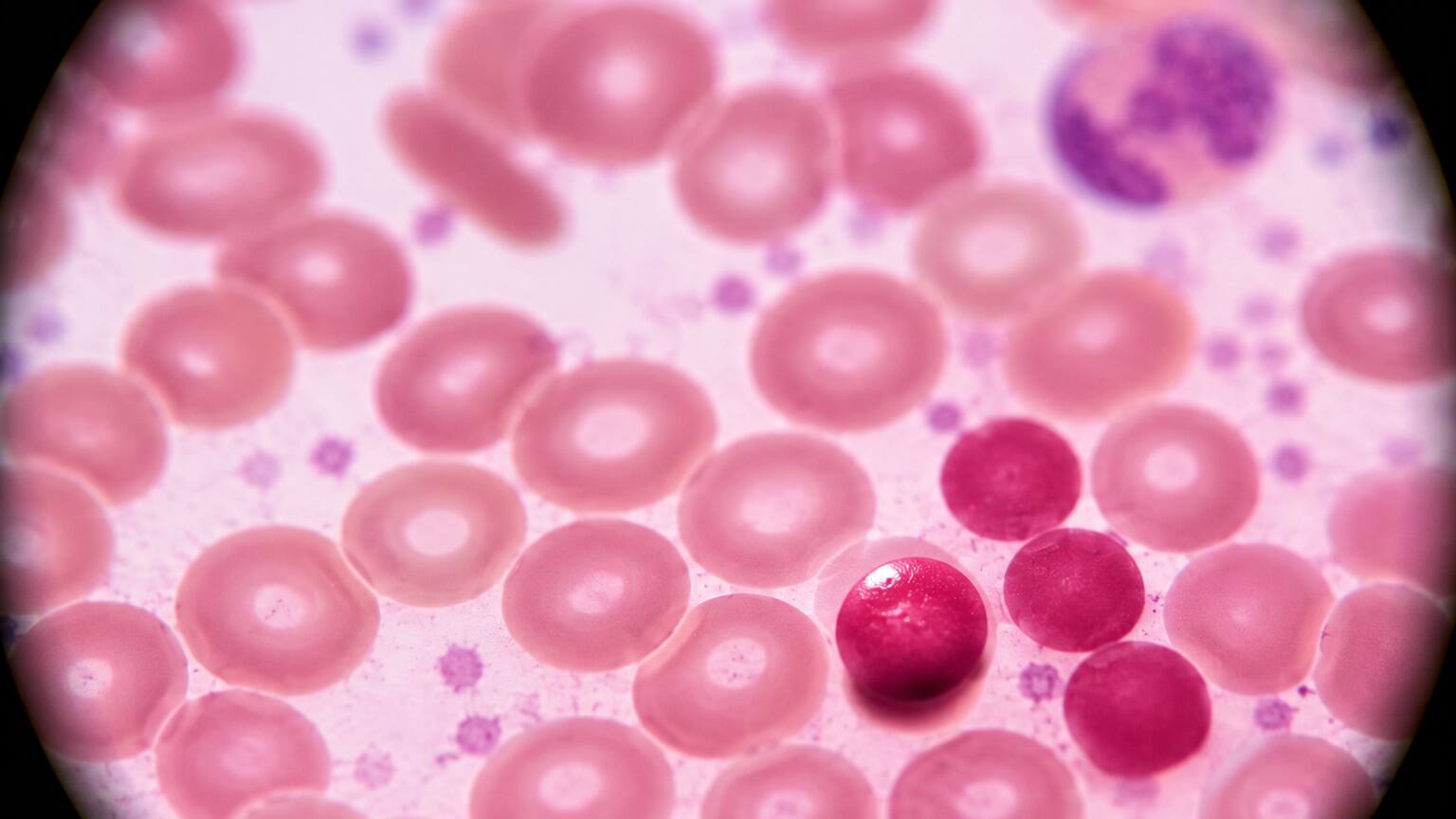
A healthy red blood cell has a very particular and recognizable shape: a flattened disc with a natural dimple on both sides, called biconcave. Under the microscope, that dimple shows up as a lighter, paler area right in the center of the cell, surrounded by a darker-staining ring — pathologists call this central pallor, and its presence is one of the first things checked when assessing red blood cell health. A spherocyte has lost that shape entirely. Instead of a flattened disc, it's collapsed into something closer to a small sphere, and because it's rounder and denser, it stains a more uniform, solid color all the way through, with no pale center visible at all. Spherocytes are also typically a bit smaller in diameter than normal red blood cells sitting right next to them on the same slide, which is part of what makes them stand out — a trained eye scanning across a field of normal, pale-centered discs immediately notices the smaller, darker, uniformly round outliers scattered among them.
How Many Is "Too Many"? Understanding the Grading on Your Report
Not every spherocyte on a slide is automatically meaningful — a rare, isolated one can sometimes be a normal variant or a preparation artifact. What matters clinically is both their presence and their quantity, which is why pathology reports typically include some indication of how many were seen, using either a numeric grading scale (commonly 1+ through 4+) or descriptive language like "occasional," "moderate," or "many/marked." A report noting occasional or 1+ spherocytes describes a small number scattered sparsely across the slide, which may still warrant follow-up but carries less immediate weight. A report describing moderate to marked spherocytosis, or a grade of 3+ to 4+, describes a slide where spherocytes make up a substantial share of the red blood cells present — a pattern strongly associated with an active, ongoing hemolytic process. In hereditary spherocytosis and autoimmune hemolytic anemia specifically, spherocytes are often essentially the only abnormal red blood cell shape seen, without the mixture of other odd shapes that some other blood disorders produce, which itself becomes a useful diagnostic clue for a pathologist scanning the slide. Some labs supplement this visual grading with an automated hyperchromia flag or cytogram from the hematology analyzer itself — a rough, machine-generated estimate of denser-than-normal cells that can prompt a manual review even before a human ever looks through the eyepiece, catching sparse spherocyte populations that might otherwise be easy to miss on a quick visual scan.
A Quick History: How Spherocytes Became a Recognized Clue
Doctors have been connecting oddly round, densely staining red blood cells to hemolytic disease for well over a century. Around the turn of the 20th century, physicians including Oskar Minkowski and, shortly after, Anatole Chauffard began systematically describing families with recurring jaundice, anemia, and an enlarged spleen, and they linked the pattern to red blood cells that looked distinctly spherical rather than the normal disc shape under early microscopy — work that laid the foundation for what we now call hereditary spherocytosis. It took several more decades of research into red blood cell membrane biology before scientists understood why the shape change happened at all, eventually tracing it to the structural proteins, like spectrin, that hold the membrane's scaffolding together. That gradual shift — from "these patients have an unusual family illness" to "this specific cell shape reflects a specific membrane defect we can now test for directly" — is a big part of why a spherocyte on a modern smear isn't just a curiosity. It's a shape with a well-understood, well-studied mechanism behind it, which is exactly what allows today's doctors to move from a microscope finding to a specific diagnosis with real confidence.
The Most Common Cause: Hereditary Spherocytosis
The condition most classically associated with spherocytes is hereditary spherocytosis, an inherited disorder affecting the proteins that give a red blood cell's outer membrane its structure and shape. Proteins called spectrin, ankyrin, band 3, and band 4.2 normally work together as a kind of internal scaffolding just beneath the cell membrane, anchoring it in its flattened, biconcave form. A genetic mutation affecting one of these proteins weakens that scaffolding, and every time the affected cell squeezes through a narrow blood vessel — especially inside the spleen — it loses a small piece of its membrane surface. Losing surface area without losing a proportional amount of hemoglobin or water forces the cell to contract into a smaller, rounder shape, which is exactly the spherocyte seen under the microscope. The spleen then recognizes these misshapen, inflexible cells as abnormal and filters them out of circulation faster than the bone marrow can replace them, producing a hemolytic anemia that can range from barely noticeable to significant, depending on the specific mutation involved and how much membrane protein function it disrupts.

Because it's genetic, hereditary spherocytosis usually runs in families, and a doctor investigating a spherocyte finding will often ask directly about relatives with unexplained anemia, jaundice as an infant, gallstones at an unusually young age, or a previous splenectomy — any of which can be a meaningful clue even before further testing. Confirming the diagnosis typically goes beyond the smear itself: an osmotic fragility test checks how easily these cells rupture when placed in a diluted salt solution, since their reduced surface area makes them more fragile under osmotic stress than normal cells, while a newer test called eosin-5-maleimide, or EMA, binding directly measures how much of a key membrane protein is present on the cell surface using a fluorescent dye and a specialized instrument called a flow cytometer. Treatment scales with severity: many people need nothing more than folic acid supplementation to support their bone marrow's increased workload, while more significantly affected individuals may eventually benefit from a splenectomy, which doesn't repair the underlying membrane defect but removes the organ responsible for destroying the fragile cells, often resolving the anemia almost entirely.
The Acquired Cause: Autoimmune Hemolytic Anemia and the Coombs Test
Spherocytes don't always mean a person was born with a membrane defect. In autoimmune hemolytic anemia, the immune system mistakenly produces antibodies that attach to otherwise completely normal red blood cells. As those antibody-coated cells pass through the spleen, resident immune cells called macrophages recognize the antibody tags and remove a bite-sized piece of the cell's membrane rather than destroying the entire cell outright. The cell survives, but having lost part of its surface, it contracts into a smaller, rounder shape — a spherocyte that, under the microscope, can look essentially identical to the inherited version. Because the visual appearance overlaps so closely, the test that actually separates these two conditions isn't done on the smear at all: it's the direct antiglobulin test, commonly called a Coombs test, which detects antibodies physically bound to the surface of red blood cells. A positive Coombs test points toward autoimmune hemolytic anemia; a negative one, combined with a suggestive family history, points back toward the inherited form. Autoimmune hemolytic anemia can appear on its own, alongside another autoimmune condition such as lupus, following certain infections, as a reaction to specific medications, or occasionally as an early sign of an underlying lymphoma — and because the underlying trigger is so different from a structural gene defect, treatment usually starts with corticosteroids to calm the immune response, rather than the folic acid or splenectomy approach used for the inherited form.
Rarer but Important Causes You Should Know About
Outside of hereditary spherocytosis and autoimmune hemolytic anemia, a handful of less common situations can also produce spherocytes on a smear, and recognizing them matters because they call for a completely different response.
Severe Burns and Transfusion Reactions
Severe thermal burns can directly damage red blood cells as they pass through heat-affected small blood vessels near the surface of the skin, shearing away bits of membrane in a way that produces spherocytes almost as a direct mechanical injury rather than an immune or genetic process. A mismatched blood transfusion works through a related but distinct mechanism: if transfused red blood cells are incompatible with the recipient's own immune system, particularly in ABO incompatibility, antibodies attack the transfused cells rapidly, and the same partial-membrane-loss process seen in autoimmune hemolytic anemia can generate spherocytes within hours of the transfusion — one of several findings that helps confirm a suspected transfusion reaction is happening.
Clostridium perfringens Sepsis — Rare but Dramatic

One of the more striking and less widely known causes is a severe bacterial infection called Clostridium perfringens sepsis, most often arising from an infection in the abdomen, such as the gallbladder or liver. This organism produces a toxin that directly attacks the red blood cell membrane, and when it takes hold, it can trigger sudden, widespread destruction of red blood cells within the bloodstream itself, producing a smear packed with small, dense microspherocytes. It's a rare cause, but a genuinely dangerous and fast-moving one — this kind of sudden, severe hemolysis in a critically ill patient is a medical emergency that requires immediate hospital-based evaluation and treatment, which is handled entirely by the critical care team rather than something to research or manage outside of that setting.
G6PD Deficiency During a Hemolytic Crisis
Glucose-6-phosphate dehydrogenase, or G6PD, deficiency is a common inherited enzyme deficiency that leaves red blood cells more vulnerable to oxidative stress — a kind of internal chemical damage that can be triggered by certain medications, infections, or even eating fava beans. During an acute hemolytic episode brought on by one of these triggers, red blood cells can show spherocytes alongside another distinctive finding called bite cells, where a visible chunk appears to have been removed from the cell's edge. Unlike hereditary spherocytosis, G6PD deficiency doesn't cause ongoing spherocyte formation at baseline — it shows up specifically during and shortly after a triggering exposure, which is an important timing clue when a doctor is putting the full picture together.
Spherocytes vs. Schistocytes — Why the Distinction Matters
Spherocytes are frequently mentioned in the same breath as another abnormal red blood cell shape, the schistocyte, and mixing the two up is an easy mistake to make since both show up in hemolytic conditions. The difference is mechanical rather than cosmetic: a schistocyte is a fragmented, jagged piece of a red blood cell — sometimes shaped like a tiny helmet or triangle — produced when a cell is physically sheared apart while forcing its way past an obstruction inside a blood vessel, the way it happens in conditions like thrombotic thrombocytopenic purpura, hemolytic uremic syndrome, or disseminated intravascular coagulation. A spherocyte, by contrast, keeps a smooth, rounded, intact outline; it isn't torn apart mechanically, it's simply lost membrane and contracted into a denser shape. Because of this difference in mechanism, the two findings point doctors down very different diagnostic paths: schistocytes push the workup toward mechanical, vessel-based causes of red blood cell destruction, while spherocytes push it toward the immune and membrane-related causes covered throughout this article. Research comparing the two has found schistocytes present in the large majority of mechanical hemolysis cases but only a small minority of immune-mediated ones, which is part of why a pathologist noting "spherocytes, no schistocytes seen" on your report is already quietly steering your workup in a specific direction before a single additional test is run.
Other Clues Pathologists Look for Alongside Spherocytes
Spherocytes rarely appear in isolation on a report, and the surrounding findings often help confirm what's going on. Polychromasia — red blood cells that stain with a slightly bluish-grey tint rather than the usual pink-red — reflects young, newly released red blood cells still containing residual genetic material, and a smear showing both spherocytes and noticeable polychromasia tells a doctor the bone marrow is actively working overtime to replace cells being destroyed, a reassuring sign that the marrow itself is responding appropriately. Howell-Jolly bodies, small round remnants of nuclear material normally removed by a healthy spleen, sometimes appear on the same slide in people who've already had a splenectomy as treatment for hereditary spherocytosis, since without a spleen there's nothing left to clear those remnants out. Pathologists also note the overall degree of anisocytosis, or variation in red blood cell size, and poikilocytosis, or variation in shape, since a slide showing spherocytes as essentially the only abnormal shape present, rather than a chaotic mixture of many different odd shapes, itself points more specifically toward hereditary spherocytosis or autoimmune hemolytic anemia rather than a broader bone marrow problem.
What Your Doctor Will Likely Do Next
Finding spherocytes on a smear is a starting point for a fairly organized workup, not an endpoint. Your doctor will typically order a reticulocyte count to see whether your bone marrow is responding appropriately by releasing extra young red blood cells, along with bilirubin, haptoglobin, and LDH levels, which together help confirm that hemolysis is genuinely occurring and give a sense of how active it is. A direct antiglobulin (Coombs) test is usually central to sorting out an acquired cause from an inherited one, and depending on what those initial results show, more specific testing — an osmotic fragility test, EMA binding, or targeted genetic testing — may follow to confirm hereditary spherocytosis specifically. Your doctor will also ask about recent infections, new medications, any recent transfusions, and your family history, since the timeline and context surrounding when spherocytes appeared is often just as informative as the spherocytes themselves.

How urgently all of this gets pursued depends heavily on how you're actually feeling and how many spherocytes were reported. A patient with occasional spherocytes, no anemia, and no symptoms may simply be monitored with a repeat CBC down the line. A patient with moderate-to-marked spherocytosis, a falling hemoglobin, fatigue, or jaundice is going to move through this workup considerably faster, sometimes within the same hospital visit, because active hemolysis at that level needs a clear explanation and, often, direct treatment. Once a specific cause is identified, treatment branches in very different directions depending on what that cause turns out to be: folic acid and, in more significant cases, splenectomy for hereditary spherocytosis; corticosteroids and sometimes other immune-suppressing medications for autoimmune hemolytic anemia; stopping the offending medication and supportive care for a G6PD-related crisis; and, for the rare Clostridium perfringens cause, urgent antibiotics alongside intensive supportive care. Because these treatment paths are so different from one another, the diagnostic step of figuring out exactly which cause applies to you isn't a formality — it's what determines everything that happens afterward.
Frequently Asked Questions
Does finding spherocytes always mean I have hereditary spherocytosis?
No. Spherocytes look essentially the same on a smear whether they come from an inherited membrane defect or an acquired cause like autoimmune hemolytic anemia, a transfusion reaction, a severe infection, or certain rare enzyme deficiencies. Additional tests, especially the Coombs test and your family history, are what actually distinguish between these possibilities.
Is a small number of spherocytes on my report something to worry about?
Not necessarily. Reports describing "occasional" or 1+ spherocytes, especially without anemia or other abnormal findings, are often followed with monitoring rather than aggressive workup. It's the combination of spherocyte quantity, your hemoglobin level, and how you're feeling that determines how seriously it's taken.
Can spherocytes go away, or is the underlying cause always permanent?
It depends entirely on the cause. Spherocytes from a resolved trigger, such as a treated infection, a completed hemolytic episode from G6PD deficiency, or successfully treated autoimmune hemolytic anemia, can disappear once the underlying process resolves. Hereditary spherocytosis, being a genetic membrane defect, doesn't go away, though its severity and the number of spherocytes seen can vary over time.
Why did my automated CBC look normal if I have spherocytes?
Automated analyzers count and size cells extremely well but aren't designed to recognize abnormal shapes the way a person examining the slide directly can. It's common for CBC numbers, including MCV and MCHC, to look only mildly abnormal or even normal while a manual smear review still reveals a meaningful number of spherocytes — which is exactly why a smear gets ordered as a follow-up rather than relied on to catch everything by itself.
What's the difference between spherocytes and schistocytes on my report?
Spherocytes are smooth, round, intact red blood cells that have lost membrane through an immune or genetic process. Schistocytes are jagged, fragmented pieces of red blood cells produced by mechanical shearing inside blood vessels, seen in conditions like TTP, HUS, or DIC. They point toward very different diagnostic paths, so a report noting one without the other is already meaningful information.
Conclusion
Spherocytes on a blood smear are a specific, visually recognizable signal that red blood cells somewhere in your body are losing their normal shape and being cleared out faster than usual — but that signal points toward several genuinely different underlying stories, from a lifelong inherited condition to a temporary reaction your doctor can identify and address directly. The number of spherocytes seen, how you're feeling, and a short list of targeted follow-up tests are what turn that one smear finding into an actual answer. If your report mentions spherocytes, the most useful next step is simply working through that follow-up testing with your doctor rather than trying to guess which cause applies to you from the word alone — and if you already have other lab results in hand, reading them together with your smear findings, rather than in isolation, usually gives the clearest overall picture of what's actually going on.
Still Not Sure What Your Results Mean?
Upload your labs and get a complete, visual, plain-language interpretation of every biomarker — delivered to your inbox in under 15 minutes.
Get My ReportThis article is for educational purposes only and does not constitute medical advice. Always consult your healthcare provider regarding your specific lab results.