
What Does the Presence of Howell-Jolly Bodies Indicate?
Howell-Jolly bodies are one of the more oddly specific findings a pathologist can report after looking at your blood smear under a microscope — small, round, dark purple dots sitting inside otherwise normal-looking red blood cells. What makes them worth understanding isn't the dot itself; it's what the dot actually is and why it's there at all. A healthy, mature red blood cell has no nucleus — it loses that nucleus deliberately during its final stage of development, specifically so it can flex and squeeze through your smallest capillaries without a rigid structure getting in the way. A Howell-Jolly body is a small leftover fragment of that nucleus, a scrap of DNA that should have been stripped out and cleared away before the cell ever entered circulation. Finding one on a smear means one of two things happened: either your body sent an unusually immature red blood cell out before that DNA fragment was fully removed, or — far more commonly — the organ specifically responsible for catching and clearing out these fragments after the fact, your spleen, isn't doing that job the way it normally would.
This one small structural detail turns out to be a surprisingly efficient diagnostic shortcut. A pathologist doesn't need an ultrasound, a surgical history, or a genetic test to raise the possibility of reduced splenic function — the clue is already sitting directly under the microscope, embedded inside a cell that would otherwise look completely unremarkable.
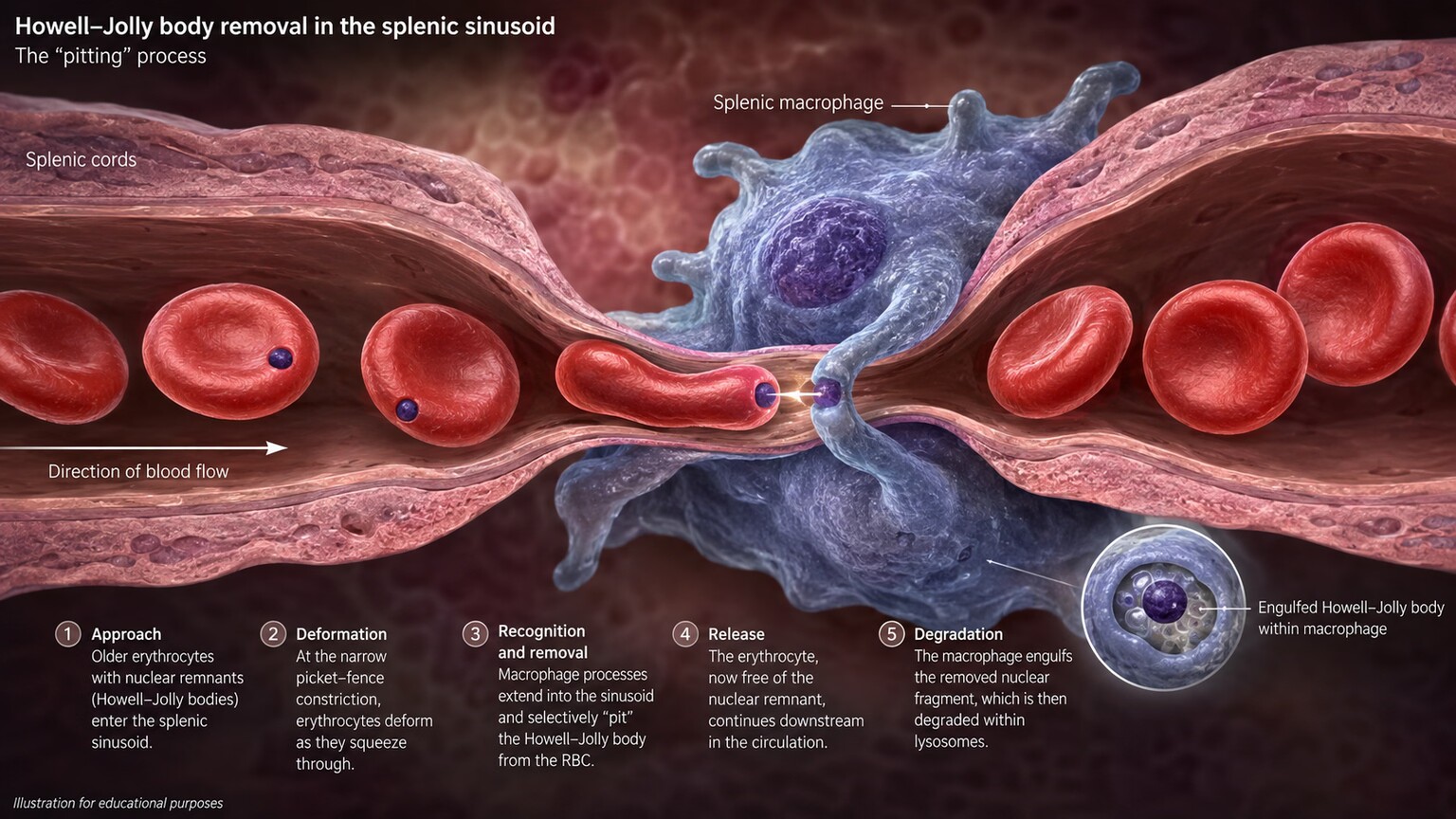
Figure 1. As red blood cells squeeze through the spleen's narrow sinusoids, specialized macrophages physically pluck out any remaining nuclear fragments in a process called "pitting" — the exact function that fails when Howell-Jolly bodies appear.
The Spleen's Quiet, Constant "Pitting" Job
To understand why Howell-Jolly bodies matter, it helps to understand a job your spleen performs continuously without you ever noticing it. Every red blood cell your bone marrow produces starts out as a nucleated cell, complete with a full nucleus like almost every other cell in your body. As it matures into the disc-shaped, flexible cell that will eventually carry oxygen through your bloodstream, it ejects that nucleus entirely — but occasionally, a small fragment of nuclear material gets left behind, clinging to the inside of the cell membrane instead of being fully expelled.
This is where your spleen comes in. As blood flows through the spleen, red blood cells are forced through an extremely narrow, winding network of vessels called sinusoids — narrower, in fact, than the red blood cells themselves, forcing each cell to physically deform and squeeze through. Specialized immune cells called macrophages line these narrow passages and perform a process hematologists call "pitting" — literally plucking any leftover nuclear fragment out of the cell membrane as it passes by, without destroying or removing the red blood cell itself, which continues on its way fully corrected. This pitting function runs constantly, cleaning up small manufacturing imperfections from your bone marrow before they ever reach general circulation in any detectable quantity.
It's worth appreciating just how specialized this filtering environment actually is, because it's part of why no other organ in the body can substitute for it. The splenic sinusoids aren't simply narrow tubes — they're lined with a discontinuous basement membrane full of small slit-like openings, deliberately engineered by nature to challenge every red blood cell passing through with a genuine physical squeeze test. A healthy, flexible, properly matured red blood cell deforms easily and slides through without issue. A red blood cell still carrying a rigid nuclear fragment gets caught at exactly this bottleneck, giving the waiting macrophages their opportunity to intervene. This isn't a passive filter catching debris that happens to float by — it's an active, mechanically enforced quality-control checkpoint that every single red blood cell in your body passes through repeatedly over its roughly four-month lifespan, which is part of why the loss of this specific checkpoint, rather than some other organ picking up the slack, produces such a consistent, reliable finding on a blood smear.
Why They Show Up: Loss of the Spleen Itself

Figure 2. Following a splenectomy — whether from trauma, disease, or a planned procedure — Howell-Jolly bodies typically appear on the very next blood smear and remain a permanent, expected finding for life.
The single clearest, most unambiguous cause of Howell-Jolly bodies is having your spleen surgically removed entirely — a splenectomy, performed for reasons ranging from traumatic injury (the spleen is one of the most commonly injured organs in blunt abdominal trauma), to certain blood disorders, to specific cancers involving the spleen itself. Without a spleen to perform its pitting function, every red blood cell that leaves the bone marrow with a small nuclear fragment simply keeps that fragment for the rest of its roughly 120-day lifespan in circulation, since nothing else in the body performs this specific cleanup task.
This means that after a splenectomy, Howell-Jolly bodies aren't a temporary or occasional finding — they become a permanent, expected feature of that person's blood smear for the rest of their life, and their presence is actually used clinically as a reliable, low-cost confirmation that a splenectomy was performed and that no meaningful amount of functioning splenic tissue remains, which matters because a small piece of spleen tissue can sometimes reimplant and regrow after trauma or surgery (a phenomenon called splenosis), and the disappearance of Howell-Jolly bodies from a previously splenectomized person's smear can actually be a clue that this has happened.
This diagnostic use runs in the other direction too, which is a genuinely useful practical detail: because Howell-Jolly bodies are such a reliable indicator of splenic status, their absence in someone with a documented total splenectomy is itself a notable, worth-investigating finding, since it suggests either splenosis (functioning splenic tissue having regrown elsewhere) or, in rare cases, that the original surgical removal may not have been fully complete. A hematologist noticing this specific mismatch between surgical history and smear findings will typically follow up with imaging to check for regrown splenic tissue, illustrating just how much diagnostic information this one small microscopic detail can carry in either direction — presence or unexpected absence.
Partial splenectomy, performed in certain situations where preserving some splenic tissue and function is possible and desirable — more common in pediatric cases or specific traumatic injuries where only part of the organ is damaged — produces a more variable picture than a complete splenectomy. Depending on how much functioning tissue remains and how effectively it can still perform the pitting function, Howell-Jolly bodies may appear only occasionally, or may not appear at all if enough splenic tissue was preserved to maintain adequate function. This variability is part of why a hematologist reviewing a smear from someone with a partial splenectomy interprets the finding, or its absence, alongside a clearer understanding of exactly how much tissue was preserved during the original procedure.
Curious what the rest of your own results are really telling you? Upload them and get a complete, plain-language breakdown in under 15 minutes.
Analyze My ResultsFunctional Asplenia: When the Spleen Is Still There but Not Working
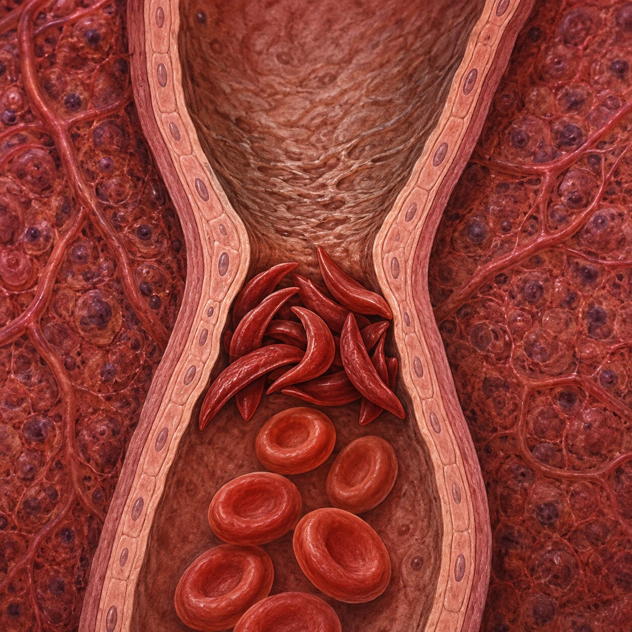
Figure 3. In sickle cell disease, rigid sickle-shaped cells repeatedly become trapped and clog the spleen's narrow vessels, gradually causing so much tissue damage that the spleen effectively stops functioning — a process called autosplenectomy.
A physically intact spleen doesn't guarantee a functioning one, and this distinction — an anatomically present spleen that has effectively stopped doing its job — is responsible for some of the more clinically important causes of Howell-Jolly bodies. Sickle cell disease is the textbook example: the same rigid, sickle-shaped red blood cells responsible for the disease's other complications repeatedly become trapped in the spleen's narrow sinusoids, causing small, repeated episodes of tissue damage and blocked blood flow. Over years, often beginning in early childhood, this cumulative damage effectively destroys the spleen's functional tissue from the inside, a process hematologists call autosplenectomy — the organ is often still physically present, sometimes even shrunken and scarred rather than surgically absent, but it no longer performs its pitting function or its other immune roles.
Celiac disease is another well-documented cause of functional asplenia, through a less fully understood but consistently observed mechanism connecting chronic intestinal inflammation to reduced splenic function over time. Other conditions capable of producing the same functional loss include certain inflammatory bowel diseases, some autoimmune conditions like lupus, amyloidosis, and advanced liver cirrhosis with significant portal hypertension. In every one of these cases, the underlying mechanism converges on the same endpoint described in the section above — a spleen that, for whatever specific reason, is no longer effectively pitting nuclear fragments out of circulating red blood cells.
The reason celiac disease specifically affects a seemingly unrelated organ like the spleen remains an area of ongoing research, but the leading explanation involves the chronic, low-grade immune activation that celiac disease produces throughout the body, not just within the gut itself. Some researchers have proposed that repeated immune signaling over years may gradually alter how splenic tissue functions at a cellular level, similar in spirit to how the chronic inflammation discussed in a different LabsFive article can quietly reshape how the liver allocates its own resources — a reminder that conditions affecting one organ system often ripple outward to affect others in ways that aren't obvious from the original diagnosis alone. What is well established, regardless of the exact mechanism, is the practical clinical pattern: Howell-Jolly bodies discovered with no other explanation should prompt consideration of celiac disease as a possible underlying cause, especially in someone who hasn't yet been screened for it, even in the complete absence of typical digestive symptoms.
A Less Common Cause: Rapid, Stressed Red Blood Cell Production
Beyond splenic causes, Howell-Jolly bodies can occasionally appear when the bone marrow is producing red blood cells unusually quickly and under significant physiological stress, releasing cells into circulation before the normal maturation and quality-control process has fully finished — essentially outpacing the entire system, spleen included. This shows up most often during a severe, actively regenerating anemia, such as a significant episode of hemolysis (red blood cells being destroyed faster than normal) or major blood loss, where the bone marrow ramps up production dramatically and sometimes releases cells slightly earlier in their development than it would under calmer circumstances.
This cause is generally less common and less clinically significant than the splenic causes described above, and it typically resolves on its own once the underlying anemia is treated and red blood cell production returns to a normal, less frantic pace. A hematologist reviewing a smear showing Howell-Jolly bodies alongside other signs of active, rapid red blood cell regeneration — a high reticulocyte count, for instance — will generally interpret the finding differently than the same dots appearing in someone with no other signs of anemia at all, where a splenic explanation becomes far more likely.
Certain nutritional deficiencies, particularly severe vitamin B12 or folate deficiency, can occasionally contribute to this same pathway through a somewhat different route: these deficiencies impair the normal, orderly maturation process red blood cells go through in the bone marrow, sometimes producing cells that are structurally abnormal in several ways at once, including incomplete nuclear ejection. In these cases, correcting the underlying deficiency through supplementation typically resolves the finding over the following weeks as bone marrow production normalizes, distinguishing this cause from the permanent, unchanging nature of a true splenic explanation described earlier in this article.
A Normal, Expected Finding in Newborns

Figure 4. A newborn's spleen hasn't yet finished developing full pitting function, which is why finding Howell-Jolly bodies on an infant's blood smear in the first weeks of life is a normal, expected finding rather than a cause for concern.
One context deserves specific mention because it's easy for a worried parent to misread: it's entirely normal for a healthy newborn's blood smear to show Howell-Jolly bodies in the first several weeks of life. A newborn's spleen, like several other organ systems, hasn't fully matured yet at birth, and its pitting function specifically takes some additional time after delivery to reach full adult-level efficiency. A pathologist reviewing a newborn's smear expects and accounts for this, and the finding typically disappears entirely as the infant's spleen matures over the following weeks to months, requiring no treatment or concern on its own.
This is a useful, concrete example of a broader theme in interpreting blood smear findings generally: the same visual finding can mean something completely different — expected and benign versus a signal of reduced organ function — depending entirely on the context surrounding it, particularly age, which is exactly why a pathologist reviewing any smear always interprets findings alongside the patient's known history rather than reading a slide in a vacuum. Premature infants, whose organ systems generally lag even further behind full-term development, can show this finding somewhat more prominently and for a somewhat longer stretch after birth than full-term babies, another detail a pathologist factors into how much weight to give the observation in that specific context.
Why This Finding Actually Matters: Infection Risk

Figure 5. Because the spleen also plays a major role in filtering certain dangerous bacteria from the blood, confirmed reduced splenic function typically triggers specific vaccination recommendations to lower the risk of severe infection.
Howell-Jolly bodies matter clinically for a reason that goes well beyond the pitting function itself: the spleen performs several other important immune jobs at the same time, and a spleen too damaged or absent to pit nuclear fragments out of red blood cells is also too damaged or absent to perform these other roles effectively. Chief among them is filtering certain encapsulated bacteria — organisms like Streptococcus pneumoniae, Haemophilus influenzae type b, and Neisseria meningitidis — directly out of the bloodstream, a job the spleen is uniquely positioned to do because of how blood flows slowly through its tissue, giving immune cells extended contact time with anything circulating in it.
Someone with confirmed absent or significantly reduced splenic function faces a genuinely elevated risk of severe, rapidly progressing infection from these specific bacteria — a risk serious enough that it shapes real, specific medical guidance once it's identified. This typically includes updated vaccination against the encapsulated bacteria mentioned above, sometimes prophylactic antibiotics kept on hand for immediate use at the first sign of fever, and clear instructions to seek emergency care promptly for any fever rather than waiting it out, since infection can progress unusually fast in this specific situation. This is exactly why Howell-Jolly bodies, despite being a small, easily overlooked dot on a slide, are taken seriously the moment a pathologist reports them — the finding itself is harmless, but what it reveals about splenic function carries real, actionable medical weight.
This elevated infection risk has a specific medical name — overwhelming post-splenectomy infection, often abbreviated OPSI — and understanding why it's treated as a genuine medical emergency helps explain why the precautions above are taken so seriously rather than treated as routine boilerplate advice. Without a functioning spleen's slow, thorough blood filtration, encapsulated bacteria that a healthy immune system would normally catch and clear within the spleen's tissue can instead multiply largely unchecked in the bloodstream, sometimes progressing from a mild-seeming fever to life-threatening sepsis within just a few hours rather than the days a similar infection might take in someone with normal splenic function. This isn't a common everyday occurrence — most people with reduced splenic function who follow their vaccination and precaution plan go years without any serious infection — but the speed at which it can occur in the rare cases it does happen is exactly why "any fever gets treated as a genuine emergency, not a wait-and-see situation" becomes a standing, permanent piece of medical advice rather than something revisited only during an active illness.
Beyond the specific encapsulated bacteria named above, people with reduced splenic function are also generally advised to take extra precautions around travel to regions with higher risk of certain parasitic infections, such as malaria, since the spleen also plays a meaningful role in clearing infected red blood cells during that specific illness. A pre-travel medical consultation becomes considerably more important for someone in this situation than it might be for the general population, and it's a detail worth proactively mentioning to a travel medicine specialist rather than assuming it will come up unprompted.
Some clinicians also recommend a specific medical identification bracelet or card for anyone with confirmed absent or severely reduced splenic function, so that emergency medical personnel encountering that person unconscious or unable to communicate can immediately understand the elevated infection risk and respond accordingly without delay. It's a small, low-effort precaution that directly addresses the exact scenario where speed of recognition matters most.
What Doctors Do Next When This Finding Appears
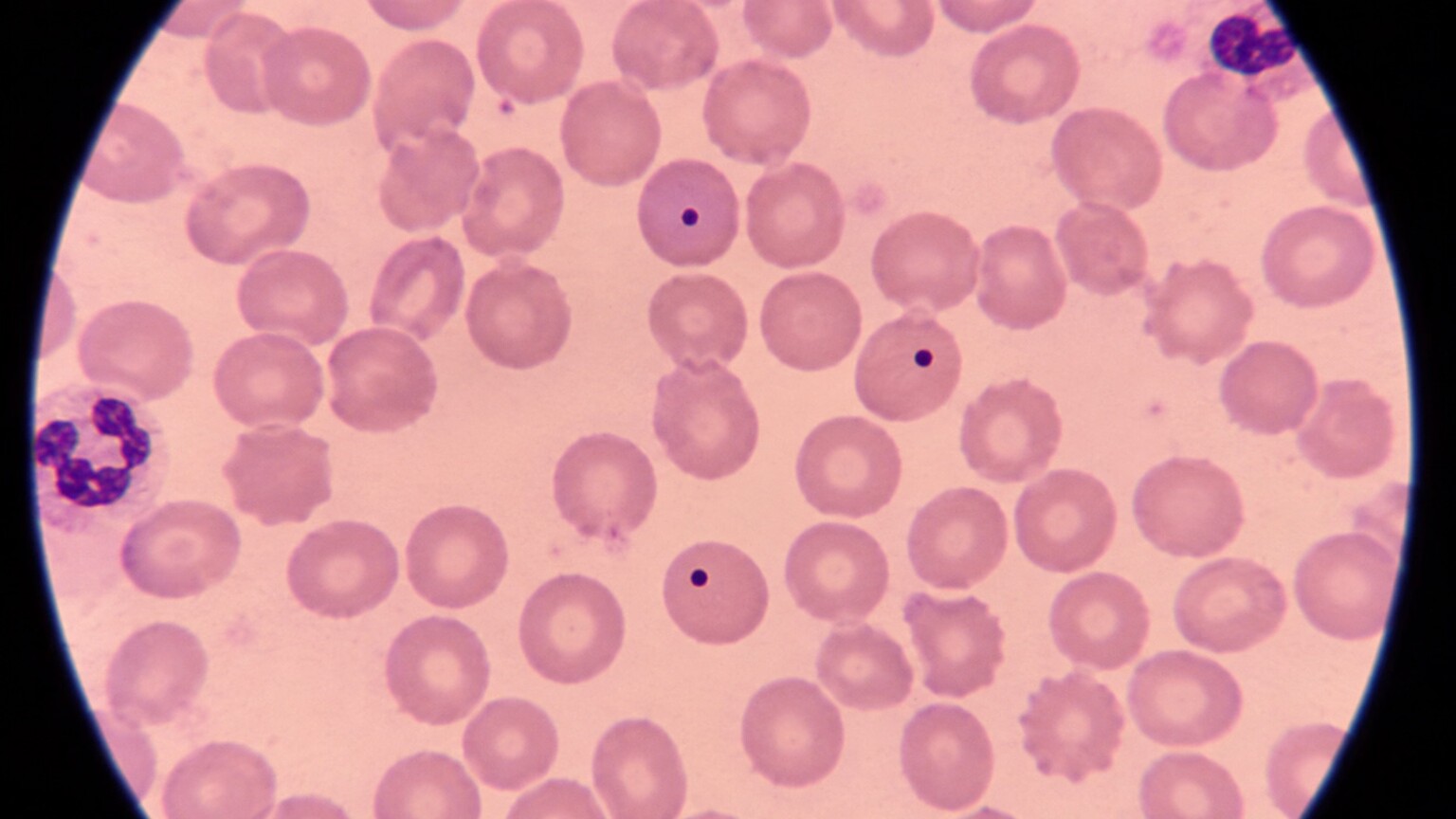
Figure 6. Once Howell-Jolly bodies are confirmed under the microscope, the follow-up workup typically starts with a simple abdominal ultrasound to check whether the spleen is anatomically present at all.
Finding Howell-Jolly bodies on a routine smear, particularly in someone with no known history of splenectomy, prompts a fairly systematic follow-up investigation rather than immediate alarm. The first, most obvious question is simply whether the spleen is physically present at all — a straightforward abdominal ultrasound can usually answer this quickly and definitively, either confirming a normal-appearing spleen (pointing toward functional rather than anatomical asplenia) or revealing a small, shrunken, scarred spleen consistent with the kind of chronic damage seen in conditions like sickle cell disease.
If the spleen is anatomically present, the next step usually involves reviewing the patient's broader medical history and other lab findings for a plausible underlying cause — known sickle cell disease or trait, symptoms or antibody testing suggestive of celiac disease, signs of chronic liver disease, or evidence of an autoimmune condition. In some cases, no clear underlying cause is identified despite reasonable investigation, and the finding is simply monitored over time, since even mild, otherwise unexplained functional asplenia still carries at least some of the infection-risk considerations described above and is worth the same precautionary conversation about vaccination and fever management.
In some cases, a doctor may also order a more direct functional test of splenic activity rather than relying solely on imaging and indirect clues. A nuclear medicine spleen scan, which uses a small amount of radioactively tagged blood cells to directly visualize how effectively the spleen is filtering and processing them in real time, can distinguish between a spleen that looks anatomically normal on ultrasound but is functionally underperforming versus one that's both structurally and functionally intact despite the presence of Howell-Jolly bodies from some other cause entirely, such as the rapid-production pathway discussed earlier. This more specialized test isn't ordered routinely for every case, but it becomes genuinely useful when the initial workup leaves real ambiguity about how much the spleen is actually contributing to the finding.
A Worked Example: Two Very Different Reasons for the Same Finding
Seeing how this reasoning plays out in two realistic but very different situations makes the whole picture click far better than discussing the mechanism alone. Picture a healthy 34-year-old with no prior medical history who has a routine annual blood count that happens to include a manual smear review, which reports occasional Howell-Jolly bodies. There's no history of surgery, no abdominal scar, no known blood disorder, and no symptoms at all. The follow-up ultrasound shows a completely normal, appropriately sized spleen. In this scenario, the finding prompts a broader look for causes of functional asplenia despite normal spleen anatomy — screening for celiac disease with antibody testing turns out to be positive, explaining the finding through the chronic intestinal inflammation pathway discussed earlier, well before the person had ever noticed a single digestive symptom serious enough to investigate on its own.
Now picture a second, very different case: a 9-year-old with known sickle cell disease, diagnosed shortly after birth through newborn screening, whose annual smear has shown Howell-Jolly bodies consistently since around age two. In this case, the finding isn't a surprise prompting new investigation at all — it's an expected, already-explained part of a known diagnosis, confirming that the autosplenectomy process described earlier has progressed as anticipated for this specific condition. The clinical response here isn't further diagnostic workup, since the cause is already well understood; it's continued adherence to the vaccination schedule and infection-precaution plan that was put in place specifically because of this known functional asplenia years earlier.
Same microscopic finding, two completely different clinical stories — one an unexpected clue that led to a new diagnosis, the other a confirmed, already-anticipated feature of a condition diagnosed in infancy. This is exactly the kind of context-dependent interpretation that makes a single dot under a microscope meaningfully different depending entirely on the person and history surrounding it, echoing the same theme raised earlier about newborns.
A third, intermediate scenario is worth mentioning too, since it's genuinely common in practice: an adult in their fifties, with no known blood disorder or autoimmune diagnosis, undergoes a routine physical that includes bloodwork, and Howell-Jolly bodies turn up unexpectedly. An ultrasound shows a spleen that's present but notably smaller than typical for their age, without any of the dramatic sickle-cell-related damage described earlier. Further questioning reveals years of heavy alcohol use and a recent diagnosis of early liver cirrhosis, which — through the portal hypertension pathway mentioned earlier in this article — has been quietly reducing effective splenic blood flow and function for longer than anyone had realized. Here, the Howell-Jolly bodies become one of the earlier, more concrete signals prompting a fuller liver workup, illustrating how this small finding can sometimes arrive well before the underlying condition driving it has otherwise announced itself.
How Howell-Jolly Bodies Are Actually Counted and Reported
It's worth understanding a bit about how this finding actually makes it onto your lab report, since the process involves more human judgment than many other blood tests you might be used to. Unlike a fully automated result like your hemoglobin or white blood cell count, which comes from a machine counting cells electronically, Howell-Jolly bodies are identified through direct visual review — either by a trained laboratory technologist or a pathologist physically examining a stained slide under a microscope, scanning across a representative portion of the smear and noting how frequently the finding appears.
Modern automated blood analyzers can sometimes flag a smear for manual review based on other unusual patterns in a sample, which is often how Howell-Jolly bodies get discovered in the first place, even in a routine test that wasn't specifically ordered to look for them. Once flagged, a human reviewer examines the slide and typically reports the finding using a rough qualitative scale — "rare," "occasional," or "frequent," for instance — rather than an exact numeric count, since the goal is to communicate a general pattern rather than a precise measurement the way a cholesterol value would be reported. A report noting "frequent" Howell-Jolly bodies generally signals a more complete, longer-standing loss of splenic function than one noting only an "occasional" or "rare" finding, though even a single confirmed observation is enough to prompt the kind of follow-up investigation described in this article.
This human element also explains why the finding is sometimes missed entirely on a routine visit — if nothing about the automated results prompts a manual smear review, and no smear review is separately, specifically requested, this particular clue simply never gets the chance to surface, since it isn't something a standard automated blood count panel reports on its own. This is part of why, in someone with a strong suspicion of reduced splenic function based on history alone, a doctor may specifically request a manual smear review rather than relying on the automated results to flag it independently.
Other Findings Often Seen Alongside Howell-Jolly Bodies
Howell-Jolly bodies rarely show up as the only unusual feature on a smear from someone with genuinely reduced splenic function, and recognizing the broader pattern helps confirm the underlying explanation. Target cells — a separate finding covered elsewhere on this site — often appear alongside Howell-Jolly bodies in people with little or no functioning spleen, since the spleen also plays a normal role in remodeling red blood cell membranes, and its absence allows both abnormalities to persist together. Pappenheimer bodies, small iron-containing granules distinct from nuclear DNA fragments, are another finding the spleen would normally pit out and can appear in the same context for a related reason.
A complete blood count accompanying the smear often shows supporting clues as well: a mildly elevated platelet count is common after splenectomy or in functional asplenia, since the spleen normally holds a meaningful reserve of platelets in reserve and helps regulate their circulating number — with that regulation gone, platelet counts frequently run somewhat higher than typical. Seeing this specific combination — Howell-Jolly bodies, target cells, and a mildly elevated platelet count together — is a recognizable, mutually reinforcing pattern that gives a pathologist and treating physician considerably more confidence in a splenic explanation than any single finding would provide alone.
White blood cell counts can shift too, though generally more modestly than platelets, since the spleen also serves as a reservoir and filtering site for certain white blood cell populations. A mild, persistent lymphocytosis — a higher-than-typical proportion of lymphocytes among the white blood cells — is sometimes observed in longstanding asplenia, adding one more small piece to the overall pattern a careful reviewer looks for when weighing how confidently to attribute a smear's findings to reduced splenic function rather than some other, unrelated explanation.
Frequently Asked Questions
Are Howell-Jolly bodies themselves dangerous?
No, the small nuclear fragments inside the red blood cells don't cause any direct harm on their own. What matters is what their presence reveals — usually reduced or absent splenic function — which does carry a genuine increased risk of certain severe bacterial infections.
Do Howell-Jolly bodies go away after a splenectomy heals?
No. Once the spleen is removed, Howell-Jolly bodies typically become a permanent, lifelong finding on blood smears, since nothing else in the body performs the pitting function that normally removes these fragments.
Can a normal-looking spleen still cause this finding?
Yes. Conditions like sickle cell disease and celiac disease can cause functional asplenia, where the spleen is anatomically present but no longer performing its normal immune and pitting functions, sometimes without any visible structural abnormality on imaging.
Is it normal for a newborn to have Howell-Jolly bodies?
Yes. A newborn's spleen hasn't fully matured at birth, and its pitting function typically reaches full efficiency over the first several weeks to months of life, making this a normal, expected, temporary finding in healthy infants.
What should I do if I'm told I have reduced splenic function?
Talk to your doctor about updated vaccination against encapsulated bacteria, whether keeping emergency antibiotics on hand makes sense for your situation, and the importance of seeking prompt medical care for any fever, since infection can progress more quickly without a fully functioning spleen.
Is overwhelming post-splenectomy infection (OPSI) common?
No, it's rare, especially in people who follow their recommended vaccination and precaution plan. But when it does occur, it can progress unusually fast, which is why any fever in someone with confirmed absent or significantly reduced splenic function is treated as a genuine medical emergency rather than something to wait out at home.
What other findings often appear on a smear alongside Howell-Jolly bodies?
Target cells and Pappenheimer bodies are commonly seen together with Howell-Jolly bodies in people with reduced splenic function, since the spleen normally clears all three from circulating red blood cells. A mildly elevated platelet count on the accompanying complete blood count is another frequently associated clue.
Conclusion
A Howell-Jolly body is a small thing to see under a microscope, but it's telling a specific, mechanistic story about a very particular organ job: your spleen's constant, quiet work of plucking leftover nuclear fragments out of red blood cells as they pass through its narrow vessels. Whether that job has stopped because the spleen was surgically removed, because it's been slowly damaged into functional silence by a condition like sickle cell disease, or because a newborn's spleen simply hasn't finished maturing yet, the underlying explanation always traces back to this same single mechanism. Understanding that connection turns a cryptic, easy-to-overlook lab finding into something genuinely actionable — a prompt to check on an organ whose job matters a lot more than its size would suggest.
If there's one takeaway worth carrying forward from this entire article, it's that the spleen's absence or dysfunction is never just a structural curiosity — it's a genuine shift in your body's infection defenses that deserves the same seriousness as any other confirmed immune deficiency. A single dot inside a red blood cell led to that realization, which is exactly the kind of outsized diagnostic value a careful, human-reviewed blood smear can still offer in an era of increasingly automated lab testing.
If a report ever comes back mentioning this specific finding — whether expected, following a splenectomy you already knew about, or genuinely unexpected on a routine check — treat it as an invitation to have a focused, specific conversation with your doctor about what it means for your particular situation, rather than a detail to skim past on the way to the rest of the report.
Still Not Sure What Your Results Mean?
Upload your labs and get a complete, visual, plain-language interpretation of every biomarker — delivered to your inbox in under 15 minutes.
Get My ReportThis article is for educational purposes only and does not constitute medical advice. Always consult your healthcare provider regarding your specific lab results.