
Why Might a Smear Show Toxic Granulation in White Cells?
If your own blood smear report happens to mention "toxic granulation," the word "toxic" quite understandably sounds alarming at first glance — but it genuinely doesn't mean your blood has been poisoned or somehow contaminated by any actual external toxin whatsoever. Toxic granulation is instead a specific, well-recognized, well-studied change in the appearance of neutrophils specifically, the single most abundant type of white blood cell found circulating in your body, in which their normally fine, pale internal granules appear coarser, darker, and more prominent under the microscope than usual. This finding is one of your body's own visible fingerprints of an active immune response, most often appearing when your bone marrow is working overtime to churn out new infection-fighting cells faster than its usual, unhurried pace — a change in production speed that leaves a distinctive mark on how those cells actually look. This article explains exactly what's happening inside a neutrophil to cause this appearance, where neutrophils actually come from and how they normally mature, why the somewhat misleading name "toxic" stuck around for over a century, what other white blood cell changes commonly travel alongside it, the conditions most often responsible, how it's distinguished from technical artifact, how it differs from age, and what it typically means for your care once a clinician sees it on your report.
What Toxic Granulation Actually Is Inside a Neutrophil
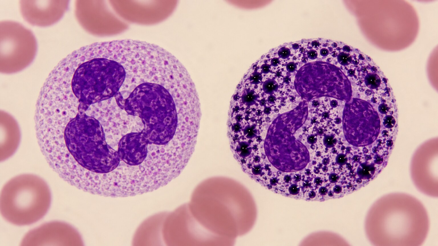
Figure 1. Toxic granulation refers to coarse, darkly staining granules inside a neutrophil's cytoplasm, visibly different from the fine, pale granules of a normally matured cell.
To understand toxic granulation, it helps to know a little about what a neutrophil actually is and what its granules are for. Neutrophils are the immune system's single most numerous first responders, constantly patrolling your bloodstream around the clock and rushing quickly toward any site of infection or injury as soon as one is detected, where they engulf and physically destroy invading bacteria before the problem can spread further. Inside each individual neutrophil, small membrane-bound sacs called granules store a genuine chemical arsenal — a mix of enzymes and antimicrobial proteins the cell strategically releases to break down and fully neutralize whatever bacterial invader it manages to capture and engulf. Under normal, everyday conditions, these granules are small in size, evenly distributed throughout the cell, and pale enough under standard laboratory staining techniques that they barely stand out visually against the rest of the cell's surrounding cytoplasm.
Toxic granulation describes a change in exactly how these granules look, not a change in what they contain in any dangerous new way. On a properly stained blood smear, affected neutrophils show granules that appear noticeably larger, considerably denser, and visibly darker overall — often described specifically by pathologists as coarse and deeply basophilic (meaning, in plain terms, that they take up a purple-blue stain far more intensely than a normal, healthy neutrophil's granules typically would). This altered staining pattern happens because the primary granules, the very first type of granule a developing neutrophil produces while still inside the bone marrow, are unusually abundant or retained in a less mature, more strongly staining form than they would be in a neutrophil that developed at its normal, unhurried pace. At the biochemical level, these primary granules are densely packed with proteins like myeloperoxidase and various proteases, and it's the sheer concentration of this protein content, combined with the fact that these granules haven't yet been diluted or replaced by the paler secondary and tertiary granules that develop later, that gives them their characteristic coarse, dark appearance under the microscope. In other words, toxic granulation is really a visible record of how a neutrophil was built, not evidence of a poison circulating in your blood.
How the Slide Is Actually Prepared and Read
Understanding the mechanics of how a blood smear gets made helps explain why toxic granulation is something only a trained eye at a microscope can reliably catch, rather than a number an automated machine simply spits out. A trained technologist places a small, precisely measured drop of blood near one end of a clean glass slide, then uses the carefully angled edge of a second slide to draw it steadily into a thin, feathered layer across the surface, deliberately aiming for a region where individual cells settle into a single, unstacked layer rather than overlapping, so that each one can later be evaluated clearly and individually under the microscope. The slide is then carefully fixed and stained, most commonly using a Wright-Giemsa stain, a specific dye combination formulated deliberately to color different cellular structures in genuinely contrasting shades — it's precisely this staining step that actually makes internal granules visible at all as distinct dark or pale spots in the first place, since entirely unstained white blood cells viewed under a standard light microscope are nearly transparent and essentially impossible to meaningfully evaluate or distinguish from one another in any useful way.
Once properly stained, the slide is first scanned at a lower magnification to survey the overall distribution of cells across its surface and locate the single ideal single-layer region best suited for detailed evaluation, then examined much more closely at high magnification, typically using a 100x oil-immersion objective lens, moving systematically and methodically across multiple separate fields of view in a consistent, repeatable scanning pattern. As the technologist encounters neutrophils along the way, they mentally note granule appearance, along with the presence of Döhle bodies, vacuolization, and any other notable features, building toward the overall impression they'll eventually summarize in the written report. This is a genuinely skilled, judgment-dependent task rather than a simple, straightforward measurement, which is a large part of why toxic granulation — quite unlike a number such as a total white blood cell count, which an automated analyzer can generate directly and instantly from a blood sample — instead depends entirely on a trained human reviewer actually looking carefully through the eyepiece and applying years of accumulated pattern recognition to what they observe on the slide in front of them.
Why It's Called "Toxic" — The Historical and Biological Reasoning

Figure 2. The term "toxic granulation" dates back over a century, coined when early hematologists assumed circulating toxins from infection were directly altering neutrophil granules.
The name "toxic granulation" is a genuine relic of early twentieth-century medicine, when pathologists first noticed this coarse-granule appearance in patients with severe bacterial infections and assumed, quite reasonably given what was known at the time, that circulating bacterial toxins were directly damaging or altering the neutrophils as they passed through infected, toxin-laden blood. That original explanation has since been revised considerably as laboratory science advanced, but the name had already become so entrenched in hematology textbooks and lab reporting conventions that it simply stuck, the way many historical medical terms persist long after the underlying science behind them has been refined or even overturned.
Modern understanding places the real explanation not in external toxins acting on mature cells, but in an accelerated and altered production process happening earlier, back in the bone marrow, before the neutrophil ever enters circulation. This is an important distinction for anyone reading their own report: seeing the word "toxic" doesn't mean a poison has been identified in your blood, and it isn't a marker your provider will investigate as a toxicology finding. It's simply the traditional, if slightly misleading, name for a specific and genuinely useful microscopic pattern — one more example, similar to how "cold sore" has nothing to do with temperature, of medical terminology that outlived the theory that originally produced it.
The Bone Marrow's Emergency Response: Why Granules Look Different During Infection
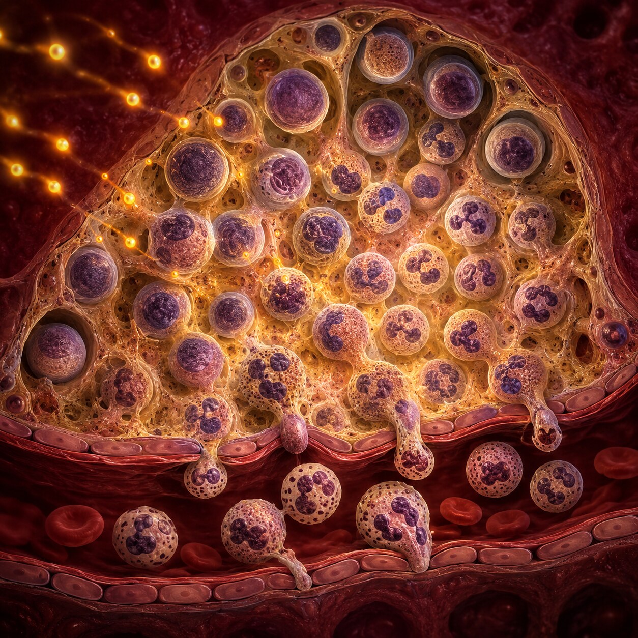
Figure 3. During active infection, chemical signals push the bone marrow to accelerate neutrophil production, shortening the normal maturation timeline and altering how granules form.
Under everyday, healthy conditions, a neutrophil takes about one to two weeks to fully mature inside the bone marrow, moving through several distinct developmental stages before it's released into your bloodstream ready for duty. During a significant bacterial infection, however, your immune system releases signaling molecules called cytokines that reach the bone marrow and effectively sound an alarm, instructing it to accelerate neutrophil production dramatically and release cells into circulation faster than usual, sometimes even before they've completed every step of their normal maturation sequence.
This accelerated, compressed production timeline is precisely where toxic granulation comes from. When neutrophils are rushed through development, the normal process of granule maturation — which typically involves granules gradually becoming smaller, more numerous, and more evenly dispersed as the cell matures — doesn't have time to fully complete. The result is a neutrophil released into circulation still carrying larger, more concentrated, darker-staining primary granules characteristic of an earlier developmental stage, essentially a visible signature of a cell that was manufactured under emergency conditions rather than through the bone marrow's usual, measured production schedule. The more severe or acute the underlying stimulus, generally speaking, the more pronounced this granulation tends to appear, which is part of why some laboratories report toxic granulation on a graded scale (mild, moderate, or marked) rather than as a simple present-or-absent finding, since capturing that spectrum of severity gives a clinician meaningfully more information than a flat yes-or-no result could, particularly when tracking how a patient's condition is evolving across serial blood draws taken over the following days of an illness.
Curious what your own white blood cell count and differential are really telling you? Upload your results and get a complete, plain-language breakdown in under 15 minutes.
Analyze My ResultsWhere Neutrophils Come From — the Bone Marrow's Normal Assembly Line
To really appreciate why acceleration changes a neutrophil's appearance so noticeably, it helps to walk through what its normal, unhurried developmental journey actually looks like. Neutrophils originate from blood-forming stem cells inside your bone marrow, the soft tissue found within the cavities of many of your bones, particularly the pelvis, spine, ribs, and breastbone in adults. From that starting stem cell, a developing neutrophil passes through a sequence of named developmental stages — myeloblast, promyelocyte, myelocyte, metamyelocyte, band, and finally segmented neutrophil — each stage marked by specific, predictable changes in the cell's size, nucleus shape, and granule content, refined step by step until the cell reaches its final, mature form.
The granules themselves are laid down in a specific order during this sequence: primary (or azurophilic) granules form earliest, during the promyelocyte stage, packed with powerful antimicrobial enzymes, while secondary and tertiary granules, containing a different set of proteins, form progressively later as the cell matures further. Under normal, unhurried conditions, by the time a neutrophil is released into circulation as a fully segmented cell, its primary granules have become smaller, less concentrated, and less prominent relative to the increasingly dominant secondary and tertiary granules, producing that fine, pale, evenly dispersed granular appearance considered normal. When production is accelerated, cells are released with a disproportionate share of those earlier, more prominent primary granules still intact and concentrated — which is, in a very literal sense, exactly what toxic granulation is: a visible snapshot of the cell's production timeline being compressed and released a step or two earlier in the sequence than it otherwise would have been.
Other Neutrophil Changes That Often Accompany Toxic Granulation
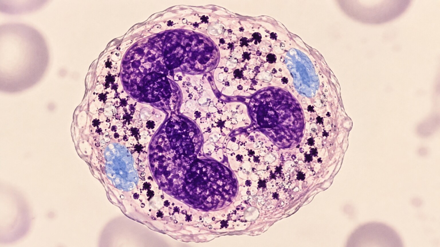
Figure 4. Toxic granulation frequently appears alongside other stress-related neutrophil changes, including pale-blue Döhle bodies and small cytoplasmic vacuoles, collectively called "toxic changes."
Toxic granulation rarely shows up as an isolated, solitary finding — it's typically one member of a small family of related neutrophil changes that pathologists collectively refer to as "toxic changes," all stemming from that same accelerated, stressed production process described above. Döhle bodies are pale, sky-blue patches sitting in the neutrophil's cytoplasm, representing remnants of a structure called rough endoplasmic reticulum, essentially a small pocket of unfinished cellular machinery that got left behind when the cell was rushed out of the bone marrow before fully clearing it. Cytoplasmic vacuolization, small clear bubble-like spaces scattered through the cytoplasm, is another commonly co-occurring change, thought to reflect the cell's own digestive and metabolic activity ramping up considerably in direct response to actively engaging with bacteria, bacterial byproducts, or the general metabolic strain placed on a cell that's working harder and faster than it normally would under everyday, non-emergency conditions.
Toxic granulation, Döhle bodies, and vacuolization can each occur alone in isolation, but seeing two or three of them appearing together in the same individual cell, or scattered consistently across many different cells on the same smear, generally strengthens the overall clinical impression that a genuine, active inflammatory or infectious process is currently underway, rather than reflecting some more benign, incidental, or borderline explanation that might otherwise be considered. Pathologists reviewing a smear typically note the presence and severity of each of these individual changes separately on the final report, since together, taken as a group, they paint a considerably more complete and textured picture of just how much genuine physiological stress the bone marrow's neutrophil production line is currently under than any single one of these individual findings could ever manage to convey entirely on its own.
What Conditions Commonly Cause Toxic Granulation

Figure 5. Bacterial infections severe enough to trigger a strong immune response are the most common overall cause of toxic granulation, though several other conditions can produce the same finding.
Bacterial infections, particularly more severe or genuinely systemic ones affecting the whole body rather than staying confined to one small area, are by far the single most common trigger for toxic granulation overall, since they reliably and consistently produce the kind of intense, sustained cytokine signaling needed to actually push the bone marrow fully into that accelerated production mode described earlier. This includes everything from significant pneumonia and urinary tract infections to abscesses and, in its most extreme and serious form, sepsis — a life-threatening, body-wide inflammatory response to infection where toxic granulation is often quite pronounced and is one of several smear findings clinicians actively watch for as supporting evidence of just how severe the underlying illness has become. Meningitis, significant skin and soft tissue infections like cellulitis, and infections following surgery are further examples from the long list of specific bacterial illnesses capable of producing this same underlying bone marrow response, since what matters biologically isn't the particular infection site so much as the overall intensity of the cytokine signal reaching the marrow. Severe burns and major tissue trauma from serious accidents or significant surgery can also trigger a genuinely comparable degree of bone marrow stress and resulting toxic granulation, since extensively damaged tissue releases many of the very same inflammatory signaling molecules that a true bacterial infection does, even in situations where no bacteria are directly involved at all in the underlying injury itself.
Certain inflammatory conditions unrelated to active infection, including some autoimmune and inflammatory diseases where the immune system generates significant inflammatory signaling even without a bacterial trigger present, along with certain medications — particularly growth factor drugs specifically used to stimulate white blood cell production in patients recovering from chemotherapy, which directly accelerate bone marrow output as their intended therapeutic effect — can also produce toxic granulation through a closely related underlying mechanism of accelerated or altered neutrophil development. Pregnancy is worth mentioning as a milder, generally benign cause as well, since some degree of toxic granulation can appear as a normal physiological finding in healthy pregnant women — likely reflecting the broader immune and hematologic adaptations pregnancy naturally produces throughout the body — illustrating that this particular finding, like so many others on a blood smear, always needs to be read within the fuller context of a person's overall clinical picture rather than being automatically assumed to signal severe infection in every single case it appears.
Distinguishing True Toxic Granulation From Artifact or Normal Variation

Figure 6. Overly acidic staining conditions can mimic true toxic granulation, which is why an experienced technologist evaluates staining quality and cell distribution before reporting the finding.
One genuinely important technical wrinkle in reporting toxic granulation is that it can be mimicked by a purely technical problem: if a smear's staining process runs slightly too acidic (an issue with the pH balance of the staining solution used), normal neutrophil granules can pick up a darker, coarser appearance that looks deceptively similar to true toxic granulation, even in a perfectly healthy person with no underlying infection at all. This is precisely why experienced laboratory technologists don't rely on the appearance of a single cell in isolation — they evaluate the pattern across the whole slide, checking whether red blood cells and other structures on the same smear also show telltale signs of a staining problem, and considering whether the granulation appears consistently across many neutrophils rather than just one or two, before confidently reporting it as a genuine finding rather than an artifact of slide preparation. Laboratories also run regular quality-control checks on their staining reagents and process specifically to catch drift toward this kind of pH-related artifact before it ever reaches a patient's actual report, treating consistent, reproducible staining as a foundational requirement of accurate blood smear interpretation rather than an incidental technical detail.
Distinguishing true toxic granulation from normal, everyday variation in neutrophil appearance likewise requires real training and experience, since some healthy people can show mild degrees of coarse granulation without any identifiable underlying cause, and the boundary between "mild toxic granulation" and simply "granules on the more prominent end of normal" isn't always perfectly sharp. This is part of why a properly performed manual smear review, done by someone trained specifically in recognizing these subtle distinctions, remains valuable even in an era of highly automated blood cell analyzers, since these particular staining and morphological nuances are exactly the kind of thing a trained human eye catches more reliably than automated counting equipment.
Toxic Granulation in Children and Newborns
Age introduces its own particular nuances worth understanding separately, since a newborn's immune system and bone marrow are still maturing and don't always behave exactly like an older child's or an adult's. In healthy newborns, especially in the first few days of life, some degree of toxic granulation and other toxic changes can appear even without a clear infection present, thought to reflect the substantial physiological transition a newborn's blood-forming system undergoes adjusting to life outside the womb — a pattern generally considered a normal variant in this specific, narrow age window rather than a reliable marker of illness the way it more consistently is in older children and adults.
At the same time, because newborns and young infants can develop serious bacterial infections that progress unusually quickly and sometimes with subtler outward symptoms than an older child or adult would show, clinicians evaluating a newborn for suspected infection generally interpret smear findings, including toxic granulation, together with a broader set of markers rather than relying on any single finding, newborn or otherwise, to rule infection in or out on its own. In older infants, children, and adolescents, toxic granulation behaves much more similarly to how it does in adults, reliably tracking with infection and inflammatory stress and fading as the underlying cause resolves, making age one more genuinely important piece of context a clinician weighs when deciding how much diagnostic weight this particular finding should carry in a specific case.
How Toxic Granulation Fits Into the Bigger Clinical Picture
Toxic granulation is almost never interpreted in isolation — it's one thread woven into a larger tapestry of findings a clinician pulls together when evaluating someone for a possible infection or significant inflammatory process. The overall white blood cell count and the neutrophil count specifically are typically reviewed alongside it, since toxic granulation appearing together with a genuinely elevated neutrophil count paints a more complete picture of an active immune response than toxic granulation alone. A "left shift" — a related but distinct finding referring to an increased number of immature neutrophil forms called bands appearing in the bloodstream — often accompanies toxic granulation for the exact same underlying reason: both reflect the bone marrow releasing cells faster than its usual, fully deliberate pace allows.
Beyond the blood smear itself, inflammatory blood markers like C-reactive protein (CRP) or procalcitonin, along with the person's actual symptoms, vital signs, and exam findings, all factor into the overall clinical assessment. In a hospital setting, particularly when sepsis is being actively considered, the combination of significant toxic granulation, a left shift, and other inflammatory markers together carries real weight in guiding how urgently a clinical team responds, since these smear findings can sometimes become apparent on quickly turned-around bloodwork even before other, slower test results like blood cultures — which can take a day or more to grow and identify a specific organism — have had time to return with a definitive answer. This layered, multi-marker approach is exactly why toxic granulation functions best as one supporting piece of evidence within a broader clinical evaluation, rather than as a stand-alone diagnostic answer on its own.
Tracking Toxic Granulation Alongside Treatment Response
Beyond its role in initially recognizing an active infectious or inflammatory process, toxic granulation carries a genuinely practical, less commonly discussed secondary use: tracking how someone is responding to treatment over the following days. In a hospitalized patient being treated for a significant infection, a clinician might request follow-up blood counts, sometimes with a repeat manual smear review, over the course of treatment — watching for the white blood cell count, band count, and degree of toxic granulation to all gradually normalize together as a reassuring sign that the infection is responding appropriately to antibiotics or other treatment being given.
Conversely, toxic granulation that remains pronounced or worsens despite several days of appropriate treatment can be one of several pieces of evidence suggesting the current treatment approach isn't adequately controlling the underlying infection, prompting a clinical team to reconsider the antibiotic choice, look harder for a source that hasn't yet been identified, or consider whether a complication like an abscess requiring drainage has developed. This tracking role illustrates something true of blood smear findings more broadly: their value isn't limited to a single snapshot at diagnosis — followed over time, they can offer a genuinely useful, relatively low-cost window into how a clinical course is actually unfolding, complementing symptoms and other lab trends rather than replacing them.
What Happens After Toxic Granulation Is Reported

Figure 7. Once toxic granulation is confirmed, clinicians typically use it as supporting evidence to guide further evaluation for infection, rather than treating it as a stand-alone diagnosis on its own.
Once toxic granulation is confirmed on a smear, the practical next step depends heavily on the person's actual symptoms and the broader clinical situation rather than the smear finding alone. In someone with a clear, already-recognized infection — a diagnosed pneumonia or urinary tract infection, for example — toxic granulation mainly serves as confirmatory evidence that the body is mounting a genuine, active immune response to that infection, information that can help a clinician judge severity alongside other findings, without necessarily changing the treatment plan on its own. In someone with fever or other concerning symptoms but no infection source yet identified, toxic granulation, especially alongside an elevated white count or left shift, can meaningfully support the decision to pursue further diagnostic workup — blood cultures, imaging, or other targeted testing — to actively search for the underlying source.
It's worth being genuinely clear about one thing throughout all of this: toxic granulation itself is never treated directly; there's no medication or targeted intervention aimed at the granules themselves, since they're simply a visible marker reflecting an underlying process happening elsewhere in the body, rather than being a problem in their own right that requires or benefits from direct treatment of any kind. As the underlying infection or inflammatory trigger resolves, whether through appropriate treatment or the body's own recovery, neutrophil production gradually returns to its normal, unhurried pace, and toxic granulation typically fades from subsequent smears over the following days as newly produced, normally matured cells replace the ones made during the acute, accelerated phase — making it a genuinely useful marker not just for identifying that an active process is underway in the first place, but occasionally for tracking the actual course of recovery over time as well, particularly in situations where repeat bloodwork is already being drawn for other reasons and a follow-up smear can be reviewed alongside it at little additional cost or inconvenience.
Toxic Granulation Compared to Other Neutrophil Findings on the Same Report
Because a blood smear report can mention several different neutrophil-related findings in the same paragraph, it's worth clearly separating toxic granulation from a couple of terms it's sometimes confused with, since each describes a genuinely different underlying phenomenon. A "left shift," mentioned earlier as a frequent companion finding, refers specifically to an increase in the number of immature neutrophil forms — bands, and in more severe cases even earlier precursors — circulating in the blood, essentially a headcount of how many not-yet-fully-mature cells have been released. Toxic granulation, by contrast, describes the internal appearance of the granules within neutrophils themselves, including ones that have technically completed their maturation and would otherwise be counted as fully segmented, mature cells. A neutrophil can show toxic granulation without being a band, and a band can show relatively normal granulation without qualifying as toxic — the two findings are related, since they share the same underlying trigger of accelerated bone marrow output, but they're measuring genuinely different things about the cells involved.
Hypersegmentation, another term that occasionally appears on the same type of report, describes the opposite kind of nuclear change — neutrophils with an unusually high number of nuclear lobes, most classically associated with vitamin B12 or folate deficiency rather than infection, and entirely unrelated to granule appearance. Seeing these various, precisely named findings listed together on one report can feel overwhelming at first glance, but each one is really answering a distinct, specific question about neutrophil production and maturation, and understanding that they're not interchangeable synonyms for "something is wrong" helps make a densely worded smear report considerably easier to parse and discuss meaningfully with a healthcare provider.
Frequently Asked Questions
Does toxic granulation mean I have a serious infection?
Not necessarily on its own. It's a supporting piece of evidence that your bone marrow has recently accelerated neutrophil production, which most often happens with infection, but the severity of the underlying cause depends on the whole clinical picture — your symptoms, your other lab values, and how pronounced the finding is — not on toxic granulation being present or absent alone.
Can toxic granulation appear without an infection?
Yes. Severe burns, major trauma, certain inflammatory conditions, some medications, and even normal pregnancy can produce toxic granulation without any active infection being present. This is exactly why the finding is always interpreted alongside your symptoms and the rest of your bloodwork rather than being assumed to mean infection by default.
Is toxic granulation the same thing as a high white blood cell count?
No, though they often occur together. A high white blood cell count is a quantity measurement — how many white cells are present — while toxic granulation is a qualitative, appearance-based finding describing how certain neutrophils look under the microscope. You can have one without the other, though seeing both together generally strengthens the overall impression of an active immune response.
Will toxic granulation show up again on my next blood test?
It depends on whether the underlying trigger is still active. As an infection or inflammatory process resolves, neutrophil production typically returns to its normal pace, and toxic granulation usually fades from later smears within days as older, accelerated cells are replaced by normally matured ones. Persistent toxic granulation on a repeat test may prompt further evaluation of an ongoing or unresolved issue.
Is toxic granulation something an automated blood analyzer can detect on its own?
Not reliably. Automated analyzers are excellent at counting cells and measuring their size, but subtle staining and morphological features like granule coarseness are typically flagged for manual review rather than confidently identified by the machine itself. This is one of several reasons a human technologist reviewing the slide directly remains an important part of a complete blood count in certain situations.
Can toxic granulation appear in newborns without meaning anything is wrong?
Yes, particularly in the first few days of life. Some degree of toxic granulation is considered a normal physiological variant in healthy newborns adjusting to life outside the womb, generally interpreted differently than the same finding would be in an older child or adult, where it more reliably tracks with an active infection or inflammatory process.
Conclusion
Toxic granulation is a well-established, genuinely useful microscopic finding — coarse, darkly staining granules inside neutrophils that reflect an accelerated, stress-driven production process happening back in the bone marrow, almost always in response to a significant infection, inflammatory trigger, or, less commonly, a benign physiological state like pregnancy. Despite its alarming-sounding name, a holdover from an outdated century-old theory, it has nothing to do with toxins circulating in your blood, and it's never treated directly — it simply serves as one valuable, supporting clue among several that helps a clinician judge how actively your immune system is responding to whatever is happening in your body. Understanding what this specific finding actually represents, and bringing it into context with your symptoms, your age, and the rest of your complete blood count and differential, is what turns an intimidating-sounding lab term into a clear, genuinely informative piece of your overall health picture, rather than a source of unnecessary worry over a word that sounds far more ominous than what it actually describes.
Still Not Sure What Your Results Mean?
Upload your labs and get a complete, visual, plain-language interpretation of every biomarker — delivered to your inbox in under 15 minutes.
Get My ReportThis article is for educational purposes only and does not constitute medical advice. Always consult your healthcare provider regarding your specific lab results.