
Why Would Sickle Cells Appear on a Blood Smear?
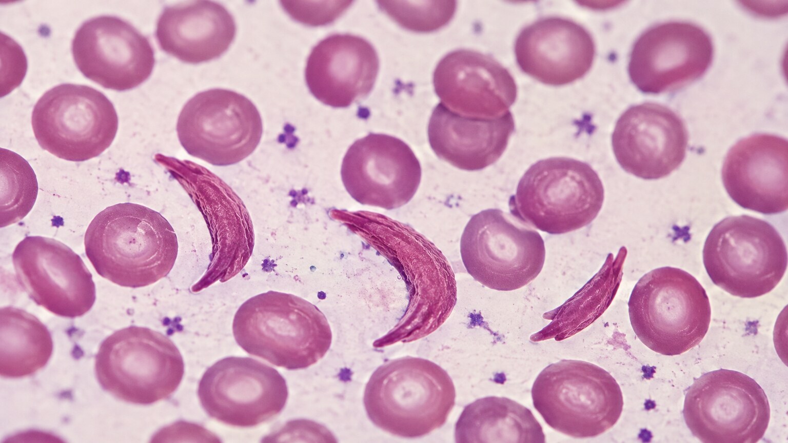
If a lab report mentions that a technologist spotted sickle cells while looking at your blood under a microscope, it means they saw red blood cells shaped like a crescent moon or an old-fashioned farming sickle, instead of the round, slightly dimpled disc every red blood cell is supposed to be. That shape isn't random damage or a smudge on the slide — it's a direct, visible sign that the hemoglobin inside those cells, the protein that carries oxygen, is a variant called hemoglobin S, and that it's behaving in a way normal hemoglobin never does. This article walks through exactly what's happening inside a red blood cell to bend it into that shape, why the finding means something very different depending on whether one or two copies of the gene are involved, and what typically happens after a smear turns up this specific finding.
What a Blood Smear Actually Shows That a Standard Count Can't
Most blood tests today run through an automated analyzer — a machine that counts and sizes thousands of cells per second and spits out numbers like your red blood cell count, hemoglobin, and hematocrit. What that machine generally can't do well is describe shape. A blood smear is the low-tech but irreplaceable answer to that gap: a single drop of blood is spread into a thin film across a glass slide, stained with a dye that colors different cell structures different shades, and then examined directly by a trained eye under a microscope. Shape, in red blood cells, is not cosmetic detail — it's one of the most information-dense things about the cell, because a healthy red blood cell has a very specific, biologically necessary shape (a flattened disc, pinched in the middle like a doughnut without the hole all the way through), and almost anything that distorts that shape is a clue pointing back to a specific cause. A sickle shape is one of the most immediately recognizable and specific of all these clues, because outside of hemoglobin S, essentially nothing else makes a red blood cell bend into that particular crescent.
The Hemoglobin Molecule Behind the Shape
To understand why the cell bends, it helps to understand what's inside it. Hemoglobin is the protein packed into every red blood cell that physically grabs oxygen in your lungs and releases it out in your tissues. Normal adult hemoglobin, called hemoglobin A, is built from a precise sequence of amino acids — the building blocks of every protein in your body — arranged in a specific three-dimensional shape that lets it pick up oxygen, carry it smoothly through your bloodstream, and let it go exactly where it's needed. Hemoglobin S is the result of a single, tiny typo in the genetic instructions for that protein: one amino acid, glutamic acid, is swapped out for a different one, valine, at one specific position on the beta-globin chain. That's the entire structural difference — one amino acid out of hundreds — but it's enough to change how the molecule behaves the moment it gives up its oxygen.
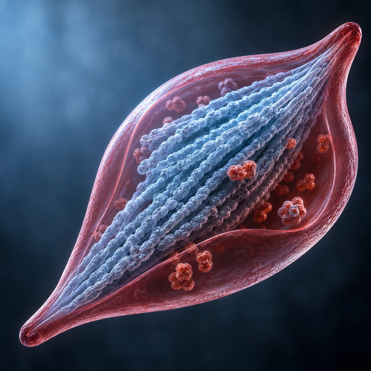
Here's the mechanism itself. When hemoglobin A releases its oxygen, it simply changes shape slightly and stays dissolved, individually, in the fluid inside the red blood cell — the way sugar stays dissolved in water. Hemoglobin S does something completely different once it gives up its oxygen: that single amino acid swap creates a sticky patch on the surface of the molecule that wasn't there before, and that sticky patch lets one deoxygenated hemoglobin S molecule latch onto the next one, which latches onto the next, and so on, building long, rigid, rod-like fibers inside the cell. Picture a jar of loose, tangled thread suddenly organizing itself into stiff, straight rods all pointing the same direction — that's roughly what happens inside the cell over the span of a second or two. Those rigid fiber bundles are physically longer than the red blood cell's own diameter, so as they grow, they push against the cell's flexible outer membrane from the inside and force it into the elongated, pointed crescent shape a smear picks up. The instant oxygen returns — as the cell circulates back through the lungs — the fibers usually fall back apart and the cell can spring back to something closer to its normal shape, at least for the first several cycles.
Why Oxygen Level Is the Trigger, Not the Cause
This is the detail that trips a lot of people up: hemoglobin S itself is present in the cell all the time, but the sickling event — the actual bending of the cell — only happens when oxygen levels drop low enough, which is precisely what happens naturally out in the body's tissues, away from the lungs, on every single lap of circulation. Anything that pushes oxygen levels even lower than usual makes sickling worse and more frequent: dehydration, which concentrates the blood and makes it harder for oxygen to move efficiently; high fever, which increases the body's oxygen demand faster than it can be delivered; cold temperatures, which cause blood vessels to constrict; high altitude, where there's simply less oxygen in the air to begin with; and intense physical exertion. This is also why a blood smear is genuinely useful here rather than just a formality — it captures a real-time snapshot of cells that have already sickled under whatever conditions existed in that person's body recently, which is a different kind of information than a purely genetic test showing what hemoglobin type is present.
It also helps to know that sickling isn't strictly all-or-nothing the first time oxygen drops — it's a probability that climbs the longer and lower oxygen stays down, and the process is reversible for a while before it isn't. Early on, as oxygen dips, only a fraction of a cell's hemoglobin S molecules polymerize, and if oxygen is restored quickly, those fibers dissolve back apart and the cell relaxes back toward its normal shape with no lasting harm. But each cycle of sickling and unsickling puts mechanical stress on the cell's outer membrane, similar to how repeatedly bending a paperclip eventually weakens the metal at the crease. After enough cycles, some cells lose the ability to fully spring back and become permanently, irreversibly sickled even once normal oxygen levels return — these are the cells most likely to become lodged in a narrow vessel and most likely to be destroyed early by the spleen or the rest of the immune system, which is a large part of why red blood cell survival is so dramatically shortened in sickle cell disease compared to a healthy 120-day lifespan.
Curious what the rest of your complete blood count is telling you? Upload your results and get a complete, plain-language breakdown in under 15 minutes.
Analyze My ResultsSickle Cell Trait vs. Sickle Cell Disease — Why One Copy Is Different From Two
Every person has two copies of the gene that codes for the beta-globin chain of hemoglobin, one inherited from each parent. What happens on a blood smear, and what it means for a person's health, depends heavily on how many of those two copies carry the hemoglobin S variant. A person with sickle cell trait has inherited one normal copy and one hemoglobin S copy. Their red blood cells contain a mix of roughly 60% normal hemoglobin A and 40% hemoglobin S, and that ratio matters enormously — with that much normal hemoglobin diluting the mix, cells generally don't sickle under everyday conditions, and a routine blood smear from someone with sickle cell trait often looks entirely normal. Sickle cell trait is usually a silent genetic characteristic, not a disease, though it can occasionally become relevant under extreme physical stress, such as severe dehydration combined with intense exertion at high altitude.
Sickle cell disease is a fundamentally different situation: it means both copies of the gene are affected, either two copies of hemoglobin S (the most common and typically most severe form) or one copy of hemoglobin S paired with a different abnormal hemoglobin variant, such as hemoglobin C. With no meaningful amount of normal hemoglobin A to dilute the mix, sickling happens constantly, under completely ordinary conditions, throughout the body every day. A blood smear from someone with sickle cell disease typically shows sickle cells readily and repeatedly, along with a cluster of other visible changes that build up over years of this ongoing process.
The Other Clues That Ride Alongside Sickle Cells on the Same Slide
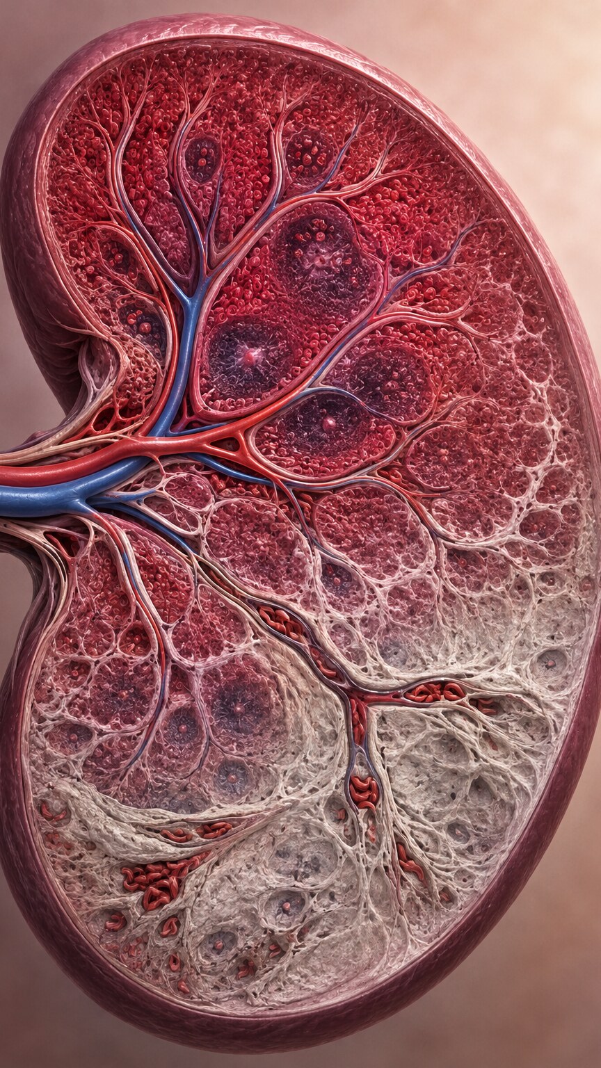
A trained eye reading a smear from someone with sickle cell disease rarely sees just sickle cells in isolation — a handful of companion findings usually show up on the same slide, and together they tell a fuller story. Howell-Jolly bodies, small round purple-staining fragments of leftover nuclear material sitting inside otherwise mature red blood cells, are one of the most telling. Normally, the spleen acts as a quality-control filter, plucking these fragments out of red blood cells before they're released into general circulation. In sickle cell disease, the spleen itself becomes one of the earliest organs damaged by the disease process: its narrow, low-oxygen blood vessels are exactly the kind of environment where sickling happens most readily, and repeated blockages inside the spleen, starting in early childhood, gradually scar and shrink it down until it stops functioning — a process doctors call autosplenectomy, since the organ essentially disables itself over time even though it's never surgically removed. Once the spleen stops filtering effectively, Howell-Jolly bodies start showing up on the smear because nothing is removing them anymore. Target cells, red blood cells with a bullseye-like ring of color in the center instead of the normal even coloring, and nucleated red blood cells, immature cells that shouldn't normally be released from the bone marrow at all, often appear alongside them, reflecting both the spleen's declining filter function and the bone marrow working overtime to replace red blood cells that are being destroyed faster than usual.
Why the Sickled Shape Causes Real, Physical Blockages
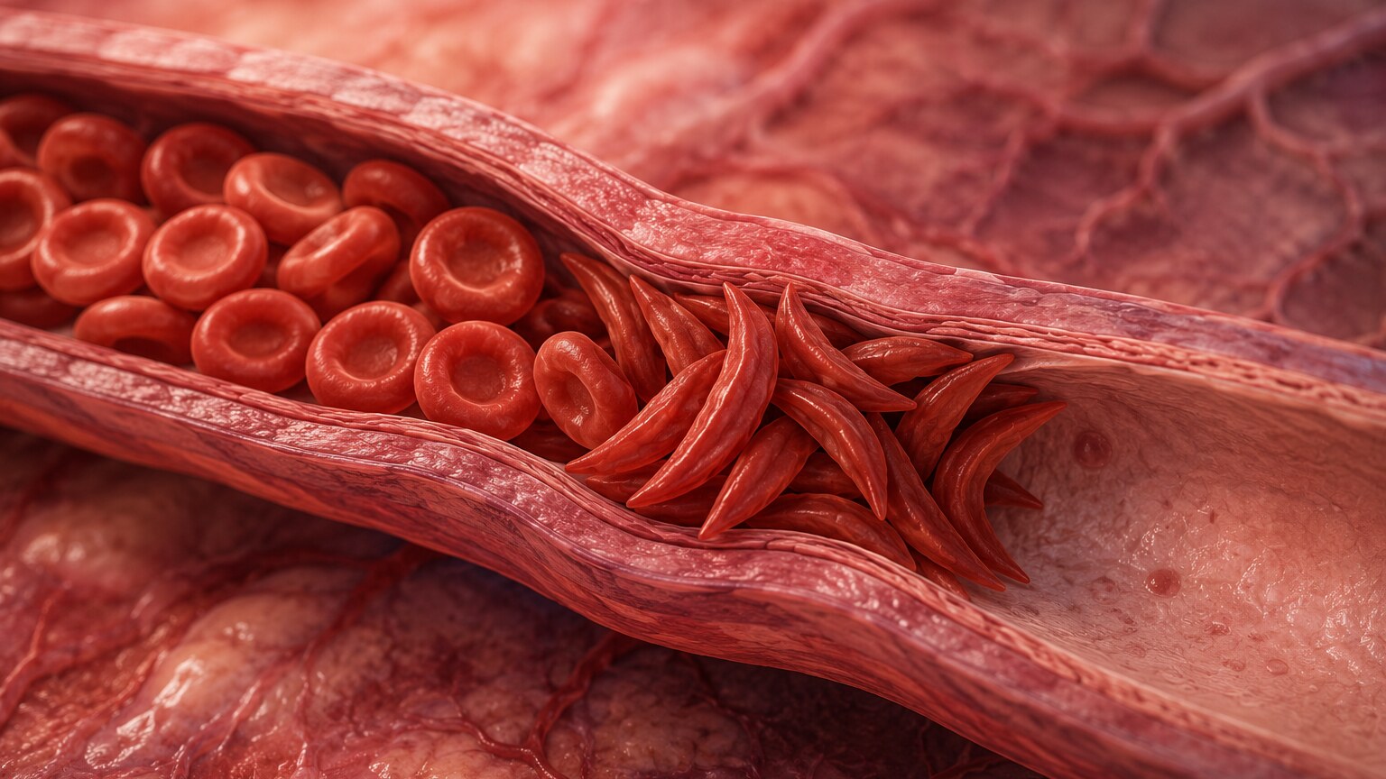
A normal red blood cell is remarkably flexible — flexible enough to fold and squeeze single-file through capillaries that are actually narrower than the cell's own resting diameter, the way a person might turn sideways to slip through a narrow doorway. A sickled cell has lost almost all of that flexibility: the rigid hemoglobin S fibers packed inside it turn what should be a soft, pliable disc into something closer to a stiff, pointed sliver that can't easily bend or fold. When enough of these rigid, oddly shaped cells try to pass through the body's smallest vessels at once, they jam against each other and against the vessel wall, physically obstructing blood flow to whatever tissue lies downstream. This is the direct mechanical basis of a vaso-occlusive crisis, the sudden, often severe episodes of pain that are the hallmark clinical event of sickle cell disease — the pain is the tissue downstream of the blockage being starved of oxygen in real time, and it can strike almost anywhere: the chest, the joints, the long bones of the arms and legs, or the abdomen. Repeated over years, this same blockage mechanism is also what damages organs beyond the spleen, including the kidneys, the eyes, and the bones, since every organ depends on that same uninterrupted capillary flow to stay healthy.
What a Pain Crisis Actually Feels Like Day to Day

For someone living with sickle cell disease, a vaso-occlusive crisis is rarely an abstract lab finding — it's a very physical, often frightening event that can begin with almost no warning. People frequently describe it as a deep, throbbing, boring pain that settles into a joint, a limb, or the lower back and doesn't respond to changing position or light stretching, unlike ordinary muscle soreness. Mild episodes might be managed at home with heat, hydration, and over-the-counter pain relief; more severe episodes routinely require emergency care and intravenous pain management, since the underlying blockage doesn't resolve just because the pain is being treated. One particularly serious variant, acute chest syndrome, involves this same blockage mechanism occurring in the small blood vessels of the lungs, producing chest pain, fever, and difficulty breathing, and it's treated as a medical emergency because it can progress quickly. Between crises, many people with sickle cell disease feel relatively well, which is part of why the disease can be so unpredictable to live with — the underlying sickling process never fully stops, but its visible, painful consequences come and go.
How Doctors Confirm What the Smear Only Suggests
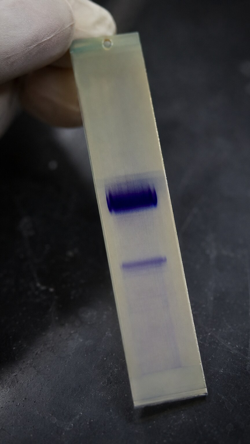
Spotting sickle cells on a smear is a strong visual clue, but it isn't, by itself, the final word on diagnosis or on distinguishing trait from disease — for that, labs turn to a test called hemoglobin electrophoresis. In this test, a blood sample is placed on a specialized gel or membrane and exposed to an electric current, which pulls the different hemoglobin types through the material at different speeds based on their size and electrical charge. Because hemoglobin A and hemoglobin S have slightly different structures, they separate into distinct, visible bands positioned at different points along the strip, and the relative darkness of each band shows roughly what percentage of a person's total hemoglobin each type makes up. This is exactly what distinguishes sickle cell trait, which shows both an A band and a smaller S band, from sickle cell disease, where the A band is faint, absent, or replaced by a different abnormal band entirely. In the United States, this distinction is now made for nearly everyone at birth: hemoglobin testing has been part of routine newborn screening since it became universal nationwide, meaning most people who have sickle cell disease or trait already know it well before adulthood, and a smear finding sickle cells later in life is more often a confirmation of an existing diagnosis than a brand-new discovery.
Managing Sickle Cell Disease: Where Treatment Actually Targets the Problem
Because the root problem is the hemoglobin S molecule's tendency to polymerize when deoxygenated, modern treatment increasingly targets that mechanism directly rather than only managing symptoms after they appear. A range of medications, some used for decades and some newer, work through different angles on this underlying process, alongside everyday preventive measures like staying well hydrated, avoiding extreme cold and high altitude when possible, and treating fevers and infections promptly. Bone marrow transplant and, more recently, gene therapy approaches that directly correct or bypass the faulty gene, offer the closest thing to a cure currently available, though they remain complex, resource-intensive options generally reserved for more severe cases. The specific treatment plan for any individual — which medications, what monitoring schedule, and how aggressively to intervene — is something a hematology team builds and adjusts over time for that patient, not a general protocol a reader should apply to their own or a family member's case.
Preventive care plays a bigger role in day-to-day management than many people expect, and the reason traces directly back to the spleen damage described earlier in this article. Because the spleen also serves as a key line of defense against certain bacteria, a child whose spleen has stopped functioning loses a meaningful part of their infection-fighting ability years before most people would think to worry about it, which is why children with sickle cell disease are typically managed with a specific preventive care and vaccination plan set by their care team, and why a routine fever in a child with sickle cell disease is treated as a more urgent situation than the same fever in a child with a normal, functioning spleen. Pain management plans are also increasingly individualized rather than one-size-fits-all: many patients and their hematology teams build a written action plan in advance, specifying what to try at home, at what point to seek urgent care, and what has and hasn't worked during past crises, so that care during a painful, frightening episode doesn't have to start from scratch every time.
Long-Term Organ Damage From Decades of Sickling
Vaso-occlusive crises get most of the attention because they're acute and painful, but a slower, quieter kind of damage builds in the background even between crises, simply from a lifetime of red blood cells passing through organs in a shape they were never meant to have. The kidneys are especially vulnerable, since they rely on an unusually slow, low-oxygen blood flow through their innermost regions to concentrate urine, which happens to be exactly the environment where hemoglobin S is most prone to polymerizing — over years, this can lead to a reduced ability to concentrate urine and, eventually, chronic kidney disease in a meaningful proportion of adults with sickle cell disease. The eyes face a related problem: sickled cells damage the small blood vessels feeding the retina, and without regular eye exams, this process, called sickle cell retinopathy, can progress silently for years before it threatens vision. Bone tissue is affected too, particularly in the hips and shoulders, where blocked blood flow to the head of the bone can cause a section of it to die from lack of oxygen, a condition called avascular necrosis that can eventually require joint replacement even in people still in their twenties or thirties.
Children with sickle cell disease face one particular risk that has reshaped how the condition is monitored early in life: stroke. Roughly 1 in 10 children with sickle cell disease will have a clinically apparent stroke by adulthood if left unmonitored, a striking figure for a pediatric population, caused by the same blockage mechanism narrowing or damaging the larger blood vessels supplying the brain. This risk is now actively screened for using a painless ultrasound technique called transcranial Doppler, which measures how fast blood is moving through vessels in the brain — unusually fast flow can signal a narrowed, high-risk vessel before a stroke ever happens, allowing doctors to start preventive blood transfusion therapy in the children who need it most. This kind of proactive, targeted screening is a major reason outcomes for children with sickle cell disease have improved substantially over the past few decades, even though the underlying molecular defect hasn't changed at all.
What This Finding Means for Family Members
Because sickle cell trait and disease are both inherited, a smear or blood test that reveals hemoglobin S in one person often opens a conversation about the rest of the family. If both parents carry sickle cell trait, each of their children has a 25% chance, with every pregnancy, of inheriting sickle cell disease, a 50% chance of inheriting trait, and a 25% chance of inheriting neither — the same odds every time, regardless of what happened with previous children. This is exactly why genetic counseling is routinely offered to people who discover they carry sickle cell trait, particularly if they're planning a family with a partner who might also carry it. It's also worth knowing that sickle cell trait and disease occur across many populations worldwide, most prominently in people with ancestry from sub-Saharan Africa, the Mediterranean, the Middle East, and parts of India — a distribution that lines up closely with regions where malaria has historically been common, since carrying one copy of hemoglobin S offers some natural protection against severe malaria, which is part of why the trait persisted and spread in those populations over many generations.
How This Finding Fits Into a Full Complete Blood Count
Sickle cells almost never show up as an isolated line item — they're one piece of a larger picture that a complete blood count (CBC) and its accompanying smear paint together. In sickle cell disease, the CBC typically shows a lower-than-normal hemoglobin and hematocrit, since sickled cells are fragile and get destroyed by the body far faster than normal red blood cells are replaced, a process called hemolysis. Where a normal red blood cell circulates for about 120 days before being retired, a sickled cell often survives only 10 to 20 days, and the bone marrow, working hard to keep up, releases immature red blood cells called reticulocytes into circulation earlier than usual — so a reticulocyte count that's higher than expected, paired with sickle cells on the smear, is a pattern that reinforces the same underlying story rather than two unrelated findings. White blood cell and platelet counts are frequently elevated as well, not because of infection or a clotting problem, but because chronic inflammation from ongoing vessel blockage keeps the bone marrow generally activated. Reading any single number on a CBC in isolation, without the smear and without this fuller context, risks missing the pattern that actually explains what's going on.
Frequently Asked Questions
Does finding sickle cells on a smear always mean someone has sickle cell disease?
Not necessarily. It usually points strongly toward sickle cell disease, since sickle cell trait rarely produces visible sickling under normal conditions. But confirming the exact diagnosis, and distinguishing trait from disease, requires a follow-up test called hemoglobin electrophoresis rather than the smear finding alone.
Can sickle cells appear on a smear even if someone has sickle cell trait, not disease?
It's uncommon but possible under extreme conditions, such as severe dehydration combined with very low oxygen levels, like high altitude paired with intense exertion. Under everyday circumstances, someone with sickle cell trait usually has a normal-looking smear because enough normal hemoglobin A is present to prevent sickling.
Why do people with sickle cell disease often have Howell-Jolly bodies on their smear too?
Howell-Jolly bodies show up once the spleen stops filtering blood effectively. In sickle cell disease, repeated blockages inside the spleen's narrow vessels gradually scar and shrink it over years, a process called autosplenectomy, which is why these leftover cell fragments start appearing on the smear once that filtering function is lost.
What triggers a sickle cell pain crisis?
Anything that lowers oxygen availability or thickens the blood can trigger one, including dehydration, fever, infection, cold exposure, high altitude, and intense physical exertion. The pain comes from rigid, sickled cells physically blocking blood flow through small vessels, starving the tissue downstream of oxygen.
Is sickle cell disease something people find out about as adults, or is it caught earlier?
In the United States, hemoglobin testing is part of routine newborn screening nationwide, so the vast majority of people with sickle cell disease or trait are identified in infancy. A smear finding sickle cells in an adult is more often confirming an already-known diagnosis than revealing a brand-new one.
Conclusion
A sickle-shaped red blood cell on a smear is one of the more specific, information-rich findings in all of laboratory medicine — it's a direct visual signature of one amino acid substitution in hemoglobin, made visible under a microscope. From that single molecular change flows everything else in this article: rigid fibers forming inside the cell when oxygen drops, cells losing the flexibility they need to pass through tiny vessels, blockages that cause real pain and organ damage over time, and a whole set of companion findings on the same slide, like Howell-Jolly bodies, that trace back to the same root cause. Whether this finding represents sickle cell trait or sickle cell disease changes the picture enormously, which is exactly why a smear finding sickle cells is typically a starting point for further testing, not an endpoint in itself — and for most people today, it's a confirmation of something already identified at birth rather than an unexpected discovery.
Still Not Sure What Your Results Mean?
Upload your labs and get a complete, visual, plain-language interpretation of every biomarker — delivered to your inbox in under 15 minutes.
Get My ReportThis article is for educational purposes only and does not constitute medical advice. Always consult your healthcare provider regarding your specific lab results.