
Low Platelet Count Combined With Abnormal Clotting, Explained
A low platelet count on its own can mean a dozen different things, and a prolonged clotting time on its own can mean a dozen more. But when both show up on the same panel at the same time — platelets running low while PT and aPTT, the two standard measures of how long blood takes to clot, are both stretched out longer than normal — that specific combination narrows the list dramatically, and it's a pattern doctors and hospital labs take very seriously. It usually points toward one of a small handful of processes where the body is either using up its clotting supplies faster than it can replace them, or has lost the organ responsible for manufacturing those supplies in the first place. This article walks through why platelets and clotting factors would drop together rather than separately, what's actually happening inside the blood vessels when that occurs, and why this particular lab pattern is treated as more urgent than either finding alone.
Why Platelets and Clotting Factors Are Usually Discussed Together
Every time you get a cut, your body seals it using two coordinated systems working almost simultaneously. Platelets, tiny cell fragments that circulate in huge numbers throughout your blood, are the first responders — they rush to the site of injury, stick to the damaged vessel wall, and clump together to form a loose, temporary plug within seconds. Right behind them comes a cascade of clotting factors, a series of proteins (numbered with Roman numerals, Factor I through Factor XIII) that activate one another in a precise sequence and ultimately weave a mesh of a protein called fibrin through and around that platelet plug, reinforcing it into a stable, durable clot. Think of the platelet plug as sandbags piled up against a breach, and the fibrin mesh as the concrete poured over them afterward — you genuinely need both, in the right order, for the seal to actually hold. Because these two systems are so tightly linked, a body that's rapidly consuming or losing one of them is very often consuming or losing the other at the same time, which is exactly why this particular lab combination tends to travel together rather than showing up in isolation.
A standard complete blood count reports your platelet number directly, typically somewhere between 150,000 and 450,000 platelets per microliter of blood in a healthy adult, and a number dropping meaningfully below that range is called thrombocytopenia. Clotting is measured differently, through two related but distinct timed tests: prothrombin time (PT), which specifically tracks one branch of the clotting cascade and is often reported alongside a standardized version called the INR, and activated partial thromboplastin time (aPTT), which tracks a different, overlapping branch of the same cascade. Both PT and aPTT measure, quite literally, how many seconds it takes a blood sample to form a visible clot once specific activating chemicals are added to it in the lab — a longer time than normal means something in that particular branch of the cascade is missing, reduced, or being actively used up faster than the body can replace it. Seeing platelets drop alongside both of these timed tests stretching out is what tells a clinician that the problem likely isn't confined to one narrow piece of the clotting system, but is instead affecting the blood's clotting capacity broadly, across multiple components at once.
The Leading Cause: Disseminated Intravascular Coagulation
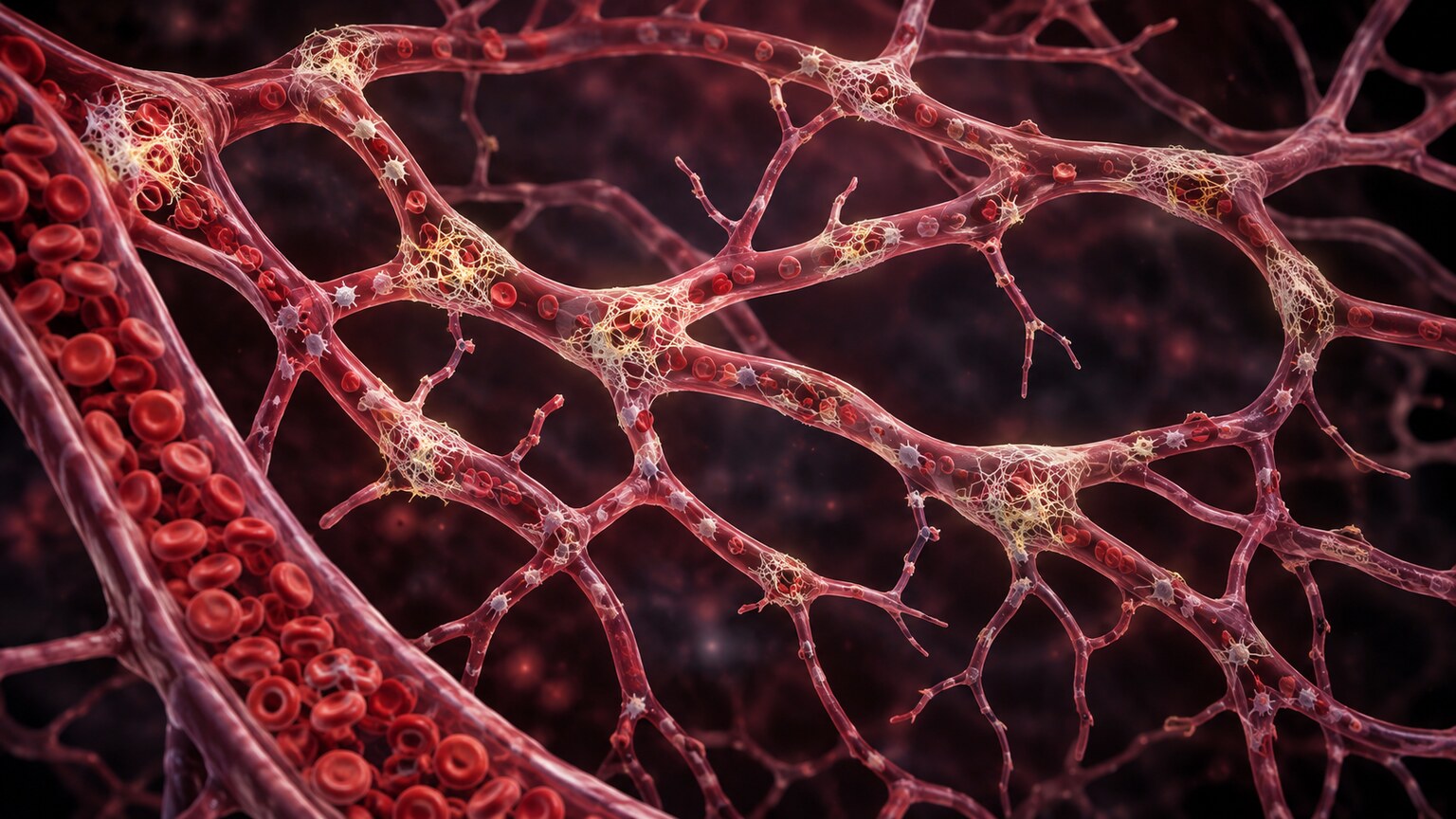
The single most important process to understand behind this lab pattern is disseminated intravascular coagulation, almost universally shortened to DIC. Despite the intimidating name, the concept is fairly direct once broken down: "disseminated" means spread throughout the entire body rather than localized to one spot, and "intravascular coagulation" means clotting happening inside the blood vessels themselves, rather than in response to an actual visible wound. In DIC, some overwhelming trigger — severe infection, major trauma, certain cancers, or a serious pregnancy complication, all discussed further below — causes the clotting cascade to switch on everywhere in the bloodstream at once, rather than being confined to one site of injury the way it's supposed to be. Tiny fibrin clots begin forming throughout the small blood vessels of multiple organs simultaneously, consuming platelets and clotting factors at a rate the body simply cannot replace fast enough. The tragic irony of DIC is that this process, driven by too much clotting, ultimately leaves the blood with too little capacity to clot at all — once platelets and clotting factors are sufficiently depleted by this widespread, inappropriate activity, the body loses the raw materials it needs to control bleeding anywhere else, including at IV sites, surgical incisions, or spontaneously through the gums and nose.
Curious what the rest of your coagulation panel is telling you? Upload your results and get a complete, plain-language breakdown in under 15 minutes.
Analyze My ResultsWhat Actually Triggers This Widespread Overreaction
DIC is never a standalone diagnosis — it's always a complication of something else severe enough to overwhelm the body's normal, contained clotting response. Severe infection, particularly sepsis from bacteria entering the bloodstream, is the most common trigger, since certain bacterial toxins directly activate the clotting cascade throughout the circulation rather than at one localized site. Major trauma and extensive tissue damage, including severe burns, can release large amounts of tissue factor, a substance that normally stays safely contained inside cells and only appears in the bloodstream when those cells are damaged, directly igniting clotting on a body-wide scale. Certain obstetric emergencies are particularly notorious triggers: placental abruption, where the placenta separates from the uterine wall before delivery, and amniotic fluid embolism, where amniotic fluid enters the mother's bloodstream, can both release enormous amounts of clot-triggering material very suddenly. Certain cancers, especially acute promyelocytic leukemia, a specific subtype of acute myeloid leukemia, are strongly associated with DIC because the malignant cells themselves release substances that activate clotting. Severe allergic reactions, certain snake bites, and major transfusion reactions round out the list of recognized triggers, but the underlying story in every case is the same: something has flooded the bloodstream with enough clot-triggering signal to overwhelm the body's normally well-contained system.
The Second Major Cause: When the Liver Itself Is Failing
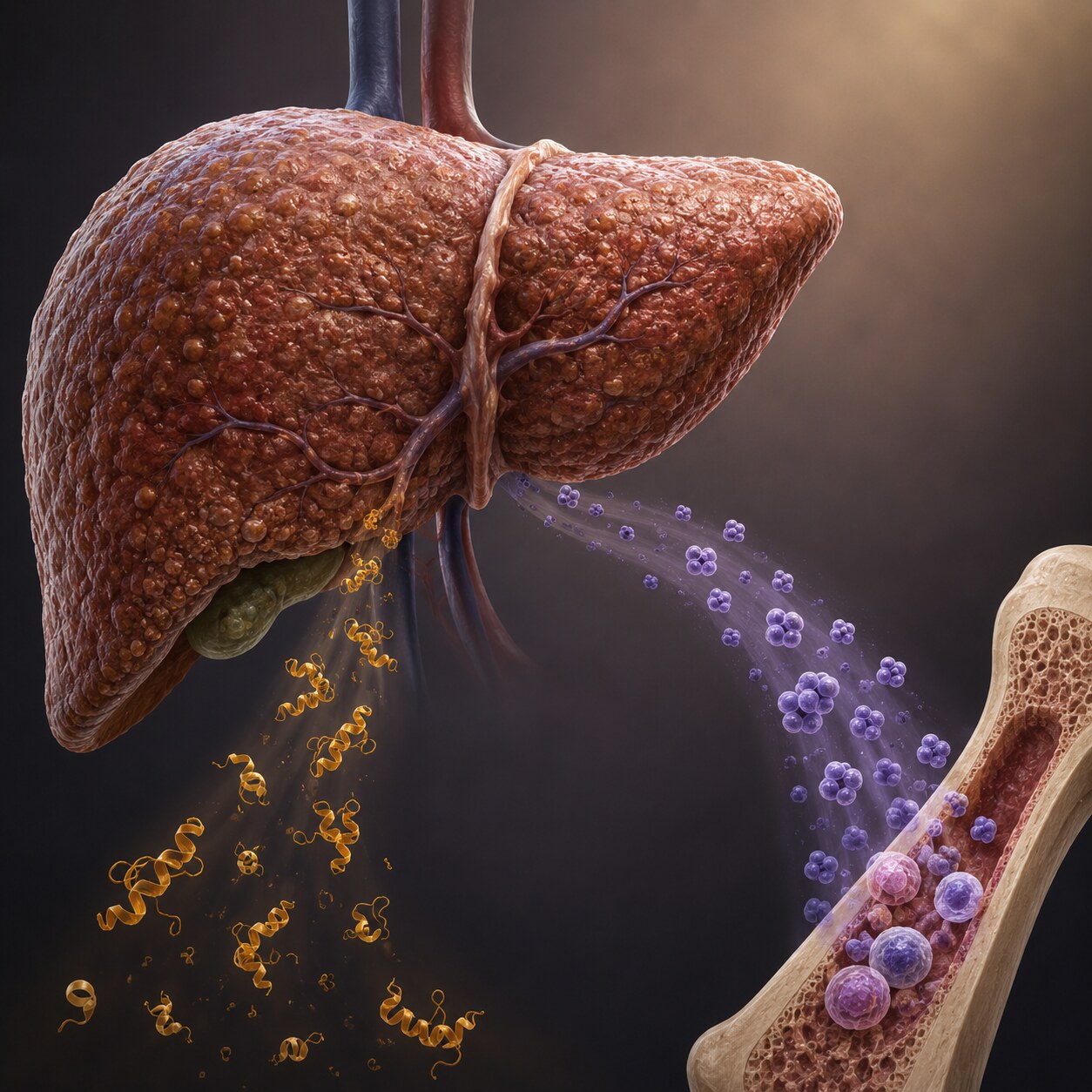
DIC isn't the only path to this lab combination, and it's worth understanding the second one in detail because the underlying mechanism is genuinely different, even though the lab numbers can look strikingly similar. Your liver is the manufacturing site for nearly every clotting factor circulating in your blood, and it's also the primary source of thrombopoietin, a hormone that signals your bone marrow to keep producing platelets at a healthy rate. In advanced liver disease — whether from cirrhosis, acute liver failure, or severe alcohol-related liver damage — the liver simply loses the manufacturing capacity to keep up with both jobs. Clotting factor levels fall because the factory producing them is failing, which shows up directly as a prolonged PT and aPTT. Platelet counts fall for a related but distinct reason: with less thrombopoietin being produced, the bone marrow receives a weaker signal to keep making platelets, and on top of that, advanced liver disease frequently causes the spleen to enlarge and trap a larger share of the platelets that are made, pulling even more of them out of general circulation. The end result on a lab report can look nearly identical to DIC — low platelets, prolonged PT and aPTT — but the mechanism is organ failure rather than a body-wide consumption process, and that distinction meaningfully changes how a doctor investigates and treats it.
There's also a slower, more chronic version of this liver-related pattern that's worth distinguishing from the acute, fast-moving picture of DIC. Someone with well-established cirrhosis often lives for years with platelet counts and clotting times sitting persistently in a mildly-to-moderately abnormal range, without any active emergency happening — their body has simply settled into a new, lower baseline of production that stays relatively stable unless something else, like a new infection or a bleeding episode, disrupts it further. This chronic baseline is part of why the same lab pattern can mean two very different things depending on the timeline: a sudden, sharp drop over hours in someone who was previously healthy is a five-alarm emergency, while a similar-looking but stable, gradual pattern in someone with known long-standing cirrhosis is something their hepatologist is often already tracking as part of routine, ongoing care rather than treating as a new crisis each time labs are drawn.
Reading the Fuller Pattern: What Separates DIC From Liver Disease
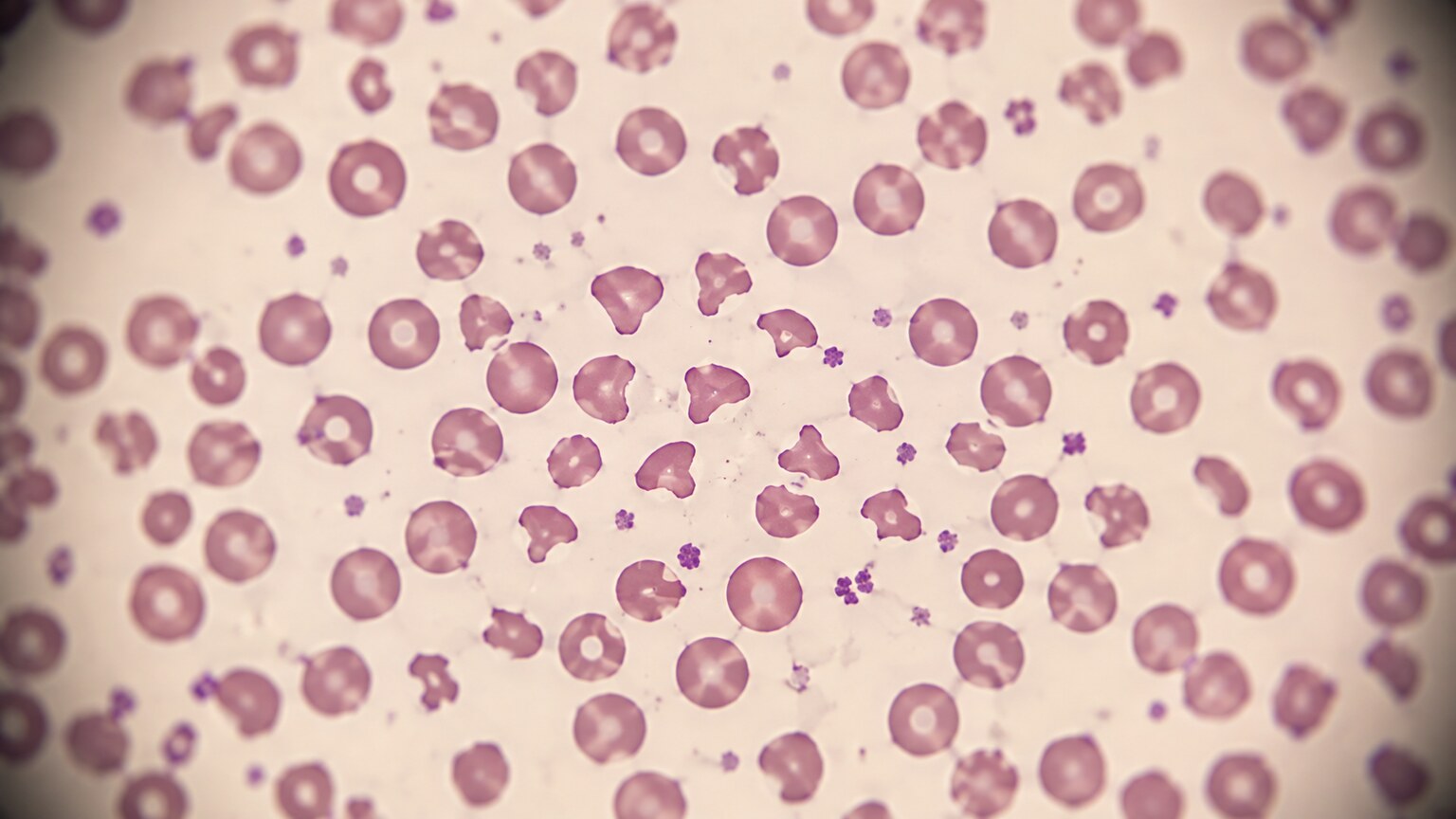
Because both conditions can produce a similar headline pair of abnormal numbers, doctors lean on a broader set of clues to tell them apart. Fibrinogen, a clotting factor that also happens to be one of the raw materials fibrin is built from, tends to be dramatically low in active DIC, since it's being consumed at the site of countless microclots throughout the body — in liver disease, fibrinogen often stays closer to normal for longer, since the liver typically has enough reserve capacity to keep producing at least baseline amounts until disease is quite advanced. D-dimer, a fragment produced specifically when fibrin clots are broken back down by the body, is usually extremely elevated in DIC, reflecting all that clot formation and breakdown happening simultaneously throughout the vasculature — it can be mildly elevated in liver disease too, but rarely to the same striking degree. A blood smear often adds another layer of evidence in DIC: the tiny fibrin strands forming throughout small vessels can physically shred red blood cells as they squeeze past, producing fragmented, jagged cell pieces called schistocytes, a finding that isn't a typical feature of liver disease acting alone. Clinical context matters just as much as any single number — a patient with known cirrhosis and no signs of infection or trauma points toward liver disease, while a patient with a raging infection, recent major trauma, or a serious pregnancy complication points strongly toward DIC.
What This Actually Looks Like in a Real Patient
The clinical picture of this lab combination is genuinely a paradox, and it's part of what makes it so unsettling to witness: a person can show signs of clotting too much and bleeding too much at the exact same time. Microclots forming throughout small vessels in the kidneys, lungs, and brain can quietly starve those organs of blood flow, contributing to organ dysfunction that might show up first as reduced urine output, confusion, or difficulty breathing — often before any bleeding is even visible. At the same time, once platelets and clotting factors are sufficiently depleted, the same patient can start oozing blood from IV insertion sites, surgical wounds, or catheter sites that would normally clot without any issue, along with spontaneous nosebleeds, bleeding gums, and small, flat, reddish-purple spots on the skin called petechiae, caused by tiny amounts of blood leaking from capillaries that no longer have enough platelets to keep them sealed. Seeing both patterns in the same person — organ dysfunction from silent clotting and visible bleeding from depleted reserves — is one of the more striking and important signs that points a care team directly toward DIC rather than a simpler, single-mechanism bleeding or clotting disorder.

These skin findings deserve a closer, more practical look, since they're often the first visible clue a patient or a family member notices before any lab work is even drawn. Petechiae are genuinely tiny, typically no larger than the head of a pin, flat rather than raised, and they don't fade or turn white when you press on them the way a normal skin flush does — that lack of blanching is actually a useful bedside clue that real bleeding under the skin, not just surface redness, is taking place. Larger confluent patches, called purpura, or deep, spreading bruises with no clear injury behind them, represent the same underlying problem playing out on a bigger scale. Because these findings can appear anywhere but tend to show up first on the lower legs, forearms, and areas where clothing or medical equipment creates friction, a sudden, unexplained crop of them in someone who is already acutely ill — septic, recently in a car accident, or shortly after a difficult delivery — is exactly the kind of detail that prompts a clinician to order platelet counts and coagulation studies immediately rather than waiting for a routine panel.
Bleeding from more than one site at once is another practical clue worth knowing, since it's genuinely uncommon outside of this kind of widespread process. A single bleeding gum after brushing too hard, or one bruise from bumping into furniture, has an obvious mechanical explanation and isn't cause for alarm on its own. Simultaneous, unprovoked bleeding from several different places — gums, an old IV site that had already stopped oozing days earlier, the nose, and new bruising all appearing within the same day or two — is a different situation entirely, since it's very difficult to explain through separate, unrelated local causes and points instead toward a body-wide problem with the blood's clotting capacity itself. Family members and bedside caregivers are often the ones who first notice this pattern precisely because they see the patient across the entire day, rather than during a single brief exam, which is part of why care teams take seriously the observations of people who spend the most time at the bedside.
How This Combination Is Actually Treated
Because DIC is always a downstream complication rather than a disease in its own right, the single most important step in treatment is identifying and aggressively treating whatever triggered it in the first place — giving antibiotics for the underlying infection, delivering the baby and addressing the placental complication, or treating the underlying leukemia, since the abnormal clotting and bleeding will generally not resolve until that root cause is brought under control. Alongside that, supportive treatment focuses on replacing what's been consumed, using blood products tailored to what a given patient's labs show they're short on. Management of DIC is genuinely complex and highly individualized — decisions about which products to give, in what amounts, and whether a medication to interrupt clotting is appropriate in a given case are made hour by hour by the treating hospital team based on the full clinical picture, not something that follows a fixed, one-size-fits-all protocol a reader could apply to their own situation.
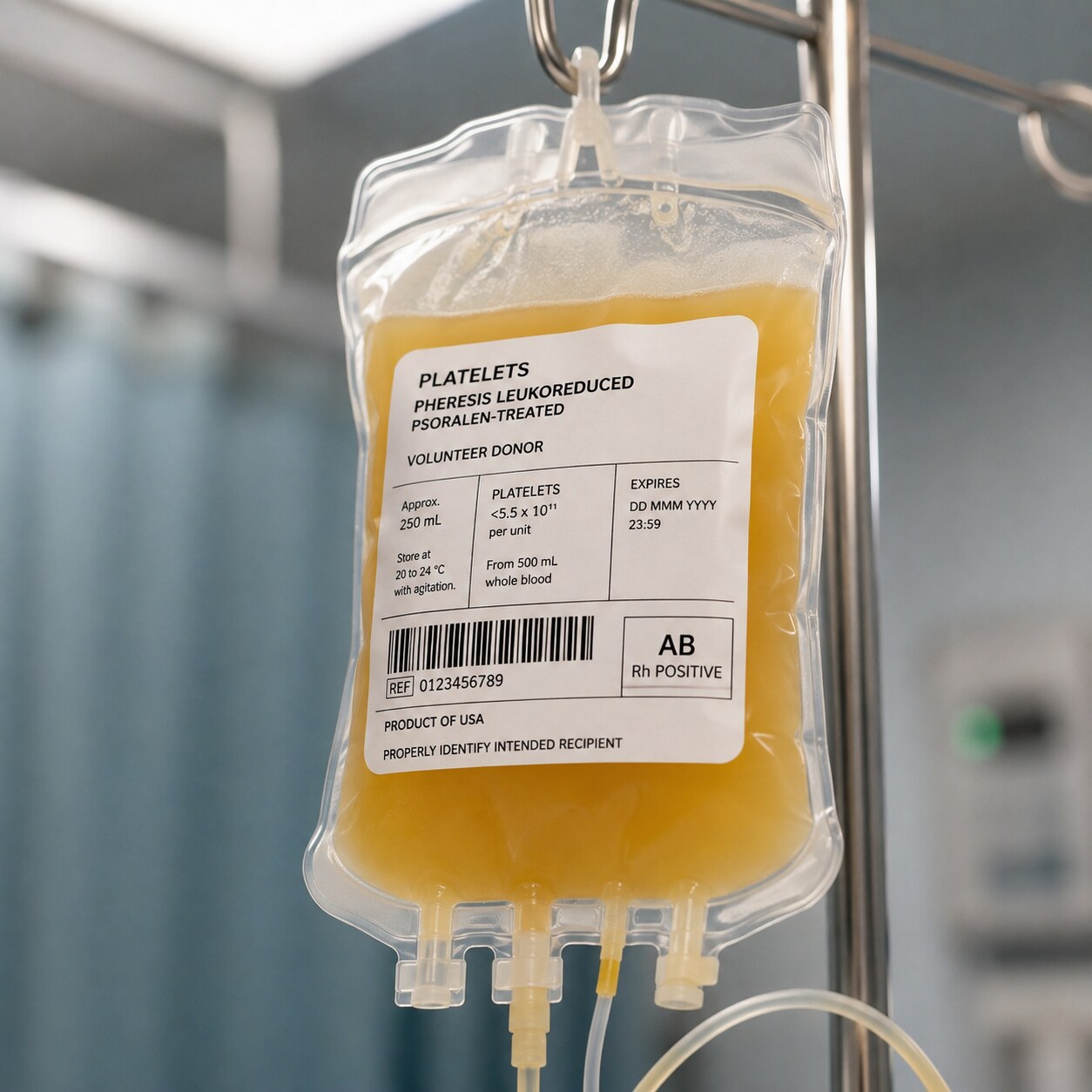
Timing matters enormously in how these replacement products are used, and doctors don't simply transfuse platelets and plasma the moment either number looks low — that decision weighs how actively someone is bleeding, whether a procedure is planned, and how the trigger itself is being treated. This is exactly why DIC is managed by a coordinated hospital team rather than through a single fixed protocol — the right blend of treating the underlying trigger and correcting the blood's clotting capacity shifts hour by hour as new lab results come back, and it is not something that can be reasoned through from outside a hospital setting.
Why Hospitals Treat This Specific Combination as a Medical Emergency
A single abnormal platelet count or a single prolonged clotting time, found in isolation on an otherwise stable person, is often something a doctor can investigate over the course of days with outpatient follow-up. This particular combination, appearing together and especially appearing suddenly in someone who is already critically ill, is treated very differently, because it signals that a large-scale, body-wide process is actively consuming the blood's clotting resources faster than they're being replaced — a process that, left unaddressed, can progress within hours to uncontrolled bleeding, widespread organ damage from microclots, or both simultaneously. Repeat lab testing in this setting typically happens every few hours rather than once a day, precisely because the numbers can shift rapidly as the underlying trigger either responds to treatment or continues to worsen. Recognizing this specific lab pattern quickly, and understanding that it reflects consumption or organ failure rather than two coincidental, unrelated findings, is exactly what allows a care team to move fast enough to actually change the outcome.
Other, Less Common Reasons This Pattern Appears
Beyond DIC and liver disease, a handful of other situations can produce this same combination, and knowing them rounds out the full picture of what a lab team considers. Massive transfusion, where someone receives a very large volume of red blood cells over a short period, most often after severe trauma or major surgical bleeding, can dilute the remaining platelets and clotting factors simply through sheer volume, since older-style red cell transfusions contain very few functional platelets or clotting proteins of their own — this is a mechanical dilution problem rather than true consumption or organ failure, and it's specifically why modern trauma protocols now give plasma and platelets alongside red cells in a balanced ratio, rather than red cells alone. Certain rare, severe forms of preeclampsia, particularly a variant called HELLP syndrome (named for Hemolysis, Elevated Liver enzymes, and Low Platelets), can also produce a related picture during pregnancy, driven by small blood vessel damage and clotting activation that overlaps mechanistically with DIC without being identical to it. Thrombotic microangiopathies, a group of rarer disorders including thrombotic thrombocytopenic purpura, cause small clots to form throughout the body in a way that also consumes platelets, though they typically do so without consuming clotting factors the way DIC does, which is one of the key lab differences that helps separate these conditions from one another.
Why Serial Testing Matters More Than Any Single Result
Because the processes behind this lab pattern move quickly and can either resolve or worsen within hours, a single snapshot in time is rarely enough to fully understand what's happening — clinicians typically rely on trends across repeated testing rather than any one result in isolation. A platelet count and fibrinogen level that are dropping rapidly across consecutive draws, even if the most recent individual numbers aren't yet dramatically abnormal, can be just as concerning as a single very low result, since the trajectory itself reveals an active, ongoing consumption process rather than a one-time abnormality. Several hospitals use a formal scoring system, developed by the International Society on Thrombosis and Haemostasis, that combines platelet count, fibrinogen, D-dimer, and PT into a single numeric score specifically to help standardize the diagnosis of DIC across different patients and different care teams, since relying on gut instinct alone for a pattern this consequential leaves too much room for delay or disagreement. This is also why someone recovering in an intensive care unit with this diagnosis will often have blood drawn far more frequently than a typical hospitalized patient — sometimes every four to six hours during the most unstable period — specifically so the care team can catch a worsening trend before it becomes a crisis rather than after.
Frequently Asked Questions
Can low platelets and abnormal clotting times happen together without it being DIC?
Yes. Advanced liver disease is the other major cause, since the liver produces both clotting factors and the hormone that regulates platelet production. Massive blood loss requiring large-volume transfusion can also dilute both platelets and clotting factors simultaneously without true DIC being present.
Is DIC always caused by a serious underlying condition?
Yes, DIC is never a standalone diagnosis — it's always a complication of something else severe enough to overwhelm the body's normal, localized clotting response, such as sepsis, major trauma, certain cancers, or serious pregnancy complications like placental abruption.
How can a person be at risk of both bleeding and clotting at the same time?
Widespread, inappropriate clotting throughout small blood vessels consumes the platelets and clotting factors the body needs to control bleeding elsewhere. Microclots can silently damage organs like the kidneys and lungs, while the resulting depletion leaves the rest of the body unable to stop bleeding from wounds, IV sites, or the skin.
What test result most strongly points toward DIC rather than liver disease?
A dramatically low fibrinogen combined with a markedly elevated D-dimer, along with schistocytes on a blood smear, points more strongly toward DIC. Liver disease alone tends to preserve fibrinogen longer and produce less extreme D-dimer elevation, though clinical context is always considered alongside the numbers.
Does this combination always require a hospital stay?
In its acute, actively developing form, yes — this lab pattern usually appears in people who are already critically ill, and it requires close monitoring, treatment of the underlying trigger, and often blood product support that can only be managed in a hospital setting.
What Recovery and Follow-Up Usually Look Like
For people who survive the acute phase of DIC, recovery tends to track closely with how the underlying trigger resolves — once the infection clears, the delivery is completed and the placenta is removed, or the leukemia responds to treatment, platelet counts and clotting times generally climb back toward normal over a period of days rather than requiring separate treatment of their own. Doctors typically watch these numbers daily during that recovery window to confirm the trend is genuinely moving in the right direction rather than plateauing at a still-abnormal level, since a platelet count or fibrinogen that stalls partway back to normal can be an early signal that the underlying trigger hasn't been fully controlled yet. For survivors of liver-disease-driven versions of this pattern, recovery depends heavily on how much of the liver's function can be restored — some causes of acute liver failure are reversible with time and supportive care, while chronic cirrhosis-related depletion tends to persist at some baseline level indefinitely, meaning ongoing monitoring becomes a permanent part of that person's care rather than a temporary post-illness checkpoint. In either case, once someone has experienced this pattern, it typically becomes a permanent part of their documented medical history, flagged for any future care team to consider quickly if similar symptoms or lab abnormalities ever reappear.
Conclusion
Low platelets and prolonged clotting times rarely arrive together by coincidence — the combination almost always reflects one of two things happening in the body: a widespread, inappropriate activation of clotting throughout small blood vessels that's using up the blood's clotting supplies faster than they can be replaced, or a failing liver that's lost the capacity to manufacture both platelets and clotting factors in the first place. Understanding which mechanism is at play changes everything about how it's treated, from what's transfused to how urgently the underlying trigger needs to be addressed. This is exactly why this specific lab pairing, more than either finding alone, is treated as a signal to look for something serious happening in the body — and why it moves so quickly from a lab report to an active hospital conversation about what's driving it.
Still Not Sure What Your Results Mean?
Upload your labs and get a complete, visual, plain-language interpretation of every biomarker — delivered to your inbox in under 15 minutes.
Get My ReportThis article is for educational purposes only and does not constitute medical advice. Always consult your healthcare provider regarding your specific lab results.