
Understanding PT and aPTT Can Help Explain Abnormal Bleeding
Prothrombin time (PT) and activated partial thromboplastin time (aPTT) are two of the most fundamental, widely used coagulation tests in all of modern medicine, and together they work by deliberately testing two genuinely separate, largely independent chains of chemical reactions your blood is fully capable of using to form a clot — two distinct chains that ultimately share the exact same ending but begin from two entirely different, unrelated triggers. PT specifically tests what's called the extrinsic pathway, activated by tissue damage from outside the blood vessel, while aPTT tests the intrinsic pathway, activated by damage occurring inside the vessel itself. Because these two separate pathways genuinely rely on entirely different clotting factors along most of their overall length, a problem affecting just one particular pathway while sparing the other one produces a distinctive, recognizable pattern — one test result reading clearly abnormal while the other reads entirely normal — that meaningfully narrows down exactly where within this complex, multi-step chain of chemical reactions something has actually gone wrong, often long before any considerably more specialized testing ever genuinely becomes necessary.

Figure 1. Clotting factor proteins activate one another in a strict sequence, each step triggering the next, until the chain finally converges on forming a stable fibrin clot.
Curious what the rest of your bloodwork is telling you? Upload your results and get a complete, plain-language breakdown in under 15 minutes.
Analyze My ResultsWhat Clotting Factors Actually Are
Before understanding what PT and aPTT measure, it helps to understand what they're actually testing. Blood clotting depends on a series of proteins called clotting factors, numbered I through XIII, most of which circulate in blood in an inactive form until they're needed. When a blood vessel is injured, a specific sequence of these factors activates one after another, each activated factor triggering the next in line, in what's collectively called the coagulation cascade — a chain reaction that amplifies rapidly, since each activated factor can switch on multiple copies of the next factor, ensuring a fast, robust response to injury. This cascade ultimately converges on converting a protein called fibrinogen into fibrin, the fibrous mesh that forms the structural backbone of a stable blood clot.
The cascade isn't actually a single, linear chain — it's traditionally described as two separate starting pathways that eventually merge into one shared final pathway. This two-pathway structure is precisely why two different screening tests exist in the first place: each test is specifically designed to trigger and measure one of the two starting pathways in isolation, allowing clinicians to determine which one, if either, is behaving abnormally.
PT: Testing the Extrinsic Pathway
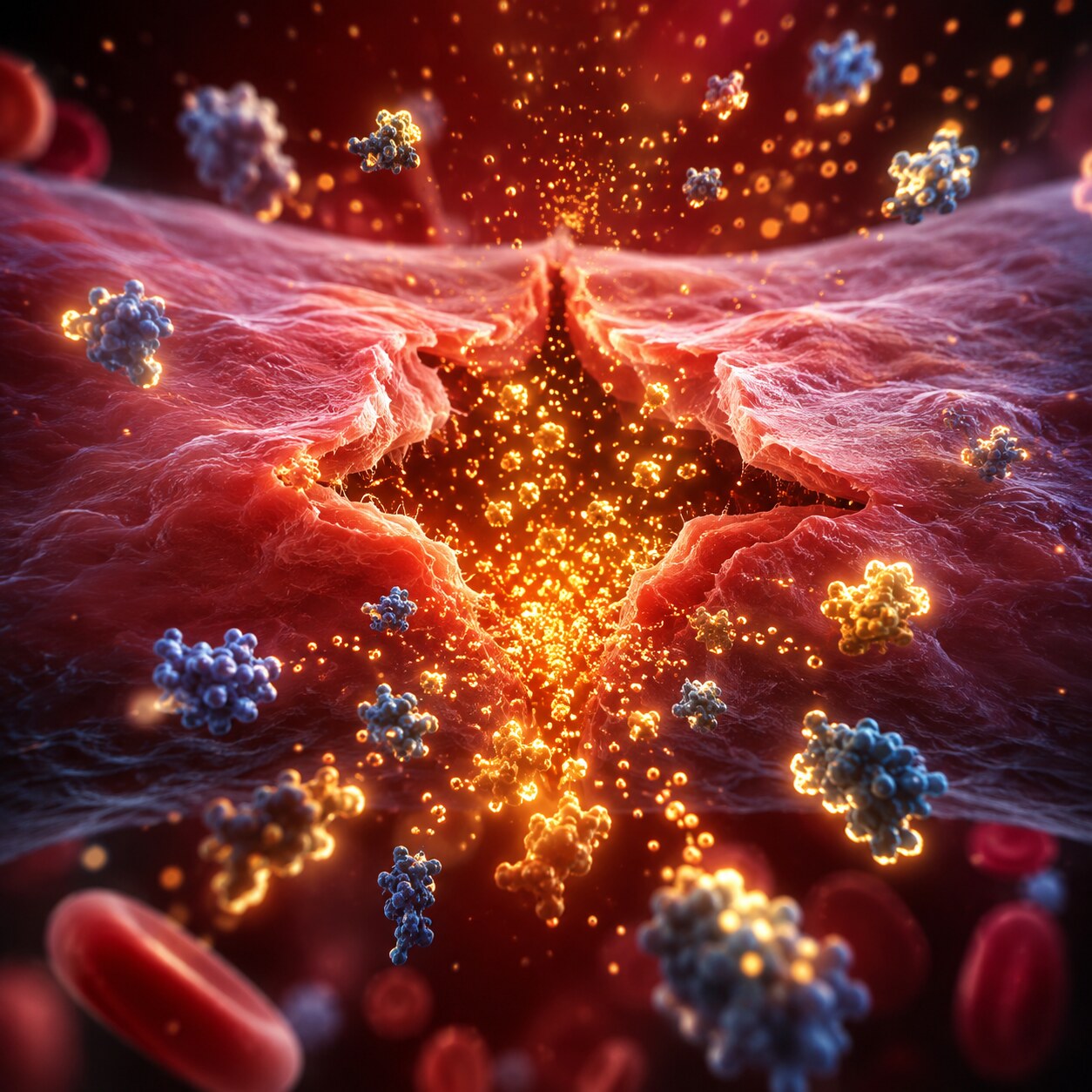
Figure 2. The extrinsic pathway, measured by PT, is triggered by tissue factor released from damaged tissue outside the blood vessel wall.
Prothrombin time measures how long it takes blood plasma to clot after adding a substance called tissue factor (along with calcium, since clotting requires calcium as a cofactor throughout the cascade) directly to the sample in a lab. Tissue factor is the natural trigger for what's called the extrinsic pathway, so named because the original activating signal comes from outside the blood vessel itself — normally released by damaged tissue surrounding an injury, but reproduced deliberately in the lab by adding it directly to the blood sample.
The extrinsic pathway relies specifically on clotting factors VII, along with the shared final pathway factors (X, V, prothrombin, and fibrinogen), meaning PT is most sensitive to deficiencies or dysfunction involving factor VII specifically, plus any problem affecting the shared final common pathway. A prolonged PT — blood taking longer than normal to clot in this specific test — points toward a problem somewhere along this particular route, while a normal PT with an abnormal aPTT points the investigation elsewhere.
aPTT: Testing the Intrinsic Pathway
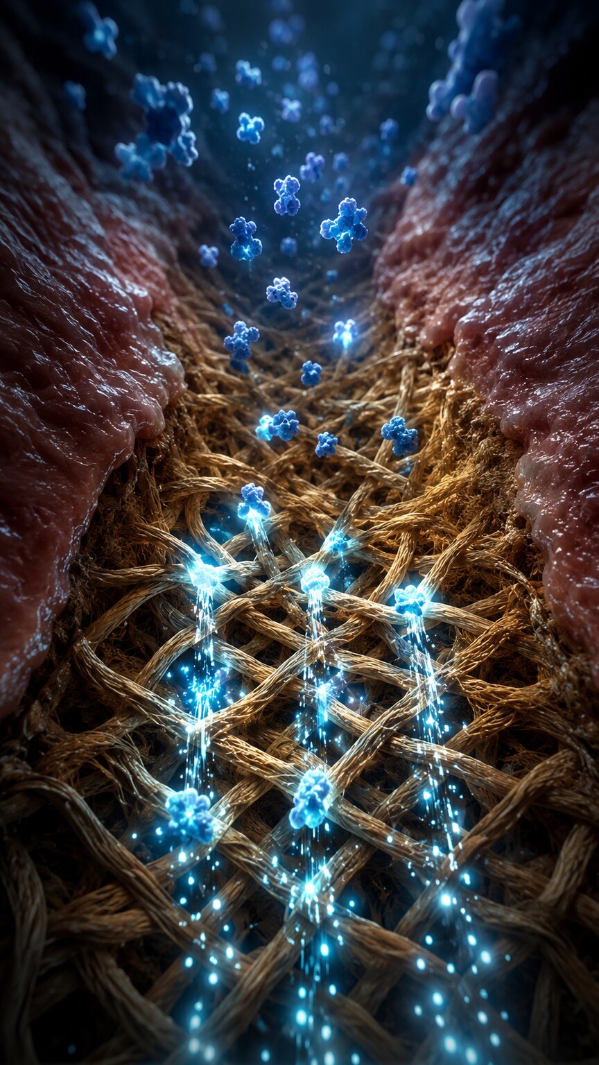
Figure 3. The intrinsic pathway, measured by aPTT, is triggered when blood itself contacts exposed collagen and other surfaces inside a damaged vessel wall.
Activated partial thromboplastin time measures how long it takes plasma to clot after adding substances that activate the intrinsic pathway instead — named because, unlike the extrinsic pathway's outside trigger, every component needed to activate this pathway is already present within the blood itself, triggered when blood contacts a damaged, roughened vessel surface (specifically exposed collagen) rather than requiring an external tissue-derived signal.
The intrinsic pathway relies on a longer chain of factors — XII, XI, IX, and VIII specifically, in addition to the same shared final pathway factors PT also depends on — making aPTT sensitive to deficiencies in any of these earlier-acting factors. This is exactly why aPTT is the test most closely associated with hemophilia, a group of inherited bleeding disorders caused specifically by deficiencies in factor VIII (hemophilia A) or factor IX (hemophilia B), both of which sit within the intrinsic pathway aPTT is designed to detect.
Why Comparing Both Results Together Is the Real Diagnostic Tool
Neither PT nor aPTT is typically ordered or interpreted entirely alone — the real diagnostic power comes from comparing the pattern between the two results together. Four broad patterns are possible, and each points investigation in a genuinely different direction. Both PT and aPTT normal generally suggests the coagulation cascade itself is functioning appropriately, though this doesn't rule out every possible bleeding disorder, since some conditions, discussed further below, don't show up on these two screening tests at all.

Figure 4. Both PT and aPTT are measured the same basic way — a specific activating reagent is added to plasma, and an automated analyzer precisely times how long clot formation takes to begin.
Isolated PT prolongation with normal aPTT points specifically toward factor VII deficiency or dysfunction, or toward early vitamin K deficiency, since factor VII has the shortest half-life of the vitamin K-dependent clotting factors and tends to drop first when vitamin K availability declines. Isolated aPTT prolongation with normal PT points toward the intrinsic-pathway-specific factors — VIII, IX, XI, or XII — making it the pattern most associated with hemophilia and also commonly seen with heparin use, a blood-thinning medication that specifically prolongs aPTT. Both PT and aPTT prolonged together points toward a problem in the shared final common pathway (factors X, V, prothrombin, or fibrinogen), toward significant liver disease (since the liver manufactures most clotting factors), toward more advanced vitamin K deficiency affecting multiple factors at once, or toward warfarin use, a different blood-thinning medication that affects multiple vitamin K-dependent factors simultaneously.
A Brief History of Why Two Separate Pathways Were Discovered
The two-pathway model of coagulation wasn't always the accepted framework — it emerged gradually through decades of laboratory observation in the mid-twentieth century, as researchers noticed that adding different substances to blood plasma produced clotting through what appeared to be genuinely distinct routes. Early researchers observed that clotting triggered by adding tissue-derived material behaved differently, and involved apparently different factors, than clotting triggered simply by exposing blood to a foreign glass surface without any added tissue material at all — an observation that eventually crystallized into the extrinsic-versus-intrinsic pathway framework still taught and used clinically today.
It's worth noting that modern coagulation research has refined this classic two-pathway model considerably, since more recent cell-based models of coagulation have shown that the two pathways aren't as cleanly separate in the living body as the traditional laboratory-based model suggests — tissue factor exposure, for instance, is now understood to also indirectly activate factor XI, blurring the strict separation the classic model describes. Despite this more nuanced modern understanding, the traditional extrinsic-and-intrinsic framework remains genuinely useful and widely taught specifically because it maps directly onto how PT and aPTT are actually performed in the lab, even if the underlying biology in a living person is somewhat more interconnected than the simplified textbook diagram suggests.
How the Test Is Actually Performed in the Lab
Both PT and aPTT are performed on blood plasma, the liquid component of blood left after red and white blood cells have been removed by centrifuging a blood sample collected into a tube containing an anticoagulant (typically sodium citrate, which binds calcium and prevents the sample from clotting prematurely before testing can begin). To run PT, a lab technician adds a reagent containing tissue factor and calcium (calcium being necessary to reverse the anticoagulant's effect and allow clotting to actually begin) to the plasma sample, then precisely times how long it takes for a visible clot to form, typically using an automated optical or mechanical clot-detection instrument rather than manual visual inspection, which was the older, less precise historical method.
aPTT follows a similar basic principle but uses a different reagent combination specifically designed to activate the intrinsic pathway instead — typically a phospholipid substance (the "partial thromboplastin" the test's name refers to) combined with a surface-activating agent and calcium. The word "activated" in the test's full name specifically distinguishes it from an older, less standardized version of the test that lacked this surface-activating component, since adding that activator significantly improved the test's reproducibility and sensitivity compared to earlier methods. Modern coagulation analyzers can run both tests on the same plasma sample within minutes, often as part of the same automated testing run, which is part of why PT and aPTT are so commonly ordered and reported together on the same lab report.
Congenital Versus Acquired: Two Genuinely Distinct, Clinically Meaningful Categories of Coagulation Problems
It's worth drawing a clear distinction between congenital and acquired causes of abnormal PT or aPTT, since this distinction shapes both how a result is interpreted and what happens next. Congenital coagulation disorders, including hemophilia and rarer inherited deficiencies of other specific clotting factors, are present from birth, caused by genetic mutations affecting the body's ability to produce a particular factor in adequate amounts or in a fully functional form. These conditions typically show a consistent, stable pattern on repeated testing over time, and often come with a family history of similar bleeding problems, since many are inherited in recognizable genetic patterns.
Acquired coagulation problems, by contrast, develop later in life due to an external cause — liver disease, vitamin K deficiency, medication effects, or an acquired inhibitor like lupus anticoagulant, all discussed throughout this article — and typically show a pattern that changes over time as the underlying cause develops, resolves, or is treated, rather than the fixed, stable pattern typical of an inherited condition. This distinction is part of why a clinician evaluating an abnormal PT or aPTT considers not just the specific test pattern, but also the timeline: a lifelong, stable abnormality points toward a congenital cause, while a new, evolving abnormality points toward something acquired and potentially treatable at its underlying source.
Why the Liver Plays Such a Genuinely Central, Often Underappreciated Role
Nearly all clotting factors are manufactured in the liver, and this connection is worth appreciating in its own right, since it means a coagulation panel can sometimes offer meaningful insight into liver health well before other, more obviously liver-focused blood tests reveal a problem. Which is exactly why significant liver disease commonly produces prolonged PT and aPTT together, reflecting broadly reduced production across multiple factors rather than a specific, isolated deficiency. PT is often used clinically as an early, sensitive indicator of liver synthetic function specifically because factor VII, which PT depends on most heavily, has such a short half-life — meaning reduced liver production shows up on PT relatively quickly compared to factors with longer half-lives, making PT a useful early warning sign of declining liver function even before more classic liver enzyme tests show dramatic changes.
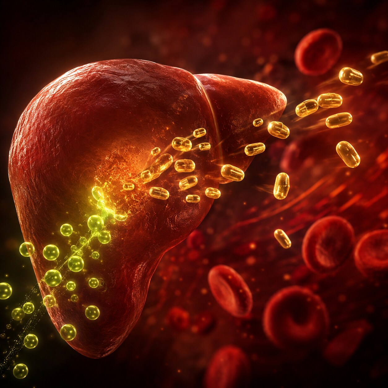
Figure 5. Vitamin K is required for the liver to complete the synthesis of several clotting factors, which is why vitamin K deficiency and liver disease both commonly disrupt PT and aPTT results.
Vitamin K plays a closely related supporting role: several clotting factors, including factor VII, require vitamin K as a cofactor for the liver to complete their synthesis into a functional form. Without adequate vitamin K, the liver still produces these factors, but in an incomplete, non-functional state — meaning vitamin K deficiency produces a coagulation abnormality through a mechanism distinct from outright liver disease, even though both conditions can produce a similar prolonged-PT pattern on testing. This shared vitamin K dependency is also exactly why warfarin, a medication that works by blocking vitamin K's role in this process, is monitored using PT specifically (reported as the INR, a standardized version of PT discussed below), rather than aPTT.
What Happens to These Tests in Newborns and Young Children
PT and aPTT behave differently in newborns compared to adults, a distinction worth understanding for anyone interpreting results from a young infant. Healthy full-term newborns are naturally born with meaningfully lower levels of several vitamin K-dependent clotting factors, including factor VII, largely because vitamin K doesn't cross the placenta efficiently during pregnancy and a newborn's gut hasn't yet developed the bacterial population that helps produce vitamin K later in life. This normal, expected physiological state produces a mildly prolonged PT in many healthy newborns compared to adult reference ranges, which is exactly why pediatric-specific reference ranges exist rather than applying adult cutoffs directly to a newborn's results.
This same vitamin K deficiency is also the reason nearly all newborns receive a vitamin K injection shortly after birth in many healthcare systems, specifically to prevent a rare but serious bleeding complication called vitamin K deficiency bleeding of the newborn, which can otherwise occur precisely because of this normal, temporary post-birth vitamin K gap. Beyond the newborn period, aPTT in particular can also run somewhat longer in healthy young children than in adults, reflecting normal developmental differences in some clotting factor levels that gradually shift toward adult patterns over the first several months to years of life — another example of why age-appropriate reference ranges matter across many different lab values, coagulation testing included.
Why PT Results Are Usually Reported as INR
PT results are frequently reported alongside a related value called the International Normalized Ratio (INR), and understanding why this conversion exists adds useful context. Raw PT values, measured in seconds, vary somewhat between different laboratories depending on the specific reagent used to trigger the test, meaning a PT of, say, 14 seconds could represent a genuinely different degree of clotting delay depending on which lab and which reagent produced that number. The INR was developed specifically to correct for this variability, applying a standardized mathematical adjustment that makes PT results genuinely comparable across different laboratories and different reagent batches, which matters enormously for people on long-term warfarin therapy who need consistent, comparable monitoring over months or years, sometimes across different labs and healthcare systems.
Hemophilia: A Concrete Illustration of an Isolated aPTT Pattern
Hemophilia offers a genuinely useful, concrete illustration of exactly how the PT-versus-aPTT pattern works in real clinical practice, since it's one of the clearest, most well-known examples of a single-pathway defect. Hemophilia A, the more common form, results from a deficiency or dysfunction of factor VIII, while hemophilia B results from a deficiency of factor IX — both factors sitting specifically within the intrinsic pathway, downstream of the initial collagen-contact trigger but upstream of the shared final common pathway. Because factor VII, the extrinsic pathway's defining factor, is completely unaffected by either condition, PT remains entirely normal in both forms of hemophilia, while aPTT becomes significantly prolonged, sometimes dramatically so in severe cases.
This isolated aPTT prolongation with a completely normal PT is such a recognizable, characteristic pattern that it often prompts specific factor VIII and factor IX activity testing even before a formal hemophilia diagnosis is confirmed, precisely because the PT/aPTT pattern itself so strongly narrows the diagnostic possibilities down to this specific category of intrinsic-pathway factor deficiency. Severity in hemophilia correlates fairly closely with how little residual factor activity remains — severe hemophilia, generally defined as less than 1% of normal factor activity, produces spontaneous bleeding into joints and muscles even without significant trauma, while mild hemophilia, with meaningfully higher residual factor activity, may only become apparent after surgery or significant injury, sometimes not diagnosed until adulthood.
How Anticoagulant Medications Deliberately Exploit These Same Pathways
Understanding the extrinsic-versus-intrinsic pathway distinction also explains why different blood-thinning medications are monitored using different tests, since each major anticoagulant class was developed to interfere with a specific point in the cascade this article has been describing throughout. Warfarin works by blocking the liver's ability to use vitamin K in synthesizing several clotting factors, predominantly affecting factor VII (given its short half-life, as discussed earlier) along with factors II, IX, and X — meaning warfarin's effect shows up primarily on PT/INR, since factor VII's rapid depletion dominates the extrinsic-pathway-sensitive PT result well before the intrinsic-pathway factors are meaningfully affected to the same degree.
Heparin, by contrast, works through an entirely different mechanism, dramatically enhancing the activity of a natural anticoagulant protein called antithrombin, which then inhibits several clotting factors, particularly thrombin and factor Xa, both located in or near the shared final common pathway but with effects that register more strongly on aPTT than PT in standard dosing, making aPTT the traditional test used to monitor unfractionated heparin therapy. Newer anticoagulant medications, including direct factor Xa inhibitors and direct thrombin inhibitors, work through yet more targeted mechanisms and often don't require routine PT/aPTT monitoring at all in typical clinical use, though these tests can still provide useful qualitative information in specific situations, including suspected overdose or before urgent surgery in someone taking one of these newer medications.
What PT and aPTT Can't Tell You
Despite their central role in coagulation screening, PT and aPTT have real limitations worth understanding. Both tests specifically measure the plasma-based clotting cascade — they don't directly assess platelet function, even though platelets play an equally essential, earlier role in normal clot formation, meaning a platelet function disorder can cause significant abnormal bleeding while both PT and aPTT remain completely normal. Von Willebrand disease, one of the most common inherited bleeding disorders, frequently produces normal or only mildly abnormal PT and aPTT despite genuinely impairing normal clotting, which is exactly why unexplained bleeding with normal PT and aPTT often prompts additional, more specific testing rather than being dismissed as unremarkable.
Mild factor deficiencies can also sometimes fail to prolong PT or aPTT enough to register as clearly abnormal, since these screening tests generally require a fairly substantial reduction in factor activity, often below roughly 30% to 40% of normal, before a measurable prolongation appears — meaning a person with a mild, partial factor deficiency can show normal screening results despite having some genuine, measurable increase in bleeding risk under specific circumstances like surgery or significant trauma.
What Happens Before Surgery: Screening Versus Predicting Bleeding Risk

Figure 6. PT and aPTT are commonly checked before surgery, though research has increasingly questioned how well these routine screening tests actually predict surgical bleeding risk in people without a personal or family bleeding history.
PT and aPTT are frequently ordered as part of routine preoperative screening before surgery, historically based on the assumption that catching an unsuspected coagulation abnormality beforehand could prevent unexpected surgical bleeding. Research over the past several decades has meaningfully complicated this assumption, however: multiple large studies have found that routine PT and aPTT screening in patients with no personal or family history suggestive of a bleeding disorder rarely identifies a clinically significant abnormality, and even when a mild abnormality is found, it often doesn't reliably predict who will actually experience excessive bleeding during surgery.
This research has led many current surgical and anesthesia guidelines to recommend a more targeted approach: reserving routine preoperative PT and aPTT for patients with a specific bleeding history, a known bleeding disorder, liver disease, or planned use of anticoagulant medication, rather than ordering the tests reflexively for every surgical patient regardless of individual risk factors. A thorough bleeding history — asking about excessive bruising, prolonged bleeding after dental work, or bleeding disorders running in the family — has actually proven more predictive of surgical bleeding risk in several studies than PT and aPTT results in a low-risk patient with no such history, an important, somewhat counterintuitive finding that has gradually reshaped how these tests are used in modern preoperative planning.
Mixing Studies: The Next Step When a Result Is Genuinely Unexplained
When PT or aPTT comes back prolonged without an obvious explanation from medication history or known liver or vitamin K status, one of the most useful next diagnostic steps is a mixing study, a relatively simple but genuinely clever laboratory technique. A mixing study combines the patient's plasma in equal parts with normal plasma from a healthy donor, then repeats the prolonged test on this mixed sample. If the mixed result corrects back to normal, it strongly suggests the original prolongation was caused by a genuine factor deficiency, since the normal plasma supplied enough of the missing factor to correct the deficiency in the mixed sample. If the prolonged result fails to correct even after mixing, it instead suggests the presence of an inhibitor — an antibody or other substance actively interfering with the clotting reaction — since a true inhibitor would continue blocking the reaction even in the presence of adequate normal factor levels supplied by the donor plasma.
This distinction matters enormously for figuring out what to do next: a factor deficiency confirmed by a correcting mixing study leads toward specific factor activity testing to identify exactly which factor is deficient, while a non-correcting result leads toward inhibitor-specific testing, including screening for lupus anticoagulant or specific factor inhibitor antibodies, conditions that carry meaningfully different clinical implications and management approaches than a straightforward factor deficiency.
Disseminated Intravascular Coagulation: When Both Tests Reflect a Systemic Emergency
At the more severe, urgent end of the clinical spectrum, both PT and aPTT play a central role in recognizing disseminated intravascular coagulation (DIC), a serious, life-threatening condition in which the coagulation cascade becomes pathologically activated throughout the entire bloodstream simultaneously, typically triggered by severe infection, major trauma, certain cancers, or serious pregnancy complications. In DIC, widespread, inappropriate clot formation throughout small blood vessels rapidly consumes clotting factors and platelets faster than the body can replace them, eventually leading to a paradoxical state where the same person is at risk of both dangerous clotting and dangerous bleeding simultaneously.
Both PT and aPTT are typically significantly prolonged in DIC, reflecting this rapid, widespread factor consumption, and are almost always interpreted alongside fibrinogen levels (often low, since fibrinogen is heavily consumed) and platelet counts (also typically low) as part of recognizing and staging this condition. DIC represents one of the clearest examples in this entire article of why PT and aPTT are so rarely interpreted in isolation — in a critically ill patient, these values are read as part of an urgent, rapidly evolving clinical picture requiring immediate correlation with the patient's overall condition, not as standalone numbers considered on their own.
Why Reference Ranges Can Differ Between Laboratories
Just as raw PT values vary between labs depending on the reagent used (the specific reason INR was developed, as discussed earlier), aPTT reference ranges also vary somewhat between different laboratories and different reagent formulations, since different commercial aPTT reagents contain different specific phospholipid and activator combinations that produce somewhat different baseline clotting times even in healthy individuals. Unlike PT, however, no equivalent universal standardization system comparable to INR has been widely adopted for aPTT, meaning aPTT results are generally interpreted against the specific reference range printed on that particular lab's own report, rather than compared directly against a result from a different laboratory using different reagents.
This is part of why someone monitoring aPTT over time, particularly during heparin therapy, is generally encouraged to have testing performed at the same laboratory consistently whenever practically possible, minimizing the reagent-related variability that could otherwise complicate interpreting a genuine trend in the underlying clotting status over time.
This same principle explains why a result flagged as abnormal at one facility might read as borderline, or even fall comfortably within range, at a different facility using a different instrument and reagent lot, without either laboratory having made any actual error. It is simply a reflection of how coagulation testing works: the clotting time itself is a relative measurement, calibrated against that specific laboratory's own population of healthy reference individuals, rather than an absolute physical quantity like a body temperature or a body weight that would read identically no matter where it was measured. For this reason, comparing an isolated PT or aPTT value against a number recalled from a previous test at an entirely different clinic, without also comparing the two laboratories' respective reference ranges, can genuinely be misleading, and a treating clinician will typically want to see the actual reference range printed alongside each result before drawing any meaningful conclusion about whether a true change has occurred.
Frequently Asked Questions
Why would a doctor order both PT and aPTT instead of just one?
Because comparing the pattern between the two results is what actually narrows down where a coagulation problem is occurring. A single test alone can't distinguish between the extrinsic and intrinsic pathways, which is why the two are almost always ordered and interpreted together.
Can normal PT and aPTT results rule out a bleeding disorder?
Not completely. These tests don't directly assess platelet function, and conditions like von Willebrand disease or mild factor deficiencies can sometimes produce normal results despite a genuine underlying bleeding tendency, which is why unexplained bleeding symptoms often warrant further evaluation even with normal screening results.
Why is INR used instead of raw PT for monitoring warfarin?
Raw PT values vary between laboratories depending on the specific reagent used, making direct comparisons unreliable. INR applies a standardized adjustment so results are genuinely comparable across different labs and over time, which is essential for consistent long-term warfarin monitoring.
Does a prolonged aPTT always mean I'm on a blood thinner?
No. While heparin therapy is a common cause of prolonged aPTT, it can also reflect factor deficiencies like hemophilia, certain antibodies that interfere with the test, or other underlying conditions, which is why a clinician considers medication history alongside other possible explanations.
Can a prolonged aPTT sometimes reflect an antibody rather than a true bleeding risk?
Yes. Lupus anticoagulant, an antibody that interferes with the aPTT test itself in the lab, prolongs the result without actually increasing bleeding risk in most cases — in fact, it's more commonly associated with an increased clotting risk, an important and somewhat counterintuitive distinction from a genuine factor deficiency.
Why don't PT and aPTT detect von Willebrand disease reliably?
Von Willebrand disease primarily affects platelet adhesion and factor VIII stability rather than the core clotting factor cascade PT and aPTT are designed to measure, which is why specific von Willebrand factor testing, rather than these two screening tests, is typically needed to diagnose it.
How to Talk to a Healthcare Provider About an Abnormal PT or aPTT Result
If a PT or aPTT result comes back outside the normal range, the most useful information to bring into a follow-up conversation isn't just the number itself, but the surrounding context: current medications, including any blood thinners or supplements, any personal or family history of bleeding or clotting problems, known liver disease, recent significant illness, and whether the abnormality was isolated to one test or affected both. This context helps a clinician quickly work through the pattern-based reasoning this entire article has been built around, rather than starting from scratch with the number alone.
It's also reasonable to ask directly whether a mixing study or specific factor activity testing is being considered as a next step, particularly if the abnormality is unexplained by an obvious cause like medication or known liver disease, and whether the result fits a recognizable pattern — isolated PT, isolated aPTT, or both prolonged together — since understanding which category applies can make sense of what further testing, if any, is likely to be recommended.
How These Tests Continue to Evolve
While PT and aPTT have remained the standard first-line coagulation screening tests for decades, research and clinical practice around them continue to evolve in meaningful ways. Point-of-care coagulation testing devices, capable of producing a PT/INR result within minutes from a small fingerstick blood sample rather than a full laboratory venous draw, have expanded access to convenient, more frequent monitoring for people on long-term warfarin therapy, including home self-testing options for appropriately trained patients. Newer, more comprehensive viscoelastic testing methods, including thromboelastography, have also gained traction in certain clinical settings, particularly major trauma and surgery, offering a more complete real-time picture of the entire clotting process — from initial clot formation through clot strength and eventual breakdown — that goes beyond what a single PT or aPTT timepoint alone can capture.
Despite these newer developments, PT and aPTT remain the foundational, most widely available coagulation screening tests worldwide, largely because they're well-validated, inexpensive, fast, and universally understood across virtually every clinical setting — a testament to how a genuinely well-designed diagnostic framework, built around a clear understanding of the two-pathway cascade this entire article has described, can remain clinically essential even as newer, more sophisticated tools continue to be developed around it.
Conclusion
PT and aPTT work by deliberately triggering two separate branches of the coagulation cascade in isolation, and the real diagnostic value comes not from either result alone but from comparing the pattern between them. An isolated abnormality showing up in just one of the two tests points specifically toward the particular factors unique to that one affected pathway, while abnormalities appearing in both tests simultaneously instead point toward the shared final common pathway, underlying liver dysfunction, overall vitamin K status, or certain medications known to affect the broader cascade as a whole. Understanding this two-pathway structure is what genuinely turns two otherwise unfamiliar clotting-time numbers into a truly informative diagnostic tool, one capable of meaningfully narrowing down the actual underlying cause of abnormal bleeding or excessive clotting long before any more specialized, targeted, and considerably more expensive testing ever becomes clinically necessary.
Still Not Sure What Your Results Mean?
Upload your labs and get a complete, visual, plain-language interpretation of every biomarker — delivered to your inbox in under 15 minutes.
Get My ReportThis article is for educational purposes only and does not constitute medical advice. Always consult your healthcare provider regarding your specific lab results.