
Why Is My D-Dimer Elevated?
If your doctor just told you your D-dimer came back elevated, the very first thing worth knowing is this: an abnormal D-dimer, on its own, does not mean you have a blood clot. D-dimer is a small protein fragment your body releases into your bloodstream whenever a blood clot is being broken down, and a positive result simply means your body has been dissolving some kind of clot somewhere, recently. That's a much broader category than "you're currently having a medical emergency." Doctors order this test specifically because it's extremely good at catching every real clot — but that same sensitivity means dozens of everyday things, from a recent surgery to being pregnant to simply getting older, can also push the number up. This guide walks through what D-dimer actually measures, the handful of situations where an elevated result really does need urgent follow-up, and the much longer list of ordinary reasons it climbs without anything dangerous going on.

Curious what the rest of your bloodwork is telling you? Upload your results and get a complete, plain-language breakdown in under 15 minutes.
Analyze My ResultsWhat D-Dimer Actually Is
To understand D-dimer, it helps to understand the two-step process your body runs every time it forms and later removes a blood clot. Step one happens whenever you're injured, even in small, invisible ways inside a blood vessel: your body builds a mesh out of a protein called fibrin, which tangles together to physically plug the injury and stop bleeding, not unlike how a spider quickly patches a torn section of its web. Step two happens once the repair is no longer needed — your body activates an enzyme called plasmin, whose entire job is to dissolve that fibrin mesh and clear it out of the way now that the vessel underneath has healed. As plasmin chops the mesh apart, one particular fragment is left behind: a small piece made of two connected sections of cross-linked fibrin, which is exactly where the name "D-dimer" comes from — a dimer being two similar pieces joined together. That fragment then circulates freely in your bloodstream, and it's precisely what a D-dimer blood test detects and measures. In other words, D-dimer isn't a sign that clotting is actively happening — it's a sign that your body has recently been breaking a clot down, which is a subtle but genuinely important distinction to hold onto as you read through the rest of this.
How the Test Is Actually Performed

Getting a D-dimer result starts with an ordinary blood draw, though the tube itself matters more than it might seem. Your blood is collected into a tube containing sodium citrate, an anticoagulant added specifically to stop the sample from clotting the moment it leaves your vein — which matters enormously here, since a sample that started clotting inside the tube would generate its own D-dimer artificially and throw off the result entirely. Once in the lab, that plasma sample runs through one of a few laboratory methods, most commonly an immunoturbidimetric assay, in which tiny particles coated with antibodies that specifically recognize D-dimer clump together in proportion to how much D-dimer is actually present, and a machine measures how cloudy the sample becomes as a result. Older or slower-turnaround labs sometimes use a related method called an ELISA, which works on a similar antibody-recognition principle but takes longer to process. Either way, the entire test typically takes well under an hour from blood draw to result, which is a big part of why it's so useful in a fast-moving emergency room, where a doctor needs an answer quickly rather than a multi-day wait.
Why This Test Gets Ordered in the First Place
D-dimer testing shows up most often in emergency rooms and urgent care settings, ordered when a doctor is trying to figure out whether someone's symptoms could be caused by a dangerous blood clot. The two conditions doctors are almost always screening for are deep vein thrombosis, or DVT, a clot that forms in one of the large veins deep inside the leg, and pulmonary embolism, or PE, which happens when part of a clot breaks loose and travels to the lungs. These aren't rare events: the CDC estimates that somewhere between 300,000 and 600,000 Americans are diagnosed with a first-time DVT or PE every year, and the risk climbs sharply with age, reaching roughly 1 in 100 people over 80. Because both conditions can be life-threatening if missed, and because their early symptoms — leg swelling, calf pain, chest discomfort, shortness of breath — overlap with dozens of far more common and far less dangerous problems, doctors need a fast, reliable way to figure out who actually needs urgent imaging and who doesn't. That's exactly the role D-dimer plays.

Before ordering it, a clinician will typically work through a structured clinical scoring tool that weighs findings from your symptoms, history, and exam to estimate how likely a clot is before any blood is even drawn. This matters because D-dimer testing is specifically validated for use in people whose clinical picture suggests a lower likelihood of a clot to begin with. When a clinician's overall assessment already points strongly toward a clot, skipping straight to imaging is usually the better path, since a positive D-dimer would be expected regardless and wouldn't change the plan, while even a normal D-dimer isn't considered reliable enough to rule out a clot in that higher-risk scenario. For patients whose clinical picture suggests lower risk, though, a normal D-dimer result is often enough, by itself, to rule out a clot and avoid a CT scan or ultrasound altogether — which is the single biggest reason this test exists: not to find clots, but to safely rule them out in people who probably don't have one. This scoring and interpretation is done by your clinician using the full clinical picture, not something meant for a patient to calculate on their own from symptoms alone.
What Counts as "Elevated"? Understanding Your Number
Most labs consider a D-dimer result under 500 nanograms per milliliter (written as ng/mL, sometimes shown as 0.50 mcg/mL) to be normal, with anything at or above that cutoff flagged as positive or elevated. That number, though, comes with an asterisk that catches a lot of people off guard: D-dimer isn't measured in a single standardized way across every lab. Some labs report results in what's called fibrinogen equivalent units, or FEU, while others use D-dimer units, or DDU — and a result of 500 in one system doesn't mean the same thing as 500 in the other, since roughly 2 FEU units equal 1 DDU unit. A lab using the DDU method might set its own normal cutoff around 230 instead of 500. This is exactly why comparing your result to a number you read online, or to a friend's result from a different hospital, can be genuinely misleading — the only number that actually matters is the reference range printed directly on your own lab report, from your own lab's specific assay.
It's also worth knowing that D-dimer isn't reported the same tidy way something like body temperature is. Different manufacturers' testing equipment can produce modestly different results even when processing the exact same blood sample, which is one more reason doctors interpret it in the context of your specific clinical situation rather than treating it as an absolute, standalone yes-or-no signal.
Why High Sensitivity, Low Specificity Matters
Two words show up constantly in any real discussion of D-dimer: sensitivity and specificity, and understanding them is the key to understanding why one elevated result rarely tells the whole story. Sensitivity describes how good a test is at catching every true case of what it's looking for — D-dimer has very high sensitivity, meaning it's excellent at flagging essentially everyone who actually has a clot, with very few clots slipping through undetected. Specificity describes the opposite quality: how good a test is at staying quiet in people who don't have the condition at all. D-dimer has notably low specificity, meaning plenty of people without any clot whatsoever still trigger a positive result. Picture a smoke detector so sensitive it never misses a real fire, but is also set off by burnt toast, steam from a hot shower, and a bit of kicked-up dust — that's essentially what a D-dimer test is doing inside your bloodstream. This tradeoff is intentional, and considered a feature rather than a flaw, in emergency medicine: when the downside of missing a real clot is potentially fatal, doctors deliberately choose a test that would rather flag ten false alarms than miss a single real emergency. It also explains why a negative D-dimer is so reassuring — a quiet smoke detector really does mean no fire — while a positive one only means "something set it off" and needs a more specific follow-up test, like an ultrasound or CT scan, to identify what that something actually was.
The Two Main Reasons: DVT and PE
When an elevated D-dimer does turn out to reflect a genuine clot, it's almost always one of these two related conditions. Deep vein thrombosis happens when a clot forms inside one of the large, deep veins of the leg, most often in the calf or thigh, frequently linked to long periods of immobility such as a hospital stay, a long-haul flight, recent surgery, or a leg injury that keeps someone off their feet. The telltale signs are usually confined to one leg: swelling that can appear suddenly, pain or tenderness that's often worse when standing or walking, and skin over the area that feels noticeably warmer than the skin around it. Some people with a genuine DVT, though, notice barely any symptoms at all, which is part of why the condition can go unrecognized until it causes a more serious complication.
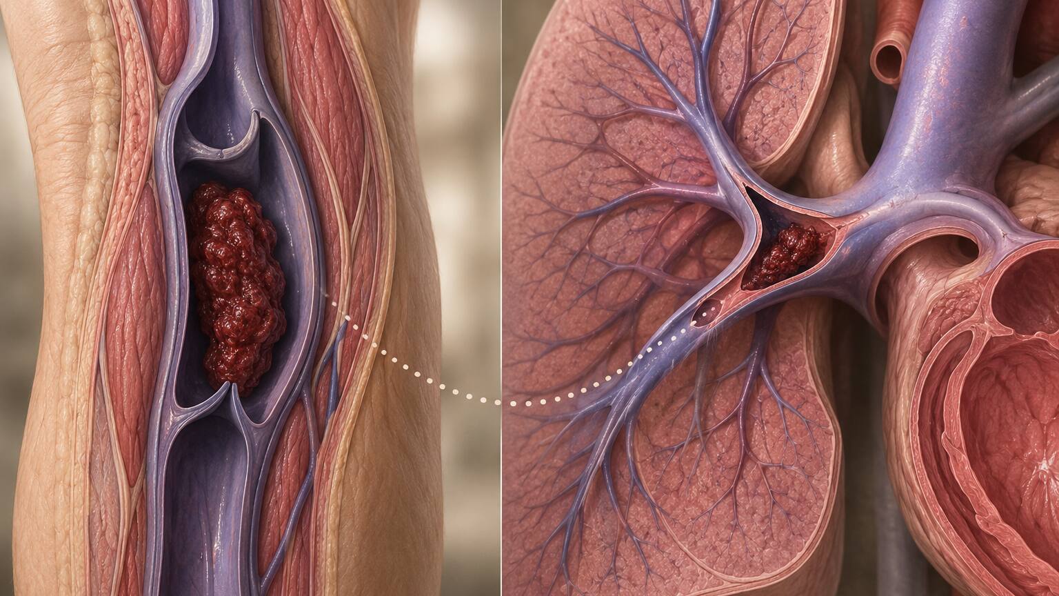
Pulmonary embolism is what happens when part of that clot breaks free, travels through the bloodstream, and lodges in one of the arteries feeding the lungs, blocking blood flow to lung tissue. This is the more urgent of the two conditions, and it's why doctors take any suspected DVT seriously even when it seems mild — treating a DVT early is one of the most effective ways to prevent it from ever becoming a PE. The hallmark symptoms of a pulmonary embolism include sudden shortness of breath, sharp chest pain that often worsens with a deep breath, a fast heartbeat, lightheadedness, and in more severe cases, coughing up blood or fainting. Because a PE can rapidly become life-threatening, any combination of sudden chest pain and breathing difficulty — especially alongside leg swelling — warrants emergency care immediately, rather than waiting to see whether it passes on its own.
Who's Most at Risk for a Real Clot
Certain factors make an actual DVT or PE meaningfully more likely, and doctors weigh these heavily when deciding how seriously to treat an elevated D-dimer. Prolonged immobility is one of the biggest — long-haul flights, hospital stays, and recovery from major surgery all slow blood flow through the leg veins enough to encourage clot formation. Obesity, smoking, and estrogen-containing birth control or hormone therapy each independently raise the odds. A personal or family history of a prior clot matters too, and sometimes points toward an inherited clotting disorder, such as Factor V Leiden, a genetic variant that makes blood more prone to clotting than usual. Active cancer remains one of the strongest risk factors of all, which is part of why doctors interpret an elevated D-dimer differently in someone currently undergoing cancer treatment than in an otherwise healthy young adult. None of these factors alone guarantee a clot is present, but they shift how much weight a doctor puts on an elevated D-dimer result and how quickly they move toward imaging.
Common Non-Clot Reasons Your D-Dimer Could Be High
Here's the reassuring part: outside the emergency room, a genuine blood clot is actually one of the less common explanations for an elevated D-dimer. Because the test reacts to any recent fibrin breakdown happening anywhere in the body, a long list of ordinary, non-dangerous situations can push the number above the cutoff.
Pregnancy, Surgery, and Physical Trauma
Pregnancy is one of the most significant everyday causes of an elevated D-dimer — a woman's level rises steadily throughout pregnancy as part of the normal hormonal and circulatory changes that prepare the body for delivery, and it often stays elevated well into the postpartum period before gradually returning to baseline. This is exactly why D-dimer testing is considered far less useful for ruling out a clot during pregnancy; an elevated result is almost expected, so doctors typically lean more heavily on ultrasound imaging in pregnant patients rather than the blood test alone. Recent surgery and any significant physical trauma, from a car accident to a bad fall, produce a similar effect for a related reason: tissue repair itself runs on the exact same fibrin-formation-and-breakdown cycle the test is built to detect, and levels can realistically stay elevated for several weeks after a major operation or injury, long after any actual bleeding risk has passed.
Infection, Inflammation, and Critical Illness

Infections and inflammatory conditions make up another major category of non-clot causes. Something as ordinary as a routine bacterial infection can nudge D-dimer upward, and more serious systemic illnesses — sepsis, and viral infections including COVID-19 — are well documented to raise it substantially, since the body's inflammatory response activates clotting-related pathways throughout the bloodstream even without a single, discrete clot ever forming. This overlap became especially well known during the COVID-19 pandemic, when doctors noticed that patients with more severe infections tended to have dramatically higher D-dimer levels, and the value was used as one of several markers to help gauge how seriously a patient's body was responding to the virus.
Several longer-term health conditions elevate D-dimer through their own distinct mechanisms, too. Active cancer is a well-established cause, since many tumors directly promote a low-grade, ongoing cycle of clotting and clot breakdown in the blood vessels feeding and surrounding them. Chronic liver disease, heart failure, and autoimmune diseases like lupus can each raise the number as well, through effects on how the body produces clotting proteins or manages ongoing inflammation. Perhaps the single most consistent non-clot contributor, though, is simply age: D-dimer creeps upward gradually over the decades even in people with no illness whatsoever, which is exactly the pattern that led researchers to develop the age-adjusted cutoffs discussed further below. Smoking and estrogen-containing oral contraceptives round out the list of common, everyday contributors worth mentioning to your doctor if your result comes back elevated.
D-Dimer in Critical Illness: Disseminated Intravascular Coagulation
Beyond DVT and PE, there's a third, more serious condition where D-dimer plays an important diagnostic role: disseminated intravascular coagulation, or DIC. DIC is a dangerous, whole-body disorder that typically develops as a complication of another severe underlying illness — sepsis, major trauma, certain cancers, or severe complications of pregnancy among them — in which the body's clotting system becomes paradoxically overactive throughout the bloodstream all at once. Small clots form in blood vessels all over the body simultaneously, which sounds like the opposite of a bleeding problem, but the process consumes clotting factors and platelets so rapidly that the body is often left unable to form clots where it actually needs to, leading to dangerous bleeding at the same time. Because DIC involves such widespread, ongoing clot formation and breakdown, D-dimer levels in this condition are typically dramatically elevated — often far higher than what's seen in an isolated DVT or PE — and the test is one of several lab values, alongside platelet count and clotting time measurements, that doctors use together to diagnose and monitor DIC in critically ill patients. This is generally a hospital-based, intensive-care-level diagnosis rather than something identified from a routine outpatient blood draw, so an extremely high D-dimer in someone who is visibly and seriously unwell carries very different weight than a modestly elevated result in someone who otherwise feels completely fine.
Why Age Changes the Picture
Because D-dimer creeps upward with age even in perfectly healthy people, using the same flat cutoff for a 25-year-old and an 85-year-old creates a real practical problem: older adults end up with positive results — and unnecessary follow-up scans — far more often than younger patients, without actually having more clots. Researchers addressed this with what's called an age-adjusted cutoff, most commonly calculated for patients over 50 as their age multiplied by 10, so a 70-year-old's cutoff becomes 700 ng/mL instead of the standard 500. Studies backing this approach have found that it substantially improves specificity in older adults — cutting down on false alarms — without missing real clots, since sensitivity stays essentially unchanged. In practice, this means a 68-year-old with a D-dimer of 620 might actually be interpreted as a normal, low-risk result once their doctor applies the age-adjusted math, even though that same number would be flagged as elevated using the standard under-50 threshold. This adjustment is only used for patients whose clinician has assessed them as lower pretest probability based on their symptoms and clinical presentation — it isn't applied to anyone whose clinical presentation already looks highly suspicious for a clot.
What Your Doctor Will Likely Do Next
An elevated D-dimer is rarely the final word — it's the detail that determines what happens next, not a diagnosis by itself. If your doctor's clinical assessment suggested a low or intermediate likelihood of a clot to begin with, and your D-dimer comes back elevated, they will typically move straight to imaging that can actually see whether a clot is present. For suspected DVT, that usually means a compression ultrasound of the leg, a painless, non-invasive scan that uses sound waves to check whether the vein compresses normally under light pressure from the ultrasound probe — a vein containing a clot won't compress the way a healthy one does. For suspected PE, the standard next step is a CT pulmonary angiogram, a specialized CT scan using injected contrast dye to directly visualize the arteries feeding the lungs and check for a blockage.

If your clinical risk was already assessed as high before the blood draw, your doctor may skip D-dimer testing altogether and proceed straight to imaging, since a test built to rule out low-probability cases adds little value once a clot already looks likely. If imaging does confirm a DVT or PE, treatment typically begins with an anticoagulant medication, often called a blood thinner, which doesn't dissolve the existing clot directly but prevents it from growing larger while your body's own natural clot-dissolving system gradually breaks it down over the following weeks to months. Depending on the size and location of the clot and your individual risk factors, treatment might last anywhere from a few months to indefinitely, and your doctor may recheck your D-dimer level later in that process, since a normal result after treatment can sometimes help gauge whether it's reasonably safe to eventually stop anticoagulation therapy.
D-Dimer's Role After Treatment: Deciding How Long to Stay on Blood Thinners
For people who've already been diagnosed and treated for a DVT or PE, D-dimer takes on a second, entirely different role months or years down the line: helping decide how long blood thinners should actually continue. This matters because indefinite anticoagulation carries its own real bleeding risks, so doctors don't want to keep someone on it longer than necessary — but stopping too early risks a second clot. Research following patients after they completed at least three months of treatment for a first, unprovoked clot found a meaningful difference in outcomes based on a single D-dimer check taken after stopping: patients with a normal result had roughly a 3.5% annual risk of the clot recurring, while those with a positive result faced a notably higher annual risk, closer to 9%. Testing D-dimer repeatedly over the following months sharpens that prediction even further — one study found that patients whose D-dimer became abnormal again by the third month after stopping and stayed abnormal had a recurrence risk approaching 27% per year, compared to under 3% per year in those whose D-dimer remained consistently normal. Because of findings like these, current clinical practice increasingly uses D-dimer as one input for this exact decision: a persistently elevated result after stopping treatment can be a reason to consider resuming indefinite anticoagulation, while a reassuringly normal one supports the case for stopping blood thinners for good.
When an Elevated D-Dimer Needs Immediate Attention
While most elevated D-dimer results ultimately trace back to something benign, certain symptoms alongside that lab result should never wait for a scheduled follow-up appointment. Sudden swelling, pain, warmth, or redness confined to one leg or arm — especially if it developed over hours rather than days — deserves prompt medical evaluation. Sudden shortness of breath, sharp chest pain that worsens with breathing, a racing heartbeat, coughing up blood, or fainting are signs of a possible pulmonary embolism and represent a genuine medical emergency; these symptoms warrant a call to emergency services or an immediate trip to the nearest emergency room, not a wait-and-see approach. If you're already aware your D-dimer came back elevated and any of these symptoms develop or worsen, tell the emergency team about that result right away, since it directly informs how urgently they'll move toward imaging.
Frequently Asked Questions
Does an elevated D-dimer mean I have a blood clot?
Not necessarily. Because D-dimer is so sensitive, plenty of things besides an actual clot can raise it — recent surgery, pregnancy, infection, and simply growing older among them. An elevated result means your doctor needs more information, usually through imaging, not that a clot has been confirmed.
Why did my D-dimer number look different from what I read online?
D-dimer can be reported in different units (FEU or DDU) with different normal cutoffs depending on the lab and equipment used. A result that looks alarming compared to a number from a different source may actually be completely normal once compared to your own lab's specific reference range.
Can a normal D-dimer completely rule out a blood clot?
In patients with a low or intermediate pretest probability based on their symptoms and risk factors, yes — a normal D-dimer is generally considered reliable enough to rule out DVT or PE without further imaging. It's less reliable as a stand-alone rule-out tool in patients whose clinical presentation already looks high-risk.
Should I be worried if I'm over 60 and my D-dimer is elevated?
Not automatically. D-dimer naturally trends higher with age, which is exactly why many doctors apply an age-adjusted cutoff for patients over 50 rather than using the same threshold used for younger adults. Your doctor can tell you whether your specific result is elevated even after that adjustment.
Will my D-dimer be rechecked after treatment for a clot?
Sometimes. If you're diagnosed with a clot and treated with blood thinners, your doctor may recheck D-dimer months into treatment as one piece of information — alongside your overall clinical picture — when deciding whether it's reasonably safe to eventually stop anticoagulation therapy.
Conclusion
An elevated D-dimer is one of the more anxiety-inducing lab results to receive, largely because of what it's testing for — but the test's own design means a positive result is, on its own, far more often a false alarm than a true emergency. It exists to catch every possible blood clot, even at the cost of flagging plenty of people who turn out to have nothing more serious than a recent surgery, a pregnancy, or simply an older birthday. What actually matters is the full picture: your symptoms, your risk factors, your Wells score, and whatever imaging your doctor orders to look directly at your blood vessels. If you're dealing with an elevated result and you're not experiencing any of the emergency warning signs described above, the most useful next step is usually just following through on whatever imaging or follow-up your doctor recommends, rather than trying to interpret the number in isolation. And if you've already been through that process before, either with a confirmed clot in your past or with a benign explanation your doctor identified, that context matters too — bring it up at your next visit, since your history shapes how any future elevated result gets interpreted.
Still Not Sure What Your Results Mean?
Upload your labs and get a complete, visual, plain-language interpretation of every biomarker — delivered to your inbox in under 15 minutes.
Get My ReportThis article is for educational purposes only and does not constitute medical advice. Always consult your healthcare provider regarding your specific lab results.