
Why Would a Doctor Order a D-Dimer Test?
A D-dimer test almost never comes out of nowhere. Before a doctor writes that order, something specific has usually already happened: a leg has swollen up overnight, a chest has started hurting with every breath, or a patient recovering from surgery has developed symptoms that don't quite add up. Unlike a cholesterol panel or a yearly checkup blood draw, D-dimer is what's called a "rule-out" test — it exists almost entirely to answer one narrow, urgent question: could this person's symptoms be caused by a dangerous blood clot? Understanding exactly which situations prompt a doctor to reach for this particular test, and the structured reasoning they go through before ordering it, can make an unexpected ER visit or urgent-care appointment feel a lot less like a mystery and a lot more like a process you can actually follow along with.

Figure 1. A clinician's physical examination of unilateral calf swelling and tenderness is typically the first step in assessing possible deep vein thrombosis, before any laboratory testing is ordered.
Already have blood work back and want to understand more than just one test? Upload your full results and get a complete, plain-language breakdown in under 15 minutes.
Analyze My ResultsWhat D-Dimer Actually Measures, in Brief
Before getting into when this test gets ordered, it helps to know what it's actually detecting, since that's exactly what shapes when a doctor reaches for it. Whenever your body forms a blood clot — a normal, protective process that happens constantly on a tiny scale, sealing off small injuries inside blood vessels you never even notice — it builds that clot out of a protein called fibrin, which links together into a mesh, not unlike the way threads weave into a patch of fabric. Once the injury underneath has healed and the clot is no longer needed, your body activates an enzyme called plasmin, whose job is to dissolve that fibrin mesh and clear it out of the bloodstream. As plasmin breaks the mesh apart, it leaves behind small leftover fragments made of two connected, cross-linked pieces of fibrin — and that fragment is D-dimer. It circulates freely in your blood afterward, which is exactly what the test picks up.
The key detail that shapes every ordering decision described in this article is this: D-dimer isn't a marker of active clotting happening right now — it's a marker that clot breakdown has happened recently, somewhere in the body. Because clot formation and breakdown is such a universal, whole-body process, tied to healing of all kinds, a huge number of situations beyond a dangerous clot can trigger a positive result. Doctors know this going in, and it directly shapes which patients they think this test is actually useful for.

Figure 2. D-dimer is a small fragment released when the enzyme plasmin breaks down cross-linked fibrin, the protein mesh that forms the structural core of a blood clot.
This also explains why a doctor almost never orders D-dimer as a first-line test the way they might order a basic metabolic panel or a complete blood count during a routine physical. It's ordered reactively, in response to a specific clinical picture that's already raised the question of a clot in the doctor's mind — never as a general screening tool for a person who feels completely fine. The sections below walk through exactly which clinical pictures do that.
One more detail matters here: D-dimer doesn't linger in the bloodstream indefinitely once it's released. It has a relatively short half-life, generally cited as somewhere in the range of six to eight hours, meaning the level a lab measures reflects clot breakdown that's happened recently — typically within the last day or two — rather than something from weeks earlier. This is part of why the timing of the blood draw relative to the onset of symptoms matters to your doctor. A patient whose leg swelling started only a few hours ago, before much fibrin breakdown has had time to occur, could theoretically still have a falsely low D-dimer despite having a real, freshly forming clot — which is one more reason the test is always interpreted together with the clinical picture rather than as a stand-alone number floating in isolation.
The Two Scenarios Behind Most D-Dimer Orders: Suspected DVT and PE
The overwhelming majority of D-dimer tests are ordered because a doctor is trying to rule in or rule out one of two related conditions: deep vein thrombosis (DVT), a clot that forms in one of the large veins deep inside the leg, or pulmonary embolism (PE), which happens when part of that clot breaks free and travels to the lungs. Roughly 300,000 to 600,000 Americans are diagnosed with a first-time DVT or PE every year according to the CDC, and both conditions can escalate quickly if missed — which is exactly why doctors take the symptoms below seriously enough to reach for a rule-out test rather than waiting to see if things improve.
What Prompts a DVT Work-Up
A doctor starts considering DVT the moment a patient describes one leg — almost always just one, since a clot forming in both legs simultaneously is unusual — that has become noticeably swollen, often within a day or two rather than gradually over weeks. That swelling is usually accompanied by a dull, aching pain or tenderness that tends to worsen with standing or walking, and the skin over the affected area frequently feels warmer to the touch than the skin on the opposite leg. Redness along the vein itself is common too. What tips a doctor off that this might specifically be a clot, rather than a muscle strain or simple swelling, is often the context surrounding it: a recent long flight or car ride, a hospital stay, recent surgery, a leg injury that kept someone off their feet, or simply being an older adult with reduced mobility. Any one of these details in the history, paired with the physical findings, is usually enough to move a doctor toward ordering D-dimer or imaging.
What Prompts a PE Work-Up
Pulmonary embolism tends to present far more dramatically, and it's this presentation that most often sends someone straight to an emergency room rather than a scheduled doctor's visit. The hallmark combination is sudden shortness of breath that comes on over minutes to hours, not days, often paired with sharp chest pain that noticeably worsens when taking a deep breath — a pattern doctors call pleuritic pain. A racing heartbeat, lightheadedness, and a sense of anxiety or impending doom are common accompanying symptoms, and in more severe cases, a patient might cough up blood-streaked sputum or actually faint. Because a PE can rapidly become life-threatening if a large enough clot blocks blood flow to the lungs, this particular combination of symptoms is one of the fastest tracks to an urgent work-up in any emergency department, and D-dimer typically enters the picture within the first hour of that visit.
What makes PE especially tricky to catch is how often it mimics other, far more common conditions. A racing heart and shortness of breath can look like a panic attack. Sharp chest pain worsened by breathing can look like a pulled muscle or costochondritis, an inflammation of the cartilage connecting the ribs to the breastbone. Even a low-grade fever sometimes accompanies a PE, muddying the picture further by suggesting a respiratory infection instead. This overlap is exactly why emergency physicians are trained to specifically ask about risk factors — a recent flight, a recent surgery, a family history of clotting problems, current use of estrogen-containing birth control — during what might otherwise look like a routine chest-pain evaluation. Those few extra questions are often what tips the scale toward ordering D-dimer rather than treating the visit as a straightforward case of anxiety or muscle strain.

Figure 3. Sudden shortness of breath and pleuritic chest pain are the hallmark symptoms that prompt an emergency evaluation for pulmonary embolism.
What a doctor is doing in both of these scenarios, before a single tube of blood is drawn, is building a clinical picture — piecing together your symptoms, your recent history, your risk factors, and your physical exam findings into an overall impression of how likely a real clot actually is. That impression, formally called pretest probability, is the single biggest factor determining whether D-dimer gets ordered at all, and it's worth understanding in detail, because it explains a lot about why some people with leg pain get a blood draw and others get sent straight for an ultrasound instead.
How Doctors Decide Who Actually Needs the Test
D-dimer is only useful as a rule-out tool in patients who are already thought to have a lower-to-moderate chance of having a real clot. That distinction matters enormously, and it's why doctors don't simply order the test on every single person who walks in with leg swelling or chest discomfort. Instead, most clinicians run through a standardized scoring tool first — most commonly the Wells score for suspected DVT or PE, sometimes the Revised Geneva score as an alternative for PE — to put a number on how strongly the clinical picture points toward a clot before deciding what to do next.
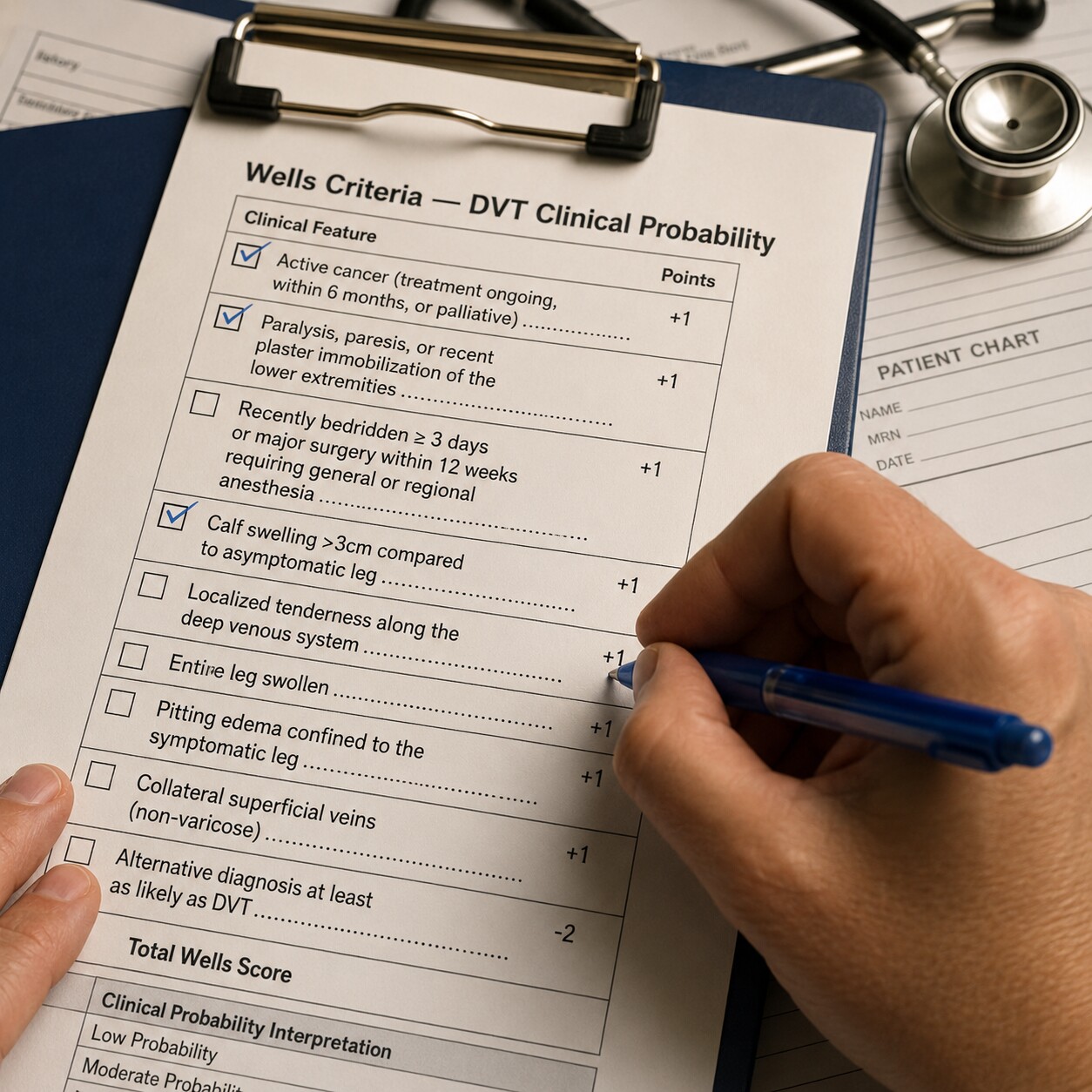
Figure 4. The Wells score assigns points to specific clinical findings — such as calf swelling over 3 centimeters or recent immobilization — to estimate the pretest probability of a deep vein thrombosis before D-dimer testing is ordered.
The Wells DVT score works by assigning one point each to a defined list of findings: active cancer, recent leg paralysis or immobilization in a cast, being bedridden for more than three days or having had major surgery within the past twelve weeks, tenderness along the deep veins, swelling of the entire leg, calf swelling more than 3 centimeters greater than the unaffected side, visible collateral superficial veins, and a previously documented DVT. A prior alternative diagnosis that's at least as likely as DVT subtracts two points. The points add up to a category — low, moderate, or high probability — and that category is what actually determines the next step. A patient scoring in the low-probability range is exactly the person D-dimer testing was designed for: if the result comes back normal, that's typically considered enough, on its own, to rule out DVT without any imaging at all. A patient who scores high-probability, on the other hand, usually skips D-dimer entirely and goes straight to an ultrasound, since even a normal D-dimer result wouldn't be trusted enough to override that level of clinical suspicion.
For suspected PE, a similar logic applies using either the Wells PE criteria or the Revised Geneva score, which weighs items like heart rate, recent surgery or fracture, prior clot history, coughing up blood, and active cancer. The Wells PE version, specifically, tallies points for clinical signs of a DVT, a heart rate above 100 beats per minute, immobilization or surgery in the past four weeks, a prior history of DVT or PE, coughing up blood, active cancer, and whether an alternative diagnosis seems less likely than PE — with the highest single weight given to the item asking whether PE is, in the clinician's judgment, the most likely diagnosis of all. Just as with DVT, the resulting probability category — not the patient's symptoms alone — is what determines whether D-dimer gets ordered as a rule-out step or whether the doctor moves directly to a CT pulmonary angiogram. This scoring process is something your clinician does using the full picture in front of them; it isn't meant to be self-calculated from symptoms at home, since several of the criteria depend on exam findings and clinical judgment a patient can't assess on their own.
This two-step approach — score first, then decide whether D-dimer adds value — is also why the test's reputation as "highly sensitive but not very specific" matters so much to how it gets used. Sensitivity means the test rarely misses a true clot; specificity means it also frequently flags people who don't have one. Ordering it indiscriminately, on every patient regardless of pretest probability, would generate huge numbers of false alarms and unnecessary follow-up scans. Reserving it for patients already screened into the lower-probability group is what keeps the test genuinely useful instead of just noisy.
A Simple Way to Picture Sensitivity and Specificity
These two words trip up almost everyone the first time a doctor uses them, so it helps to have a concrete picture in mind. Imagine an extremely sensitive smoke detector — one engineered so it will never, under any circumstance, fail to sound the alarm during an actual fire. That same sensitivity means it also goes off from a bit of steam in the bathroom, from smoke drifting in from a neighbor's grill, or from slightly burnt toast. D-dimer behaves almost exactly like that detector: doctors have deliberately chosen a test that would rather sound the alarm ten times unnecessarily than stay silent once during a genuine emergency, because the cost of missing a real DVT or PE — potentially fatal — is so much higher than the cost of ordering one extra ultrasound that turns out normal. This is precisely why a positive D-dimer, on its own, carries so much less weight than a negative one. A quiet detector really does mean no fire. A sounding one just means something set it off, and it takes a second, more specific tool — an ultrasound, a CT scan — to figure out what that something actually was.
Beyond DVT and PE: Other Situations That Lead to a D-Dimer Order
While suspected DVT and PE account for the large majority of D-dimer orders, several other clinical situations lead doctors to reach for this same test, each for a different reason.
Monitoring Critically Ill Patients for DIC

Figure 5. In critically ill patients, serial D-dimer measurements help clinicians track disseminated intravascular coagulation alongside platelet counts and clotting times.
In an intensive care setting, D-dimer gets ordered for an entirely different reason than in the ER: not to catch a single, discrete clot, but to help monitor a whole-body clotting disorder called disseminated intravascular coagulation, or DIC. DIC typically develops as a complication of another severe illness — sepsis, major trauma, certain cancers, or severe pregnancy complications among them — and it causes the clotting system to become paradoxically overactive throughout the entire bloodstream at once, forming small clots everywhere while simultaneously using up the clotting factors and platelets the body needs to stop bleeding elsewhere. Because DIC involves constant, widespread clot formation and breakdown, D-dimer levels run dramatically high, and doctors order it repeatedly, alongside platelet counts and clotting-time measurements, to track how a critically ill patient's condition is evolving day to day. This is a hospital-based, ICU-level use of the test, entirely separate from the single-draw rule-out testing described above.
Because DIC is a moving target rather than a single yes-or-no diagnosis, a single D-dimer reading in this setting doesn't carry much meaning on its own — what a critical care team is actually watching is the trend across repeated draws, often several per day during the sickest stretch of an ICU admission. A D-dimer that keeps climbing alongside a falling platelet count paints a very different picture than one that's already sky-high but holding steady, and that distinction can shape decisions about blood product transfusions, treatment of the underlying illness driving the DIC, and how aggressively the care team needs to intervene. This is one of the clearest examples of how the same test can be ordered for almost opposite reasons depending on the setting — a single, decisive rule-out draw in the ER versus a repeated, trend-watching tool at the bedside in the ICU.
Pregnancy Complications
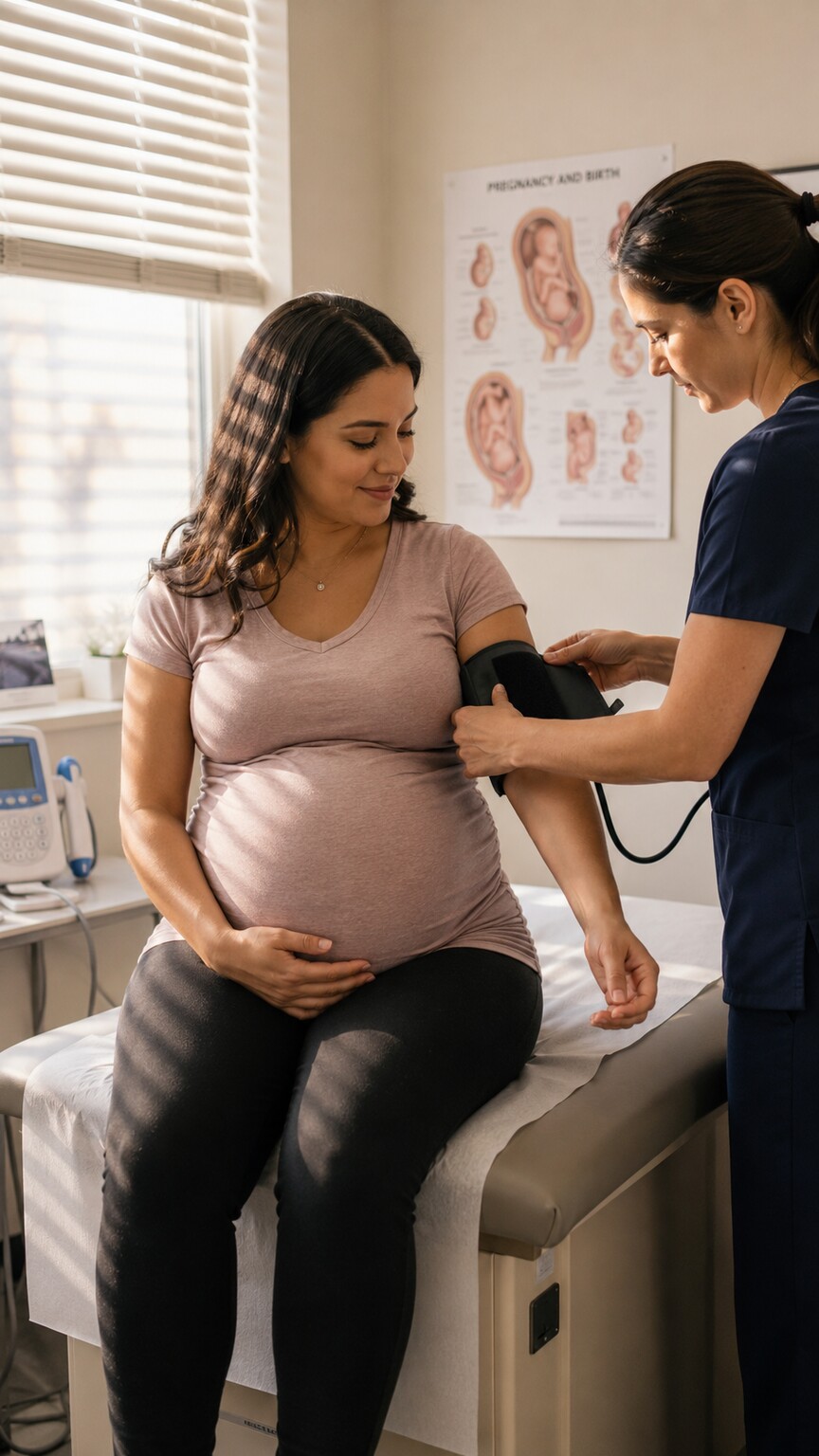
Figure 6. Because normal pregnancy already elevates D-dimer, doctors weigh the test differently when evaluating a pregnant patient for a possible clot or a pregnancy-related clotting complication.
Pregnancy itself substantially raises the risk of blood clots — the combination of increased clotting factors, reduced blood flow from an enlarging uterus pressing on pelvic veins, and reduced mobility late in pregnancy all push clot risk higher, which is exactly why a pregnant patient describing leg swelling or shortness of breath gets taken seriously. The complicating factor is that a pregnant woman's baseline D-dimer level rises steadily through pregnancy as a completely normal physiological change, and can stay elevated well into the weeks after delivery. Because of this, D-dimer alone is considered a much less reliable rule-out tool during pregnancy, and a doctor evaluating a pregnant patient for a suspected clot typically leans more heavily on ultrasound imaging. Still, D-dimer sometimes gets ordered here as one data point among several, and it plays a distinct role in the work-up of preeclampsia and its more severe form, HELLP syndrome, where abnormal clotting activity can be part of the broader picture a doctor is trying to piece together alongside blood pressure, liver enzymes, and platelet counts.
Patients With Active Cancer and Unexplained Symptoms
Cancer is one of the strongest known risk factors for blood clots, since many tumors directly promote a low-grade, ongoing cycle of clot formation and breakdown in the vessels feeding and surrounding them. Because of this, a doctor treating a patient with active cancer tends to have a much lower threshold for ordering D-dimer when that patient reports new leg swelling, unexplained shortness of breath, or vague chest discomfort — symptoms that might be dismissed as unremarkable in an otherwise healthy young adult carry more weight in this context. The reasoning works somewhat in reverse here too: because cancer itself often elevates D-dimer independent of any new clot, doctors interpret a positive result in a cancer patient more cautiously, often moving toward imaging regardless of the number, rather than relying on D-dimer as a stand-alone decision point the way they might in a low-risk younger patient.
Deciding Whether to Stop Long-Term Blood Thinners
For patients who've already been treated for a DVT or PE in the past and have been on anticoagulation medication for months or years, D-dimer resurfaces in a completely different role: helping decide whether it's reasonably safe to eventually stop. Because staying on blood thinners indefinitely carries its own real bleeding risks, doctors don't want to continue them longer than necessary, but stopping prematurely risks a second clot. Research following patients after they'd completed treatment for a first, unprovoked clot found that a normal D-dimer checked after stopping anticoagulation was associated with a meaningfully lower annual risk of the clot returning compared to a positive result. Because of findings like this, a doctor managing a patient's long-term anticoagulation plan may order D-dimer specifically at the point of considering discontinuation, using the result as one input, alongside the patient's overall history and risk factors, in that decision.
Unexplained Symptoms After Surgery
Recovery from any major surgery already causes a temporary, expected rise in D-dimer, since tissue repair itself runs on the same fibrin-formation-and-breakdown cycle the test detects — so a doctor generally wouldn't order it just because someone recently had an operation. What changes the calculation is a specific new symptom during recovery: sudden calf swelling that looks disproportionate to the expected post-surgical swelling, new shortness of breath, or chest pain in the days or weeks following a procedure. Because major surgery is itself one of the strongest risk factors for DVT and PE — reduced mobility during recovery slows blood flow through the leg veins — a doctor evaluating a post-surgical patient with these new symptoms is often working through the same Wells-score-based reasoning described earlier, just with a patient whose baseline risk is already elevated by the recent operation itself.
Orthopedic procedures in particular — hip replacements, knee replacements, and major spinal surgery — carry some of the highest post-operative clot risk of any surgical category, which is why patients recovering from these procedures are often placed on preventive blood thinners for weeks after discharge, precisely to avoid ever reaching the point where a doctor needs to order D-dimer for a new symptom in the first place. When prevention doesn't fully eliminate the risk and a concerning symptom shows up anyway, the same evaluation process described throughout this article — history, exam, a probability score, and then D-dimer or imaging depending on where that score lands — applies just as it would in any other patient, regardless of how recently they left the operating room.
What Actually Happens Once the Test Is Ordered
Once a doctor decides D-dimer testing is the right next step, the process itself is quick and low-key compared to the reasoning that led up to it. A phlebotomist or nurse draws blood from a vein, typically in the arm, into a tube containing sodium citrate, an anticoagulant added specifically to keep the sample itself from clotting inside the tube before it ever reaches the lab — a detail that matters, since a sample that started clotting on its own would generate artificial D-dimer and throw off the result. That tube is then spun down and processed, most often using an immunoturbidimetric assay, in which antibody-coated particles clump together in proportion to how much D-dimer is present, with a machine measuring how cloudy the resulting sample becomes.
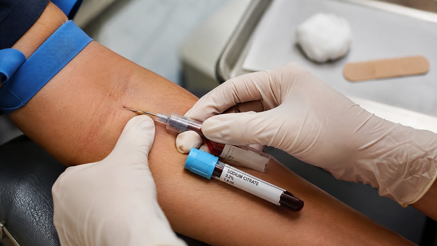
Figure 7. D-dimer testing requires blood collected into a sodium citrate tube, which prevents the sample from clotting before it reaches the laboratory.
What makes D-dimer especially practical in an emergency setting is speed: results are typically available well under an hour after the blood draw, which is a large part of why it's become such a routine part of ER work-ups for suspected clots rather than something that requires an overnight wait. Many hospitals also have access to point-of-care D-dimer analyzers — smaller, faster devices kept right in the emergency department rather than in the central lab — which can shorten that turnaround even further in a busy ER where every extra minute matters for a patient in real discomfort. A doctor can often have your result back, discuss it with you, and either send you home or move you toward imaging, all within the same visit.
There's no special preparation required on your end. You don't need to fast beforehand, and there's no need to stop any regular medications unless your doctor specifically tells you otherwise — in fact, if you're already taking a blood thinner, it's worth mentioning that clearly, since anticoagulant therapy can itself lower D-dimer levels and is something your doctor will factor into how they interpret the result. The blood draw itself typically takes less than a couple of minutes, and most people feel nothing more than the brief pinch of the needle, similar to any other routine blood test.
Why This Test Isn't Ordered for Every Ache and Pain
Given how common leg pain, calf cramps, and occasional shortness of breath are in everyday life, it's fair to wonder why every patient describing these symptoms doesn't just get a D-dimer test as a precaution. The honest answer is that doing so would actually make care worse, not better. Because the test flags so many non-clot causes — infection, inflammation, recent injury, older age, pregnancy, and dozens of other ordinary conditions — testing everyone regardless of pretest probability would generate large numbers of false positives, and each one of those typically leads to a follow-up ultrasound or CT scan that the patient didn't actually need. That means real costs, real radiation exposure in the case of CT imaging, real anxiety while waiting on results, and real strain on imaging departments that could otherwise be seeing patients who genuinely need urgent scans.
This is exactly the reasoning behind the structured, score-first approach described earlier: it isn't bureaucratic caution, it's what keeps a highly sensitive test from becoming a source of unnecessary alarm. A doctor who examines a patient with mild, several-week-old calf soreness and no other risk factors, and decides D-dimer testing isn't warranted, isn't dismissing the complaint — they're applying the same evidence-based reasoning that determines when the test genuinely adds value versus when it would only add noise.
This philosophy shows up across medicine under the banner of "Choosing Wisely," an initiative built around the idea that more testing isn't automatically better care, and that unnecessary tests carry their own downstream harms even when the test itself is simple and low-risk. In the specific case of D-dimer, professional emergency medicine societies have explicitly recommended against ordering it in patients with a low pretest probability of PE who also meet a separate checklist called the PERC rule — eight criteria including age under 50, a normal heart rate, and no recent surgery or hormone use — since patients who clear that checklist have such a low baseline risk that even the small number of false positives D-dimer would generate isn't worth the downstream imaging it triggers. In other words, sometimes the most evidence-based decision a doctor can make is not ordering the test at all, and that decision is every bit as deliberate as the decision to order it.
What the Result Can — and Can't — Tell Your Doctor
It's worth setting expectations correctly going in: a D-dimer result is never the final word on its own, in either direction. A normal result in a patient already assessed as low-probability is genuinely reassuring and, per current clinical guidelines, is typically enough by itself to rule out DVT or PE without further imaging. An elevated result, on the other hand, doesn't confirm a clot — it simply means more investigation, usually imaging, is warranted to find out what actually caused it. If you want a deeper breakdown of what a positive result specifically means and the long list of non-clot reasons it can happen, that's covered in detail in our companion article on elevated D-dimer results. The test's entire value lies in what it does well: helping a doctor decide, quickly and reliably, who can safely skip a scan and who needs one right away.
Frequently Asked Questions
Can I request a D-dimer test myself if I'm worried about a clot?
You can certainly raise the concern with your doctor, but whether the test is actually useful in your situation depends on your pretest probability, which your doctor determines through your symptoms, history, and a physical exam. If your clinical picture already looks high-risk, your doctor may recommend skipping straight to imaging instead, since D-dimer wouldn't be considered reliable enough to rule anything out in that scenario.
Does every patient with chest pain get a D-dimer test?
No. Chest pain has many causes besides pulmonary embolism, including cardiac, musculoskeletal, and gastrointestinal ones. A doctor uses your specific symptom pattern, exam findings, and risk factors — often formalized through the Wells or Geneva scoring criteria — to decide whether a clot is a strong enough possibility to warrant D-dimer testing or other clot-specific work-up.
Why did the ER order both a D-dimer and imaging at the same time?
This sometimes happens when a patient's pretest probability falls in a moderate or higher range, where a clinician isn't confident a normal D-dimer alone would be reliable enough to rule out a clot, or when the clinical picture is urgent enough that waiting for one result before ordering the next isn't appropriate given how quickly a real clot could become dangerous.
Is D-dimer testing part of a routine annual physical?
No. It isn't a general screening test and isn't included in standard preventive bloodwork. It's ordered specifically in response to symptoms or a clinical situation that raises concern for an active clot, ongoing clotting disorder, or a decision about stopping long-term anticoagulation — never as a baseline check in someone without a specific reason to test.
If my D-dimer test comes back normal, am I completely in the clear?
In a patient your doctor has already assessed as low or intermediate probability, a normal D-dimer is generally considered reliable enough to rule out DVT or PE without further imaging. It's a different story in a high-probability patient, where a normal result alone wouldn't be trusted to override strong clinical suspicion, and further testing would still be pursued.
Conclusion
A D-dimer order is rarely arbitrary — it's the product of a doctor weighing your specific symptoms, your history, and a structured probability assessment before deciding a quick blood test is the most useful next step. If you take away one thing from all of this, let it be that the order itself is a piece of good news in disguise: it means a clinician is actively working to rule the dangerous possibility out, using a test specifically chosen because a normal result can end the search safely, without you needing to sit through a CT scan or an ultrasound you very likely don't need. Most of the time, that decision traces back to one of two scenarios: concerning leg symptoms that raise the question of a DVT, or sudden breathing or chest symptoms that raise the question of a PE. Beyond those two, the test also plays quieter but important roles in monitoring critically ill patients, working through pregnancy-related complications, managing cancer patients, and helping decide when it's safe to stop long-term blood thinners. In every one of these situations, the test itself is simple — a routine blood draw processed within the hour — but the reasoning behind ordering it is anything but casual. If you've been told you're getting this test, it usually means your doctor has already done a fair amount of clinical thinking on your behalf, and the result is simply the next data point in a process built around ruling out the most dangerous possibility first.
Still Not Sure What Your Results Mean?
Upload your labs and get a complete, visual, plain-language interpretation of every biomarker — delivered to your inbox in under 15 minutes.
Get My ReportThis article is for educational purposes only and does not constitute medical advice. Always consult your healthcare provider regarding your specific lab results.