
Lab Artifact or Real Finding? Making Sense of a High MCHC Result
Two entirely different explanations produce the exact same flagged number on your CBC, and telling them apart is really what this article is about. MCHC stands for mean corpuscular hemoglobin concentration — a measure of how tightly hemoglobin, the iron-rich protein that carries oxygen, is packed into each red blood cell — and it's one of the least common CBC values to actually come back abnormal. Most of the time, an elevated result traces back to something mundane: mild dehydration on the morning of your draw, or a quirk in how the lab's automated analyzer processed your sample. But in a smaller share of cases, it's an early signal pointing toward a genuine red blood cell shape disorder, either inherited or acquired later through an immune process. This article is a working guide to distinguishing those two possibilities — what actually separates a harmless artifact from a result worth investigating further.
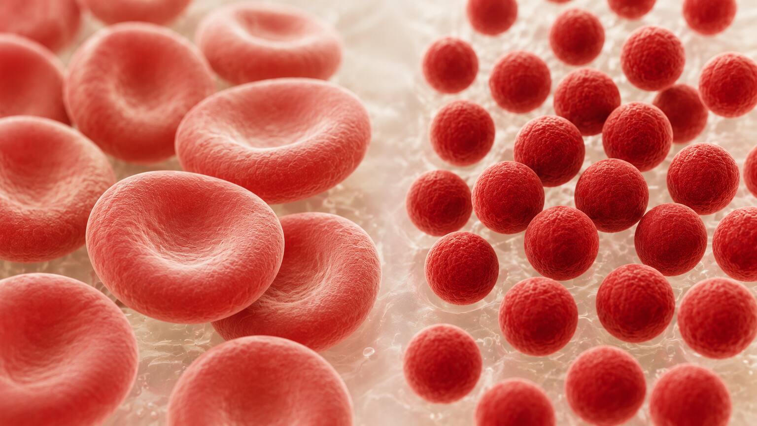
Already have an MCHC number from a recent test, but not sure if it's actually good or bad for you? Just plug it in and see instantly.
🧮 Try the Free CalculatorWhat MCHC Actually Measures (And Why It's Different From MCH and MCV)
Every red blood cell in your body is essentially a tiny, flexible sac built to carry hemoglobin from your lungs out to the rest of your body. Three separate numbers on your CBC describe different aspects of that cargo, and they're easy to mix up because their names all start with "M" and "C." MCV, or mean corpuscular volume, tells you the average size of your red blood cells — think of it as the size of the sac itself. MCH, or mean corpuscular hemoglobin, tells you the average total weight of hemoglobin inside one of those cells, regardless of how big or small the cell happens to be. MCHC takes it one step further and asks a slightly different question: how crowded is that hemoglobin inside the space it actually has to work with? It's a concentration, not a raw amount — the same relationship between the number of coffee grounds in a cup and how strong that cup of coffee actually tastes. Two cups can hold the exact same number of grounds, but if one cup is smaller, that coffee will taste noticeably stronger. MCHC is measuring exactly that kind of density inside your red blood cells.
This distinction matters clinically because a cell can be small and still be normally concentrated, or small and unusually concentrated, and those two situations point toward completely different explanations. Most of the time, when something goes wrong with red blood cell production, MCV and MCH move together while MCHC stays put in its normal range — this is what happens in classic iron-deficiency anemia, for instance, where cells become both smaller and paler, but proportionally so. A high MCHC specifically means the ratio itself is off, not just the total amount of hemoglobin or the total size of the cell. That narrower, more specific signal is exactly why an elevated MCHC gets its own investigation rather than being lumped in with more common CBC abnormalities.
How Your Lab Calculates This Number
Unlike some CBC values that a machine measures directly by shining a laser through individual cells, MCHC is a calculated figure. Your lab arrives at it using a simple formula: your hemoglobin level, divided by your hematocrit, multiplied by one hundred. Hemoglobin is the total amount of that oxygen-carrying protein in a given volume of your blood, while hematocrit is the percentage of your blood's total volume that's actually made up of red blood cells rather than liquid plasma. Dividing one by the other and scaling the result gives a number that describes concentration specifically — how much hemoglobin exists per unit of "red blood cell space," independent of how much total blood you have or how many cells are floating in it. Modern hematology analyzers run this calculation automatically, instantly, every single time a CBC is processed, which is why MCHC appears on essentially every basic blood panel without anyone having to request it separately. The fact that it's calculated rather than measured directly turns out to matter quite a bit, because anything that throws off the hemoglobin or hematocrit inputs — even something with nothing to do with your actual health — can throw off the MCHC that comes out the other end. Keep that detail in mind; it becomes the key to understanding several of the causes further down.
What Counts as "High"? The Normal Range for MCHC
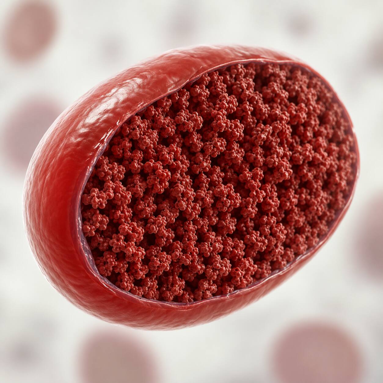
Most laboratories consider a normal MCHC to fall somewhere between 32 and 36 grams per deciliter (g/dL), though you may occasionally see a slightly different range, such as 31.5 to 35.7 g/dL, depending on the specific analyzer and reference population your lab uses. Anything reported above that upper cutoff — typically above 36 g/dL — gets flagged as high. One detail that sets MCHC apart from several other CBC values is that this range doesn't meaningfully shift with age or between men and women, the way reference ranges for hemoglobin or hematocrit do. That's because MCHC describes a ratio internal to the red blood cell itself, a piece of basic cell biology that stays remarkably constant across a healthy adult population, rather than a measurement affected by body size, muscle mass, or hormonal differences. In practice, this makes an abnormal MCHC a fairly clean signal: when it's elevated, something specific is going on, either with your red blood cells themselves or with how the sample was handled, rather than a value simply drifting because of who you are.
It's also worth knowing that a low MCHC is far more common in everyday practice than a high one. Ordinary iron deficiency, the most frequent cause of anemia worldwide, tends to push MCHC down or leave it unchanged, never up. That asymmetry is part of why a high reading tends to prompt a slightly different, more targeted line of questioning from your doctor than a low one would.
Why Doctors Look at MCV and MCHC Together
MCHC rarely tells its full story alone — it's almost always interpreted side by side with MCV, the measure of your red blood cells' average size, because the combination of the two paints a much sharper picture than either number does by itself. In the far more common causes of anemia, like iron deficiency, MCV and MCHC tend to move in the same direction: cells become smaller and, because they're producing less hemoglobin overall, less densely packed too, so MCHC often stays normal or even dips slightly rather than rising. A high MCHC paired with a low or low-normal MCV is a distinctly different and much less common pattern, and it's precisely the pattern seen in conditions where a cell has lost membrane surface area faster than it's lost hemoglobin or water — shrinking the sac around the same cargo, rather than shrinking the cargo along with the sac. That specific combination is what makes hereditary spherocytosis and related membrane disorders stand out from the far more common, purely nutritional causes of small red blood cells, and it's often the detail that first makes a hematologist suspect a shape-related problem rather than reaching for the more routine explanations.
Red cell distribution width, or RDW — a third value reported on the same panel that measures how much your red blood cells vary in size from one another — often stays close to normal in hereditary spherocytosis too, which can feel a little counterintuitive. You might expect a "sick" population of cells to look more varied, not less, but spherocytosis tends to affect cells fairly uniformly rather than producing a wide mixture of shapes and sizes. Taken together, a low-to-normal MCV, a high MCHC, and an unremarkable RDW form a specific enough pattern that many hematologists will ask for a blood smear before ordering anything more elaborate, since actually seeing spherocytes under a microscope confirms in seconds what the numbers alone can only suggest.
The Most Common Real Cause: Hereditary Spherocytosis
When a high MCHC does reflect genuine biology rather than an artifact, the condition doctors think of first is hereditary spherocytosis, an inherited disorder of the red blood cell's outer membrane. A normal red blood cell has a distinctive shape — a flattened disc with a dimple on each side, called biconcave — held in place by a scaffolding of structural proteins just beneath the cell's surface, most notably one called spectrin, working alongside partner proteins named ankyrin, band 3, and band 4.2. In hereditary spherocytosis, a genetic mutation weakens one of these proteins, and the membrane gradually loses small pieces of itself every time the cell squeezes through narrow blood vessels, especially inside the spleen. Losing surface area without losing much of its hemoglobin or water content forces the cell to collapse into a smaller, rounder shape — a sphere instead of a disc, which is where the condition gets its name. A sphere-shaped cell can hold the same hemoglobin as a disc-shaped one, but in noticeably less physical volume, which is exactly the geometry that pushes MCHC upward. Some research also points to a second, compounding mechanism: the same membrane changes let more sodium leak into the cell and more potassium leak out, quietly dehydrating the cell's interior and concentrating its contents even further.
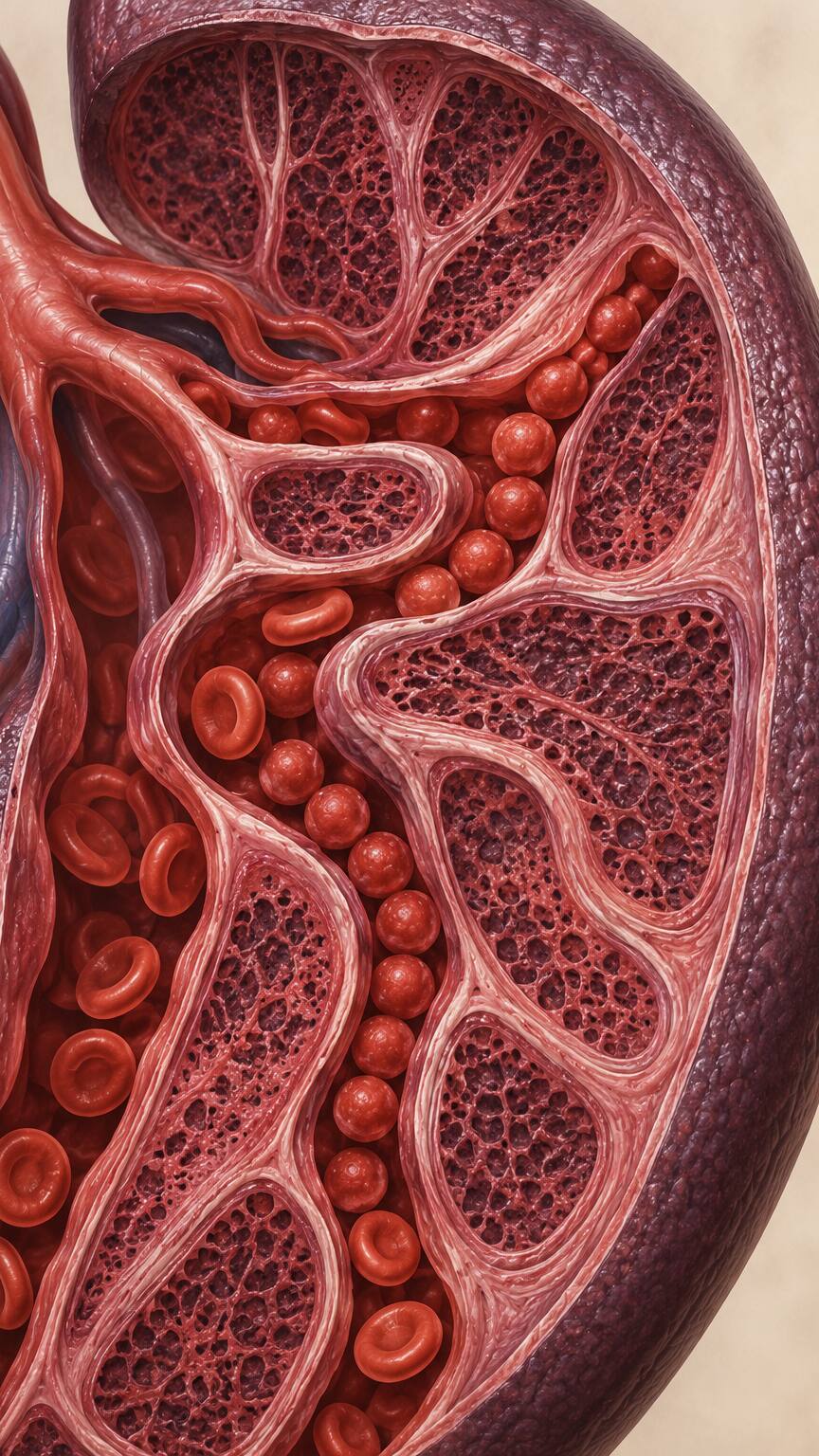
The spleen's job is to filter aging or damaged red blood cells out of circulation, and it's very good at recognizing spherocytes as abnormal, since they've lost the flexibility a healthy red blood cell needs to squeeze through the spleen's narrowest passages. That recognition comes at a cost: the spleen ends up destroying spherocytes faster than the bone marrow can replace them, producing a form of anemia called hemolytic anemia, in which red blood cells are broken down prematurely. The downstream effects show up as a fairly recognizable pattern — fatigue and paleness from the anemia itself, yellowing of the skin and eyes (jaundice) from the bilirubin pigment released as those cells break apart, and sometimes a noticeably enlarged spleen from all the extra filtering work. Because bilirubin turnover stays elevated for years, people with hereditary spherocytosis also develop pigment gallstones far more often than average, sometimes as early as their teens or twenties.
Hereditary spherocytosis runs in families in most cases, most commonly passed down in a pattern where inheriting just one copy of the affected gene from either parent is enough to cause the condition — which is why a family history of unexplained jaundice, anemia, gallstones at a young age, or a relative who had their spleen removed is a genuinely meaningful clue. Diagnosis typically starts with a technologist actually looking at your blood under a microscope, where spherocytes have a very particular appearance: round, uniformly dark-staining cells missing the pale center a normal biconcave cell shows under magnification. From there, more specific tests can confirm the diagnosis, including an osmotic fragility test, which checks how easily these cells rupture when placed in a diluted salt solution, and eosin-5-maleimide (EMA) binding, a more modern test that measures how much of the key membrane protein is actually present on the cell's surface. Treatment depends heavily on severity: mild cases may need nothing more than folic acid supplements to support the bone marrow's higher-than-normal demand for new red blood cells, while more significant cases may eventually need a splenectomy — surgical removal of the spleen — which doesn't fix the underlying membrane defect but removes the organ responsible for destroying the fragile cells, often resolving the anemia almost completely.
Hereditary spherocytosis is the most common inherited cause of hemolytic anemia among people of Northern European descent, affecting roughly 1 in 2,000 to 1 in 5,000 people in that population, though it appears less frequently in other ethnic backgrounds. Severity varies considerably from one family to the next, and even between relatives who carry the exact same mutation — some people live with a barely noticeable, well-compensated anemia their entire lives and only find out about the condition incidentally, when a routine CBC turns up a high MCHC, while others develop symptoms severe enough in early childhood that a splenectomy becomes medically necessary well before adulthood.
The Acquired Version: Autoimmune Hemolytic Anemia
Not everyone who develops spherocytes was born with a faulty membrane gene. In autoimmune hemolytic anemia, the immune system mistakenly produces antibodies that attach themselves to otherwise completely normal red blood cells. When those antibody-coated cells pass through the spleen, resident immune cells called macrophages recognize the antibody flags and take a partial bite out of the cell's membrane rather than destroying the whole cell outright. The cell survives that encounter, but it's lost a piece of its surface in the process — and just like in hereditary spherocytosis, losing membrane without losing hemoglobin forces the cell into a smaller, rounder, more densely packed shape. Under a microscope and on a CBC, the resulting spherocytes can look essentially identical to the inherited version, and MCHC rises for the exact same geometric reason.
The key test that tells these two conditions apart is the direct antiglobulin test, often called a Coombs test, which specifically detects antibodies stuck to the surface of red blood cells — a finding that's present in autoimmune hemolytic anemia and absent in the inherited disorder. Autoimmune hemolytic anemia can appear on its own, alongside another autoimmune condition like lupus, following certain infections, as a reaction to specific medications, or occasionally as an early sign of an underlying lymphoma. Because the trigger is so different from a structural gene defect, the treatment looks completely different too, typically starting with corticosteroids to calm the immune system rather than folic acid or spleen surgery, though a splenectomy remains an option in cases that don't respond well to medication alone. Getting this distinction right matters beyond just picking the correct treatment — a correct diagnosis also determines whether other family members should be screened for an inherited condition, or whether the focus should instead turn to identifying and treating whatever triggered the immune system in the first place.
Another Real Cause: Dehydrated Red Blood Cells in Sickle Cell Disease
Hereditary spherocytosis isn't the only condition where red blood cells lose water and become denser than normal — sickle cell disease produces a strikingly similar effect through an entirely different mechanism. Sickle cell disease is caused by an inherited variant of hemoglobin itself, called hemoglobin S, that distorts into rigid, crescent-shaped rods when oxygen levels drop, which is what gives sickle-shaped cells their name and their tendency to get stuck in small blood vessels. Less widely known is that the same underlying process damages the channels in the cell's membrane that normally keep potassium inside and water balanced, causing the cell to leak potassium and lose water at the same time. A red blood cell that's lost water without losing hemoglobin becomes more concentrated by definition, and in sickle cell disease this creates a specific subset of red blood cells that researchers describe as "hyperdense" — dense enough to register an MCHC well above the standard cutoff, sometimes above 46 g/dL, a level the reference range doesn't even account for in most other conditions.
These extra-dense, dehydrated cells matter beyond just the lab number: they're more prone to the rigid sickling process in the first place, since a more concentrated solution of hemoglobin S polymerizes more readily, and research has linked a larger dense-cell population to worse disease complications overall. For someone already diagnosed with sickle cell disease, a rising MCHC over time can be a meaningful data point for their hematologist to track, quite separate from how it would be interpreted in someone without that diagnosis.
When It's Not Real Biology at All — Lab Artifacts That Mimic a High MCHC
Not every high MCHC traces back to something happening inside your body. Because MCHC is a calculated ratio built from other measurements, anything that distorts those underlying numbers — even something with nothing to do with your red blood cells' actual health — can push the final figure above the normal range. Two of the most common culprits are simple to explain and, more importantly, simple to fix.
Dehydration and Other Causes of a "Concentrated" Sample
When you're dehydrated, the liquid portion of your blood — the plasma — shrinks in volume while your actual number of red blood cells stays exactly the same. That shift concentrates nearly everything measured in a blood sample, including hemoglobin and hematocrit, and it can be enough to nudge a borderline MCHC just over the 36 g/dL cutoff without anything actually being wrong with your red blood cells. This kind of hemoconcentration is usually mild after ordinary dehydration and tends to resolve within a day or two of drinking normally again, which is why a doctor who suspects it will often simply ask you to rehydrate and repeat the test rather than pursuing anything further. A much more extreme version of the same phenomenon shows up in people with severe burns, where large amounts of plasma are lost through damaged skin over a short period of time. That fluid loss can push hematocrit, hemoglobin, and MCHC all upward together, sometimes dramatically, purely as a reflection of how concentrated the remaining blood has become — a pattern clinicians treating burn patients are specifically trained to expect and account for during fluid resuscitation.

This is also part of why a doctor faced with an isolated high MCHC and no other red flags will often reach for the simplest explanation first, rather than assuming the worst. A quick review of how much you'd had to drink that morning, whether you'd exercised heavily beforehand, or whether you'd been sick with vomiting or diarrhea in the days before your blood draw can go a long way toward explaining a mildly elevated result without any further testing at all.
Cold Agglutinins — When the Sample Itself Gets Too Cold
A less familiar but genuinely common cause of a falsely high MCHC involves antibodies called cold agglutinins, which cause red blood cells to clump together specifically when blood cools below body temperature — and a tube of blood sitting at room temperature on its way to the lab is exactly cold enough to trigger that clumping in people who happen to carry these antibodies. Automated hematology analyzers count and size cells one at a time as they pass through a narrow channel, and a clump of several stuck-together red blood cells gets misread as a single, unusually large cell. That misreading artificially lowers the total red blood cell count the machine reports, since dozens of real cells get counted as just a handful of "giant" ones.
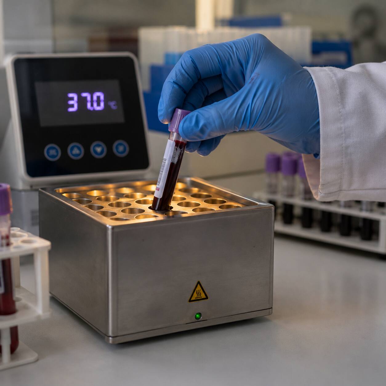
Here's where the math goes sideways: hemoglobin itself is measured after the lab deliberately breaks open, or lyses, every red blood cell in the sample, a step that dissolves any clumps completely and isn't affected by cold agglutinins at all. So the lab ends up with an accurate hemoglobin reading paired with an artificially low, clumping-distorted red blood cell count and hematocrit — and when that distorted hematocrit gets divided into the correct hemoglobin figure, the resulting MCHC comes out falsely elevated, sometimes dramatically so. The fix is refreshingly simple: warming the blood sample back up to normal body temperature, about 98.6°F (37°C), for roughly an hour causes the clumps to disperse, and repeating the CBC on the rewarmed sample typically produces a completely normal result. Most people with cold agglutinins have no symptoms whatsoever and only discover them because of this exact lab quirk. A smaller number have an actual condition called cold agglutinin disease, in which these antibodies are present at high enough levels to cause real problems in cold weather, including a bluish discoloration of the fingers and toes and, in more significant cases, an ongoing hemolytic anemia of their own.
Symptoms That May (or May Not) Come With a High MCHC
Whether a high MCHC comes with any symptoms at all depends entirely on what's actually driving it, not on the number itself. If the cause is dehydration or a cold sample sitting in a collection tube, there's usually nothing to feel beyond ordinary thirst — the MCHC is simply reporting a lab artifact, not a change in how your body is functioning. If the underlying cause is a genuine hemolytic process, like hereditary spherocytosis or autoimmune hemolytic anemia, the symptoms tend to track with how much red blood cell destruction is actually happening. Mild cases may cause nothing more than occasional fatigue that's easy to write off as an ordinary busy week. More active hemolysis tends to produce a more recognizable cluster: paleness, tiredness that doesn't improve with rest, yellowing of the skin or the whites of the eyes, darker-than-usual urine from the extra bilirubin your kidneys are filtering out, and sometimes a dull ache or fullness in the upper left abdomen where an enlarged spleen sits. None of these symptoms are unique to a high MCHC specifically, but their presence or absence gives a doctor a genuinely useful clue about how urgently to pursue the underlying cause, and how far to look beyond the number itself before drawing any conclusions.
What Your Doctor Will Likely Do Next
Because lab artifacts are so common with this particular value, the first and simplest step is usually to repeat the CBC, sometimes specifically asking the lab to rewarm the sample first if cold agglutinins are suspected based on the pattern of results. If the repeat test still shows a high MCHC, the next move is typically a manual review of your blood smear, where a trained technologist looks directly at your red blood cells under a microscope rather than relying only on the automated counts — this is genuinely the single best way to spot spherocytes and distinguish a real shape abnormality from a counting artifact. From there, a few additional tests help narrow things down: a reticulocyte count checks whether your bone marrow is churning out extra young red blood cells to compensate for ones being destroyed early, which is a hallmark of active hemolysis; haptoglobin and LDH levels help confirm hemolysis is actually happening, since haptoglobin tends to drop and LDH tends to rise when red blood cells are breaking down faster than normal; and a direct antiglobulin, or Coombs, test checks specifically for the antibody-coating pattern seen in autoimmune hemolytic anemia.

Your doctor will likely also ask about your family history — unexplained anemia, jaundice as a newborn, early gallstones, or a relative who had a splenectomy — since that single conversation often points toward hereditary spherocytosis faster than any individual lab test. Taken together, this workup is rarely long or complicated; for most people, it resolves within one or two follow-up visits, ending either with a clear diagnosis or, just as often, with reassurance that the original result was nothing more than a temporary quirk of the sample.
Frequently Asked Questions
Is a high MCHC dangerous by itself?
No. MCHC is a calculated ratio, not a disease or a diagnosis — it's a signal that prompts your doctor to look further, not something that's treated directly. The underlying cause it points to, if any, is what actually determines whether treatment is needed.
Can drinking more water bring a high MCHC back down?
If dehydration turns out to be the confirmed cause, rehydrating can bring a mildly elevated MCHC back into the normal range within a day or two on a repeat test. But whether dehydration is actually the explanation for your result isn't something to assume on your own — a doctor needs to confirm that before drinking more water is treated as the fix, since a red blood cell shape disorder like hereditary spherocytosis or autoimmune hemolytic anemia can produce the same elevated number, and drinking water won't change that.
Does a high MCHC always mean I have hereditary spherocytosis?
Not at all. A meaningful share of high MCHC results turn out to be lab artifacts from dehydration or cold agglutinins, and among the results that do reflect real biology, autoimmune hemolytic anemia can produce a nearly identical picture. A blood smear review and a few follow-up tests are what actually sort out which explanation applies.
Why did my MCHC come back normal when the test was repeated?
This is a common and genuinely reassuring outcome. It usually confirms that the original high reading was a temporary artifact — most often dehydration on the day of the first draw, or a cold agglutinin effect from the sample sitting at room temperature — rather than an ongoing red blood cell problem that needs further workup.
Conclusion
A high MCHC is one of the rarer flags to show up on a CBC, and in a large share of cases, it turns out to be nothing more than a snapshot of mild dehydration or a sample that clumped up on its way to the lab — both situations a simple repeat test usually resolves. When it does reflect real biology, it's most often tracing back to red blood cells that have become smaller and rounder than they should be, whether from an inherited membrane condition like hereditary spherocytosis, an immune system that's mistakenly begun attacking otherwise healthy cells, or the cell dehydration seen in sickle cell disease. None of these explanations require guesswork on your part. A repeat CBC, a look at your blood smear under a microscope, and a conversation about your family history and how you've been feeling will almost always point clearly toward the right answer, and toward the right next step.
Still Not Sure What Your Results Mean?
Upload your labs and get a complete, visual, plain-language interpretation of every biomarker — delivered to your inbox in under 15 minutes.
Get My ReportThis article is for educational purposes only and does not constitute medical advice. Always consult your healthcare provider regarding your specific lab results.