
What Does a High MCV Mean on a Blood Test?
A high MCV on a complete blood count means your red blood cells are, on average, larger than they should be — a finding doctors call macrocytosis. MCV stands for mean corpuscular volume, and it's one of the automatic calculations your lab runs every single time blood is drawn for a CBC, whether or not anyone specifically asked for it. Seeing it flagged high can be unsettling, especially paired with unfamiliar words like "macrocytic" on your report, but an elevated MCV is not, by itself, a diagnosis of anything — it's a clue. Sometimes that clue traces back to something as simple and fixable as a vitamin deficiency or a few too many drinks a week. Other times it's the first hint of a thyroid problem, a liver issue, a medication side effect, or, less commonly, a bone marrow disorder that deserves a closer look. This guide walks through exactly what MCV measures, why your red blood cells might be running larger than average, and what your doctor will likely do to figure out which explanation actually applies to you.

Figure 1. In megaloblastic macrocytosis, impaired DNA synthesis causes a developing red blood cell's nucleus to mature more slowly than its cytoplasm, producing an abnormally large cell before it ever leaves the bone marrow.
Already have an MCV number from a recent test, but not sure if it's actually good or bad for you? Just plug it in and see instantly.
🧮 Try the Free CalculatorWhat MCV Actually Measures
Every time a hematology analyzer processes your blood, it doesn't just count how many red blood cells you have — it also measures the size of each one individually, hundreds at a time, as they stream single-file through a narrow sensing channel. MCV is simply the average of all those individual measurements, expressed in femtoliters (fL), a unit of volume so small that a single red blood cell typically measures somewhere around 90 femtoliters, or roughly one ten-trillionth of a teaspoon. Think of MCV as the answer to a very specific question: if you lined up a thousand of your own red blood cells and asked how much space the "typical" one takes up, what would the answer be? It's not a count, and it's not a measure of how much oxygen-carrying hemoglobin those cells contain — those are separate numbers on the same panel. MCV describes size, full stop, and it does so with remarkable precision because the machine is directly measuring physical volume, not estimating or calculating it from other values the way a few of its CBC neighbors do.
Red blood cells aren't produced all at once and left alone — they're manufactured continuously in your bone marrow, maturing over about a week before being released into circulation, where they then live for roughly 120 days doing the work of carrying oxygen. During that week of maturation, a young cell called an erythroblast gradually shrinks, ejects its nucleus, and packs itself with hemoglobin before it's ready to leave the marrow as a reticulocyte and, shortly after, a fully mature red blood cell. Anything that disrupts this maturation process — whether it speeds a cell out the door too early, slows down one particular step while others continue normally, or damages the marrow's ability to build cells correctly in the first place — can change the final size of the cell that ends up in your bloodstream. That's the common thread running through nearly every cause of a high MCV: something, somewhere, has interfered with the normally tightly choreographed process of building a red blood cell from scratch.
What Counts as "High"? The Normal Range for MCV
Most laboratories consider a normal MCV to fall somewhere between 80 and 100 femtoliters (fL), and unlike quite a few other values on a CBC, this range doesn't meaningfully shift by sex or by age once you're past early childhood — a 25-year-old and an 80-year-old are held to essentially the same reference interval. You may occasionally see a lab report a very slightly different cutoff, such as 79 to 97 fL rather than 80 to 100, depending on which reference population and analyzer that particular lab uses, but the difference is minor and doesn't change how a meaningfully high result should be interpreted. Anything reported above the lab's upper limit — in practice, usually anywhere from about 100 to 105 fL — is classified as macrocytosis. Mild elevations, in the 101 to 110 fL range, are the most common pattern seen in everyday practice and are frequently explained by benign, easily identified causes. Values climbing well past 110 or 115 fL are less common and more often point toward a true vitamin deficiency, active bone marrow disease, or a significant amount of young reticulocytes flooding into the bloodstream, and they tend to prompt a more thorough workup sooner rather than later.
It's worth knowing upfront that a high MCV is considerably less common in the general population than a low one. Iron deficiency, the single most frequent cause of an abnormal CBC worldwide, pushes MCV down, not up — which is part of why an elevated MCV, when it does show up, tends to get its own distinct line of questioning from a physician rather than being folded into the far more routine workup for small, iron-starved red blood cells.
The Key Distinction: Megaloblastic vs. Non-Megaloblastic Macrocytosis
Once a high MCV is confirmed, doctors don't jump straight to a list of possible causes — they first sort the finding into one of two broad biological categories, because the sorting itself narrows the list of suspects dramatically. The first category, megaloblastic macrocytosis, happens when something interferes specifically with a cell's ability to synthesize DNA while it's still developing in the bone marrow. DNA synthesis is a slower, more delicate process than the rest of a cell's maturation, so when it's disrupted, the cell's nucleus falls behind schedule while the surrounding cytoplasm keeps maturing on its usual timeline. The mismatch — cytoplasm ready to go, nucleus still lagging — produces an unusually large, structurally abnormal cell, and it leaves a very specific fingerprint on a blood smear: neutrophils (a type of white blood cell examined on the same slide) with an unusually high number of nuclear lobes, a finding called hypersegmentation, along with oval-shaped rather than round macrocytes. Vitamin B12 deficiency and folate deficiency are, by a wide margin, the two most common causes of this pattern, because both vitamins are essential ingredients in the specific biochemical pathway your cells use to build new DNA.
The second category, non-megaloblastic macrocytosis, covers every other mechanism that produces large red blood cells without that specific DNA-synthesis defect — and it's a broader, more varied group. Heavy alcohol use, liver disease, an underactive thyroid, certain medications, a bone marrow actively churning out large numbers of young reticulocytes in response to blood loss or cell destruction elsewhere in the body, and primary bone marrow disorders like myelodysplastic syndrome all fall into this bucket, each through its own distinct mechanism. Reviewing a blood smear under a microscope is usually the fastest way to tell these two categories apart, since hypersegmented neutrophils are a fairly specific tell for the megaloblastic pattern and their absence shifts suspicion toward the non-megaloblastic list instead. From here, walking through each individual cause in more detail is the most useful way to understand where your own result might fit.
Vitamin B12 Deficiency

Figure 2. A smooth, sore, beefy-red tongue — atrophic glossitis — is a classic physical sign of vitamin B12 or folate deficiency, caused by the same impaired cell turnover that enlarges red blood cells.
Vitamin B12 is essential for a chemical reaction your cells rely on to build new DNA, and without enough of it, developing red blood cells in your bone marrow can't complete their maturation on schedule, producing the large, oval macrocytes described above. The most common cause of true B12 deficiency in adults isn't diet — it's pernicious anemia, an autoimmune condition in which the immune system attacks the stomach's parietal cells, the cells responsible for producing intrinsic factor, a protein your small intestine absolutely requires to absorb B12 from food. Without intrinsic factor, B12 passes straight through the digestive tract unabsorbed no matter how much of it you eat. Other causes include a strictly plant-based diet without supplementation (since B12 occurs naturally almost exclusively in animal products), gastric bypass surgery or other procedures that remove the stomach tissue producing intrinsic factor, and conditions like Crohn's disease or celiac disease that damage the specific stretch of small intestine — the terminal ileum — where B12 absorption actually happens. Long-term use of certain common medications is also increasingly recognized as a contributor: metformin, one of the most widely prescribed diabetes medications, is known to reduce B12 absorption with years of continuous use, and long-term acid-suppressing drugs like proton pump inhibitors reduce the stomach acid needed to release B12 from the protein it's bound to in food.
What sets B12 deficiency apart from nearly every other cause of macrocytosis on this list is its potential to damage the nervous system, sometimes irreversibly, and sometimes before the anemia itself becomes obvious on a CBC. B12 plays a direct role in maintaining the protective myelin sheath around nerve fibers, and deficiency can produce a distinctive pattern of nerve damage called subacute combined degeneration, affecting both the sensory pathways that relay position sense and the motor pathways controlling movement. In practice, this shows up as tingling or numbness that typically starts in the feet and hands, an unsteady, wide-based gait, difficulty with fine coordination, and in more advanced cases, memory problems or mood changes that are sometimes mistaken for an unrelated cognitive or psychiatric issue, particularly in older adults. Glossitis — a smooth, sore, reddened tongue that's lost its normal surface texture — is another classic physical sign, caused by the same underlying disruption to rapidly dividing cells that also affects the lining of the tongue. Because nerve damage from B12 deficiency can become permanent if it goes uncorrected for too long, doctors treat a suspected deficiency with real urgency, even when the macrocytosis itself looks mild.
Folate Deficiency
Folate, also known as vitamin B9, works in the same DNA-synthesis pathway as B12 and produces a nearly identical megaloblastic picture on a blood smear — oval macrocytes and hypersegmented neutrophils included — which is exactly why the two are almost always tested together rather than in isolation. The biggest practical difference between the two deficiencies is how quickly each one develops: your body stores several years' worth of B12 in the liver, but it only keeps a few months' supply of folate on hand, since folate is used up quickly by any process involving rapid cell division. That makes folate deficiency develop noticeably faster whenever intake drops or demand rises. Poor dietary intake is a common driver on its own, particularly in people who eat few leafy greens, legumes, or fortified grains, but demand also rises sharply during pregnancy, when a developing fetus's rapid cell division consumes maternal folate at a much higher rate — which is exactly why folic acid supplementation is standard, well-publicized advice for anyone trying to conceive. Chronic heavy alcohol use is another major contributor, interfering with folate through more than one route at once: it reduces dietary folate intake in people who eat poorly, damages the small intestine lining in ways that impair folate absorption, and directly disrupts how the liver processes and recycles the folate that is absorbed. A handful of medications also interfere with folate metabolism directly, most notably methotrexate, a drug used for cancer treatment and for autoimmune conditions like rheumatoid arthritis, along with certain anticonvulsants and sulfasalazine, used for inflammatory bowel disease.
Unlike B12 deficiency, folate deficiency on its own does not cause the same nerve damage — the neurological risk really is specific to B12 — but the two deficiencies frequently travel together, particularly in people with alcohol use disorder or malabsorption conditions, which is one more reason clinicians rarely test one without the other. One more detail worth knowing: taking a folic acid supplement without also correcting a coexisting B12 deficiency can actually make things more confusing rather than less, because folic acid can correct the anemia and normalize the blood count on paper while doing nothing to stop the B12-related nerve damage happening underneath — a phenomenon called "masking" that's part of why doctors insist on checking both levels before treating either one on its own.
Alcohol Use and Liver Disease
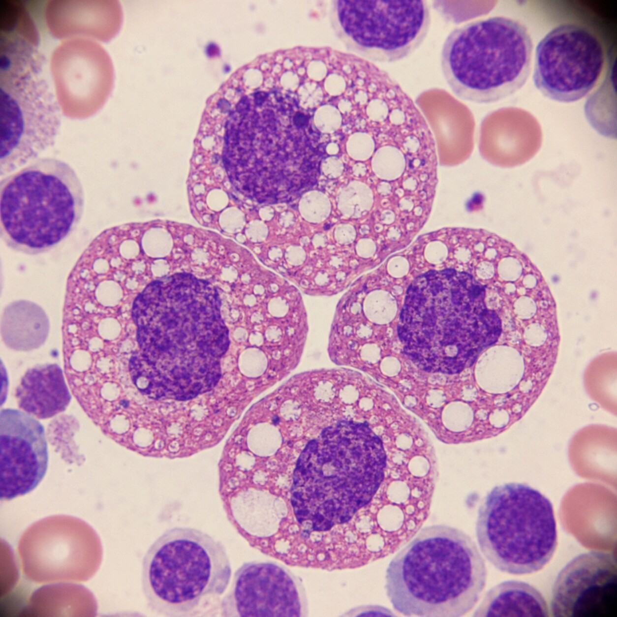
Figure 3. Chronic alcohol exposure has a direct toxic effect on the bone marrow, producing visible vacuoles — small fluid-filled gaps — inside developing red blood cell precursors even before anemia appears on a CBC.
Heavy alcohol use is one of the single most common causes of a mildly elevated MCV, and unlike most of the other causes on this list, it doesn't necessarily route through a vitamin deficiency at all — alcohol has a direct toxic effect on the bone marrow, independent of nutrition. Under a microscope, bone marrow samples from people who drink heavily often show developing red blood cells riddled with small vacuoles, fluid-filled gaps inside the cell that reflect direct ethanol-related damage to the machinery a cell needs to mature correctly. This effect can show up within a matter of weeks of heavy drinking and, encouragingly, tends to reverse within about two to four months of cutting back or stopping, since bone marrow cells turn over relatively quickly. Because alcohol frequently also interferes with folate absorption and intake, as covered above, a heavy drinker's macrocytosis is often a mix of two overlapping mechanisms working at once rather than a single clean cause — which is part of why this particular pattern rarely climbs to extreme MCV values on its own; alcohol alone typically pushes MCV into the low-to-mid 100s, and a much higher number in a person who drinks heavily should prompt a look for a coexisting folate or B12 deficiency rather than being chalked up to alcohol by default.
Liver disease produces macrocytosis through an entirely different route: the composition of a red blood cell's outer membrane. A healthy liver plays a central role in regulating the balance of cholesterol and phospholipids circulating in the blood, and both of those substances are drawn into a red blood cell's membrane from the surrounding plasma throughout the cell's 120-day lifespan. When liver function is impaired — from cirrhosis, chronic hepatitis, or fatty liver disease — that lipid balance shifts, and red blood cell membranes end up incorporating extra surface area as they circulate, causing the cells to swell larger than normal over time. This mechanism explains why liver disease and heavy alcohol use so often show up together as overlapping causes of the same lab finding, since ongoing heavy drinking is itself one of the leading causes of liver damage in the first place — making the two causes less like separate entries on a checklist and more like two ends of the same clinical picture in a lot of real-world cases.
Hypothyroidism
An underactive thyroid is a less intuitive but well-documented cause of macrocytosis, showing up in a meaningful minority of people diagnosed with hypothyroidism, particularly when the condition has gone unnoticed or untreated for a while. The exact mechanism isn't as fully mapped out as it is for B12 or folate deficiency, but thyroid hormone is known to play a role in regulating how quickly bone marrow cells divide and mature, and a shortage of it appears to slow the maturation process down in a way that leaves cells slightly larger than they should be by the time they're released into circulation. Hypothyroidism is also frequently linked to pernicious anemia and other autoimmune conditions that independently cause B12 deficiency, since autoimmune diseases often cluster together in the same person — so when a high MCV and an underactive thyroid appear on the same set of labs, a doctor will typically check B12 levels as well, rather than assuming the thyroid alone explains the whole picture. The reassuring part of this particular cause is that it tends to resolve on its own once thyroid hormone levels are corrected with standard replacement therapy, usually over the course of a few months, without needing any treatment directed at the blood count itself.
Medications That Can Raise MCV
A number of prescription drugs are recognized causes of macrocytosis, and reviewing a complete medication list is one of the first, simplest steps in working up an unexplained high MCV — because unlike a vitamin deficiency or a marrow disorder, a medication-related cause is often resolved simply by adjusting or switching the drug, under a doctor's guidance. Chemotherapy drugs that interfere with DNA synthesis, such as hydroxyurea and methotrexate, are among the most predictable culprits, since disrupting DNA synthesis is precisely the mechanism these drugs are designed around to slow cancer cell growth — the same disruption that produces the classic megaloblastic pattern with B12 or folate deficiency shows up here for a completely different reason. Certain antiretroviral medications used to treat HIV, most notably zidovudine (also known as AZT), are well known for raising MCV, to the point that a rising MCV is sometimes even used clinically as an informal marker of medication adherence, since the effect only appears with consistent use. Anticonvulsant medications, including phenytoin, can interfere with folate metabolism and produce a similar picture. Azathioprine, an immunosuppressant used for autoimmune conditions and after organ transplants, and metformin, through the B12-depletion mechanism discussed earlier, round out the list of medications most commonly flagged when a high MCV shows up on someone's routine bloodwork. None of this means a necessary medication should be stopped without medical guidance — in most cases, the macrocytosis is a benign, well-understood side effect that simply gets monitored rather than treated.
Reticulocytosis: When Macrocytosis Is a Sign of a Healthy Marrow Working Hard
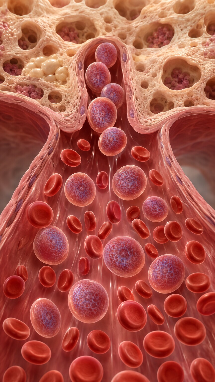
Figure 4. Reticulocytes — newly released red blood cells that still carry residual RNA — are naturally larger than fully mature red blood cells, so a surge of them into circulation can raise the average MCV on its own.
Not every cause of a high MCV points toward a problem — sometimes it's a sign that your bone marrow is doing exactly what it's supposed to do, just at a faster pace than usual. Reticulocytes, the newly minted red blood cells released from the marrow just before they reach full maturity, are measurably larger than the older, fully mature red blood cells that make up most of a typical bloodstream, since they haven't yet finished shrinking down to their final size. Under normal circumstances, reticulocytes make up only a small fraction of your total red blood cell population, too small to meaningfully shift the average MCV. But when something increases the demand for new red blood cells — significant blood loss from an injury, surgery, or heavy menstrual bleeding, or hemolysis, the premature destruction of existing red blood cells seen in certain autoimmune or inherited conditions — the bone marrow ramps up production and releases a much larger wave of these oversized young cells into circulation. That flood of larger reticulocytes can be enough to pull the overall MCV average upward, sometimes noticeably, even though nothing is actually wrong with how the cells themselves are being built.
This is exactly why a reticulocyte count is one of the very first tests a doctor orders when working up an unexplained high MCV: a high reticulocyte count alongside the elevated MCV points strongly toward the bone marrow actively compensating for something — blood loss or cell destruction — rather than a primary production problem inside the marrow itself. It reframes the entire diagnostic conversation, shifting the question away from "why are the cells being built wrong" and toward "what's causing the body to lose or destroy red blood cells fast enough that the marrow has to work this hard to keep up." Once that underlying trigger — a source of bleeding, a hemolytic process, or a recovery period following a recent transfusion — is identified and addressed, the elevated MCV typically settles back toward normal on its own as the surge of new reticulocytes tapers off.
Myelodysplastic Syndrome and Other Bone Marrow Disorders

Figure 5. When macrocytosis appears without a clear nutritional, hormonal, or medication-related explanation — especially alongside other abnormal blood counts — a bone marrow biopsy can directly examine how red blood cells are being produced.
After the more common causes above have been ruled out, an unexplained high MCV — particularly one that persists for months, appears in someone over age 60, or shows up alongside other abnormal counts on the same CBC, such as a low white blood cell or platelet count — raises the possibility of myelodysplastic syndrome, often shortened to MDS. MDS is a group of disorders in which the bone marrow's stem cells, the master cells responsible for producing all of your blood cells, develop genetic changes that cause them to produce cells that are structurally abnormal and, ultimately, less effective at their job. Macrocytosis is one of the most common and often earliest laboratory clues of MDS, sometimes showing up on routine bloodwork years before any other symptom appears. MDS is more of a spectrum than a single fixed diagnosis: some people live with a very mild, slow-moving form for years with only minor blood count changes to show for it, while other forms progress more significantly over time and, in a portion of cases, can eventually develop into acute myeloid leukemia, which is part of why doctors take a persistent, unexplained macrocytosis seriously rather than dismissing it.
Confirming or ruling out MDS ultimately requires a bone marrow biopsy — a procedure in which a small sample of marrow tissue, typically taken from the back of the hip bone, is examined directly under a microscope for the specific dysplastic changes, meaning abnormally shaped or immature cells, that define the condition. This step is only pursued after the simpler, more common explanations for macrocytosis have already been checked and excluded, since MDS is a comparatively uncommon cause overall, and the large majority of people with a high MCV never need a bone marrow biopsy at all. When it is pursued, it's precisely because the pattern of findings — persistent macrocytosis, other abnormal counts, an older age at onset, no identified nutritional or medication cause — has already pointed the workup in that specific direction, rather than being an early, reflexive step taken for every mildly elevated result.
Lab Artifacts That Can Falsely Raise MCV
Not every high MCV traces back to real biology happening inside your body. Because hematology analyzers measure cell size as blood flows through a narrow sensing channel, anything that causes red blood cells to clump together before or during that measurement can distort the result. Cold agglutinins — antibodies that cause red blood cells to stick together specifically when a blood sample cools below body temperature, which happens easily during ordinary transport from the blood draw to the lab — are a well-recognized cause of this kind of artifact. When several red blood cells clump into a single mass, the analyzer's sensor reads that clump as one unusually large cell rather than several normal ones, artificially inflating the average size it reports. Severe hyperglycemia, extremely high blood sugar, has also been reported to cause a similar false elevation in some cases, related to how swollen red blood cells become in a very concentrated glucose environment. In both situations, the fix is straightforward: warming a cold-agglutinin-affected sample back to body temperature before re-running the analysis, or simply repeating the test once blood sugar is back under control, typically produces an accurate, normal result. This is one more reason a single high MCV reading, especially a mild one with no other red flags, is often followed by a simple repeat test before anyone assumes something more significant is going on.
Symptoms That May (or May Not) Come With a High MCV
Whether a high MCV produces any noticeable symptoms at all depends almost entirely on what's actually driving it and whether it's accompanying true anemia — a drop in hemoglobin, not just a size change in the cells themselves. Mild macrocytosis without anemia, which describes a large share of alcohol-related and medication-related cases, frequently causes no symptoms whatsoever and is only discovered incidentally on routine bloodwork. When macrocytosis is significant enough to come with true anemia, the symptoms tend to be the same general ones seen in any anemia: persistent fatigue that doesn't improve with rest, noticeable paleness, shortness of breath with activity that wouldn't normally cause it, a faster-than-usual heartbeat, and difficulty concentrating. B12 deficiency stands apart from the rest of this list because of its added neurological symptoms — tingling or numbness in the hands and feet, balance and coordination problems, and in more advanced or prolonged cases, memory difficulties — which can appear even before the anemia itself becomes pronounced, making them a genuinely important detail to mention to a doctor even if they seem unrelated to a routine blood test result. Jaundice, a yellowing of the skin and the whites of the eyes, along with darker-than-usual urine, points toward a hemolytic process driving reticulocytosis rather than a nutritional or thyroid cause. None of these symptoms are unique enough to pin down a specific cause on their own, but together with the lab pattern itself, they give a physician a genuinely useful head start on where to focus first.
What Your Doctor Will Likely Do Next
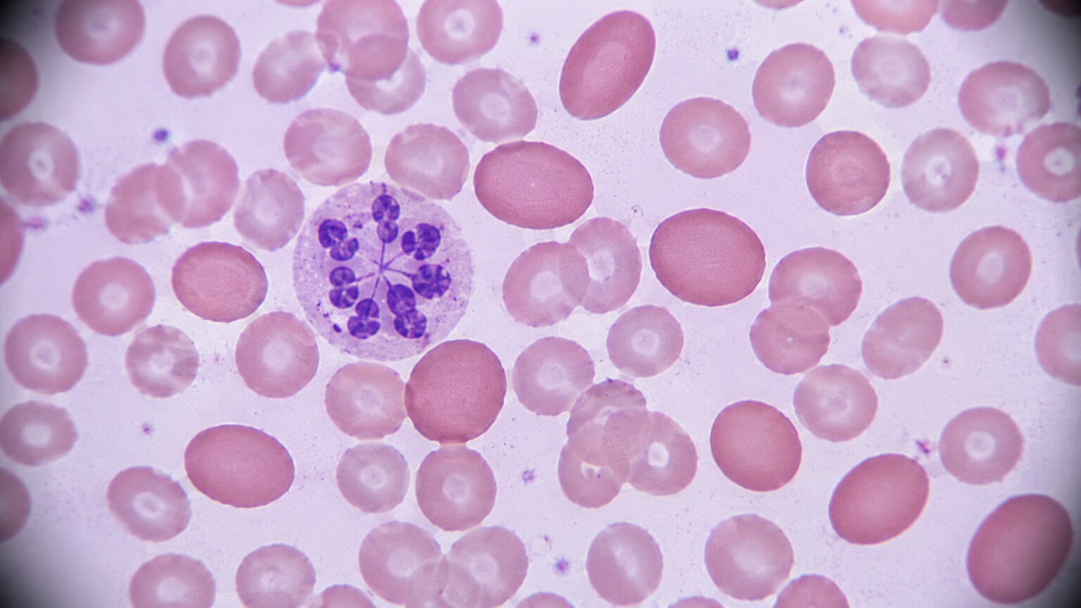
Figure 6. A peripheral blood smear lets a technologist see red blood cell shape directly — oval macrocytes and hypersegmented neutrophils point toward B12 or folate deficiency, while round macrocytes suggest a different underlying cause.
A high MCV rarely leads straight to a single test — it typically opens a short, logical sequence of follow-up tests, each one narrowing the list of possible explanations. The workup usually starts with a peripheral blood smear, where a trained technologist examines your red blood cells directly under a microscope rather than relying only on the automated numbers; the presence or absence of hypersegmented neutrophils and oval-shaped macrocytes is often the single fastest way to sort a megaloblastic cause from a non-megaloblastic one. A reticulocyte count comes next, checking whether the bone marrow is actively compensating for blood loss or cell destruction elsewhere in the body. From there, B12 and folate levels are checked directly, though for B12 specifically, many clinicians will also check methylmalonic acid and homocysteine — two substances that build up in the blood when B12 is genuinely deficient at the cellular level, offering a more sensitive read than the B12 blood level alone, particularly in borderline cases. A thyroid-stimulating hormone (TSH) test screens for hypothyroidism, and liver function tests, alongside an honest conversation about alcohol intake, screen for the alcohol- and liver-related causes. A careful review of every medication you're currently taking rounds out the standard first pass. If all of these come back unremarkable and the macrocytosis persists, especially alongside other abnormal blood counts, a referral to a hematologist and a bone marrow biopsy become the next reasonable step to evaluate for MDS or another primary marrow disorder.
For most people, this entire process resolves within one or two follow-up visits and a single round of bloodwork, ending either with a clear, treatable explanation — most commonly a vitamin deficiency, alcohol use, a medication, or a thyroid issue — or with reassurance that a mildly elevated result reflects nothing more serious than a lab artifact or an unremarkable variation worth simply monitoring on a future CBC.
Frequently Asked Questions
Does a high MCV always mean I have anemia?
No. MCV describes the size of your red blood cells, not how many you have or how much hemoglobin they're carrying. It's entirely possible to have a high MCV with a completely normal hemoglobin level — a pattern sometimes called isolated macrocytosis — particularly with mild alcohol use, certain medications, or early thyroid changes, none of which have yet caused true anemia.
Can a high MCV be harmless and go away on its own?
Yes, in a meaningful number of cases. Mild macrocytosis linked to moderate alcohol intake, a temporary lab artifact from a cold sample, or a medication that's later adjusted often resolves on a repeat test without needing any specific treatment. That said, "harmless" should be a conclusion your doctor reaches after appropriate testing, not an assumption made from the number alone.
How is a high MCV treated?
There's no treatment aimed at the MCV number itself — treatment always targets the underlying cause. That might mean B12 injections or oral supplementation, folic acid, cutting back on alcohol, adjusting a medication, correcting a thyroid problem with hormone replacement, or, in the case of active bleeding or hemolysis, treating whatever's driving the reticulocyte surge. The MCV typically normalizes once the root cause is addressed.
Can a high MCV be an early sign of cancer?
Directly, no — a high MCV itself isn't a cancer marker, and the overwhelming majority of high MCV results trace back to nutritional, alcohol-related, thyroid, or medication causes that have nothing to do with cancer. That said, a persistent, unexplained macrocytosis with no clear cause, especially in someone over 60 or alongside other abnormal blood counts, is one of the patterns that can prompt evaluation for myelodysplastic syndrome, which is why doctors don't ignore a high MCV that doesn't have an obvious explanation.
Conclusion
A high MCV is a single, precise measurement — the average size of your red blood cells — but the list of things that can push it above normal is genuinely wide, ranging from an easily corrected vitamin deficiency to a medication side effect to, less commonly, a bone marrow disorder that needs closer attention. The good news is that this isn't a mystery doctors solve by guesswork: a blood smear, a reticulocyte count, B12 and folate levels, thyroid and liver testing, and an honest review of alcohol use and medications will identify the cause in the overwhelming majority of cases, usually within a visit or two. If your own CBC came back with a flagged MCV, the most useful next step isn't to assume the worst from the number alone — it's to bring the full picture, including any symptoms you've noticed and everything you're currently taking or drinking, to a conversation with your healthcare provider, who can walk through exactly which of these explanations fits your situation.
Still Not Sure What Your Results Mean?
Upload your labs and get a complete, visual, plain-language interpretation of every biomarker — delivered to your inbox in under 15 minutes.
Get My ReportThis article is for educational purposes only and does not constitute medical advice. Always consult your healthcare provider regarding your specific lab results.