
What Does Low MCH Indicate About Your Blood?
A low MCH means the average red blood cell in your body is carrying less hemoglobin than it should — not necessarily fewer red blood cells overall, but each individual cell coming up short on the specific protein responsible for carrying oxygen. This shortfall traces back to one of a fairly short list of specific causes: not enough iron reaching the bone marrow to build hemoglobin properly, a genetic condition affecting hemoglobin production from birth, ongoing inflammation quietly suppressing how efficiently the body uses the iron it has, or, less commonly, a toxic exposure directly interfering with the hemoglobin assembly process itself. This article walks through exactly what MCH is actually measuring, each of these specific causes in turn, and how doctors distinguish between them once a low result shows up on a routine complete blood count.
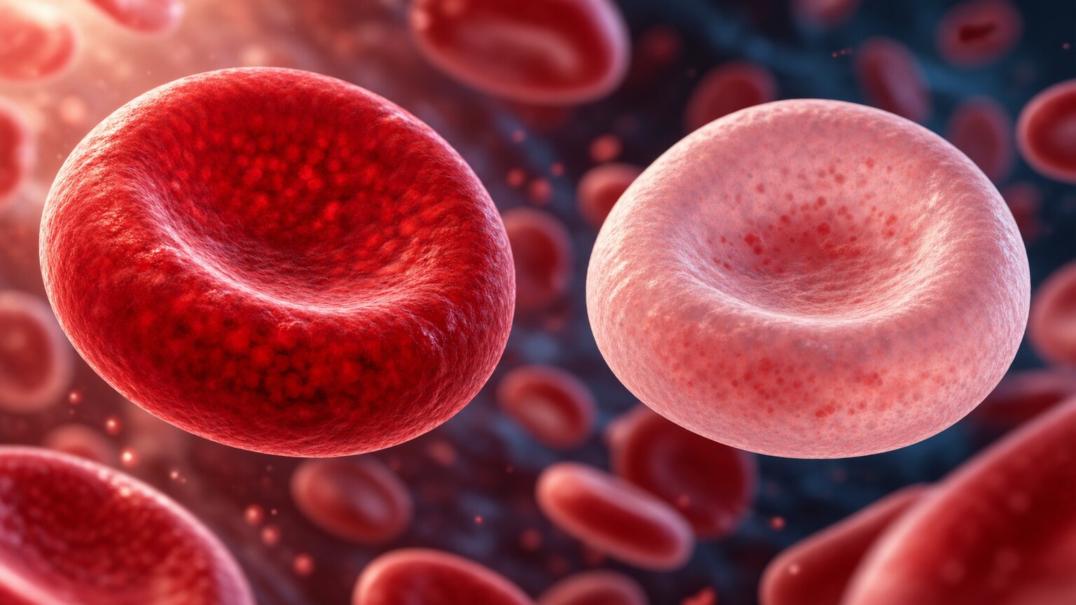
Figure 1. A low MCH reflects red blood cells carrying visibly less hemoglobin than normal, giving each cell a paler appearance under the microscope compared to a properly loaded, deeply pigmented cell.
What MCH Actually Calculates
Mean corpuscular hemoglobin, or MCH, is calculated by dividing your total hemoglobin measurement by your total red blood cell count, producing the average weight of hemoglobin packed inside a single red blood cell, regardless of that cell's overall size. This distinguishes it from MCV, which measures cell size, and from MCHC, which measures hemoglobin concentration relative to the cell's volume — MCH specifically answers "how much hemoglobin, on average, does one cell actually contain," independent of how large or small that cell happens to be.
Because MCH is a direct byproduct of these two other measurements rather than an independently measured value, it tends to move in the same direction as MCV in most conditions — smaller cells generally hold less hemoglobin in absolute terms, simply because there's less physical space inside them. This is exactly why low MCH so often appears on a report alongside a low MCV, both reflecting the same underlying process of red blood cells being built smaller and lighter than they should be.
It's worth being precise about what a "normal" MCH range actually looks like, since labs vary slightly in their reference intervals but most set the lower boundary somewhere around 27 picograms per cell. A value that sits just barely below that threshold, in someone with no symptoms and a completely unremarkable rest of the CBC, often warrants nothing more than a routine repeat test in a few months rather than an immediate workup — labs occasionally drift a point or two below the cutoff due to nothing more than normal day-to-day physiological variation or a minor lab-to-lab calibration difference. A value that's meaningfully below the reference range, particularly alongside a low MCV or a low hemoglobin, is the pattern that actually warrants the kind of systematic evaluation described in the rest of this article.
It's also useful to understand why MCH exists as a separate value at all, rather than clinicians simply relying on MCV and hemoglobin alone. In practice, MCH adds confirmatory weight to a picture that MCV alone can sometimes present ambiguously — a borderline-low MCV paired with a clearly low MCH strengthens the case that hemoglobin production is genuinely compromised, rather than the smaller cell size being an isolated finding without functional consequence. Laboratories report MCH precisely because, taken together with MCV and MCHC, it lets a single blood draw distinguish between several fundamentally different processes without requiring a more invasive test as a first step.
Iron Deficiency: By Far the Most Common Cause
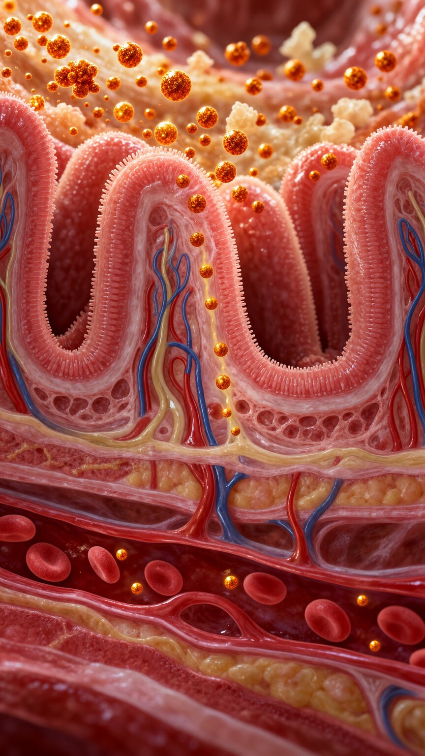
Figure 2. Iron deficiency limits how much raw material the bone marrow has available to build hemoglobin, producing red blood cells with a lower-than-normal hemoglobin content per cell.
Iron is the central component of heme, the molecule at the core of hemoglobin responsible for actually binding oxygen, and without enough iron reaching the bone marrow, developing red blood cells simply can't assemble as much hemoglobin as they normally would before being released into circulation. This is why iron deficiency is overwhelmingly the most common cause of a low MCH, accounting for the majority of cases seen in routine clinical practice.
Iron deficiency itself develops for several distinct reasons: inadequate dietary intake, particularly in people following diets low in iron-rich foods; reduced absorption, whether from a digestive condition affecting the intestinal lining or from certain medications that interfere with iron uptake; or ongoing blood loss, which removes iron along with the red blood cells themselves — heavy menstrual periods, slow gastrointestinal bleeding, or frequent blood donation are among the more common sources. Because blood loss is such a significant contributor, unexplained iron deficiency in someone without an obvious dietary explanation often prompts a search for a hidden source of bleeding, particularly in the digestive tract.
The process also unfolds in recognizable stages rather than appearing all at once. The body first draws down its stored iron reserves, reflected in a falling ferritin level while the CBC itself can still look completely normal — this early stage is sometimes called latent iron deficiency, and it can persist for weeks or months before red blood cell production actually starts to suffer. Only once those reserves are meaningfully depleted does the bone marrow start producing red blood cells with measurably less hemoglobin, which is the point at which MCH and MCV begin drifting downward on a routine CBC. This staged progression is exactly why someone can have genuinely low iron stores for a considerable stretch of time before it ever shows up as a low MCH — and conversely, why a normal MCH doesn't fully rule out an early, developing iron deficiency if the clinical suspicion is otherwise strong.
Certain groups face a meaningfully higher baseline risk of developing iron-deficiency-driven low MCH, which is worth knowing if you fall into one of them. Menstruating individuals, particularly those with heavy or prolonged periods, lose iron on a monthly basis that dietary intake doesn't always fully replace. Pregnant individuals require substantially more iron to support the expanding blood volume and the developing fetus, which is why routine prenatal iron supplementation is standard practice in most pregnancy care protocols. People who have undergone bariatric surgery often absorb iron less efficiently afterward, since the surgery frequently bypasses the section of the small intestine most responsible for iron uptake. And endurance athletes, somewhat counterintuitively, can develop a mild degree of iron deficiency through a combination of gastrointestinal blood loss from repetitive impact, iron loss in sweat, and a foot-strike-related mechanical breakdown of red blood cells during prolonged, high-impact training.
Curious what your own CBC or iron panel is really telling you? Upload your results and get a complete, plain-language breakdown in under 15 minutes.
Analyze My ResultsThalassemia Trait: A Genetic Cause Often Mistaken for Iron Deficiency
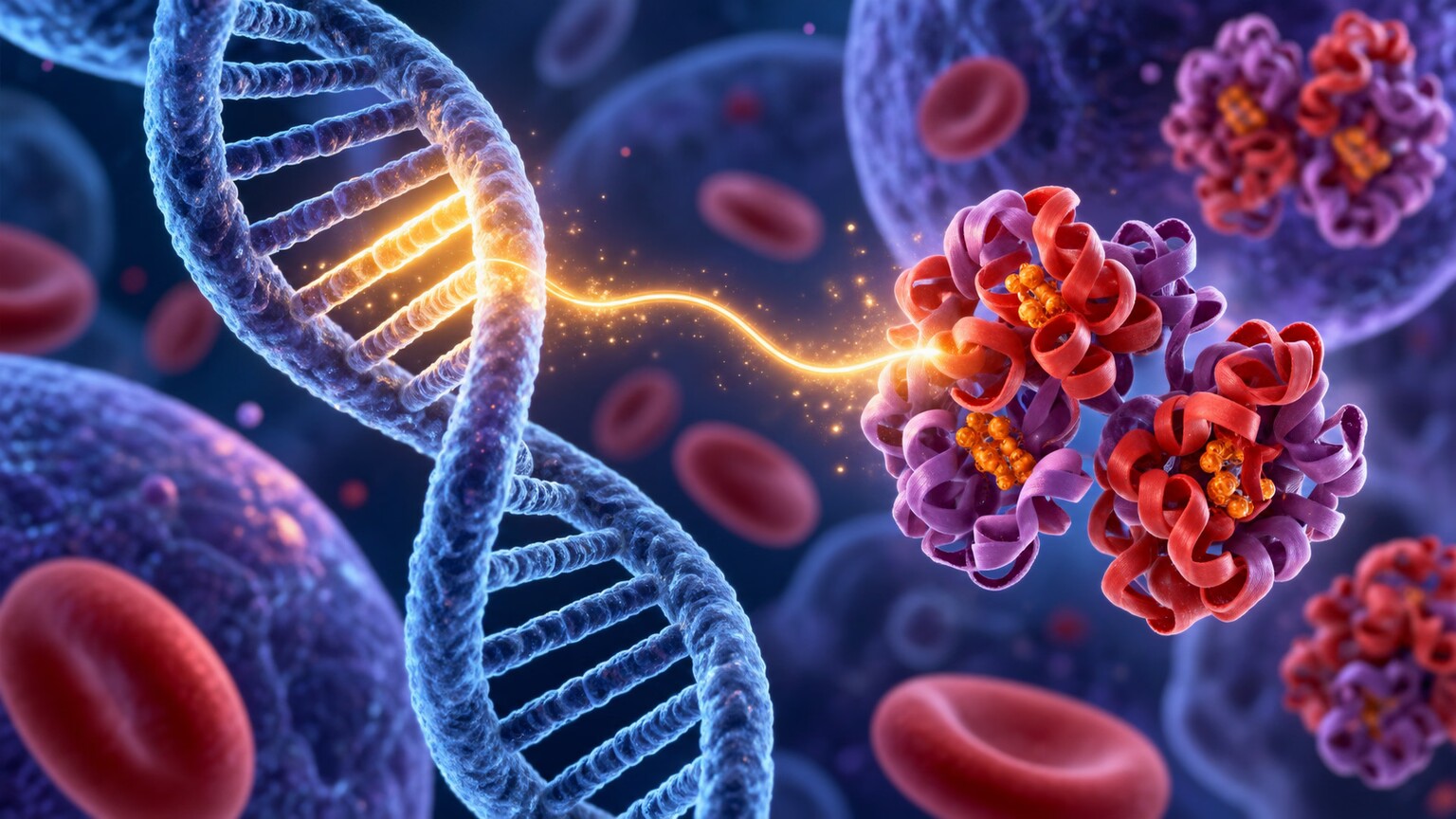
Figure 3. Thalassemia trait involves an inherited reduction in production of one of hemoglobin's protein chains, resulting in smaller, lower-hemoglobin red blood cells present from birth rather than developing over time.
Thalassemia trait is a genetic condition in which one of the genes responsible for producing a specific protein chain within hemoglobin doesn't function at full capacity, resulting in red blood cells that are inherently smaller and carry less hemoglobin than normal — producing a low MCH that's been present since birth rather than developing gradually over time the way iron deficiency does. People carrying thalassemia trait are usually otherwise healthy and often have no symptoms at all, discovering the finding incidentally on a routine blood test.
Distinguishing thalassemia trait from iron deficiency matters enormously in practice, since the two conditions can look remarkably similar on a basic CBC — both typically show low MCH alongside low MCV — but require completely different management. Iron deficiency responds to iron supplementation; thalassemia trait does not, and giving iron supplements to someone with thalassemia trait who doesn't actually need them provides no benefit and can potentially contribute to iron overload over time if continued unnecessarily. One useful early clue that favors thalassemia trait over iron deficiency is a red blood cell count that's normal or even elevated despite the low MCH and MCV, since thalassemia trait often produces a higher number of smaller cells rather than a normal number of properly sized ones running low on iron.
Thalassemia trait comes in a few different forms depending on which of the two main hemoglobin protein chains is affected. Alpha-thalassemia trait involves a reduced supply of the alpha chain and is especially common among people of Southeast Asian, African, and Mediterranean descent; beta-thalassemia trait involves the beta chain instead and is more frequently seen in people of Mediterranean, Middle Eastern, and South Asian descent, though neither form is strictly confined to these populations. Because both are inherited in an autosomal pattern, a parent carrying the trait has roughly a fifty percent chance of passing the affected gene to each child, which is why genetic counseling is often recommended for couples where one or both partners carry a known thalassemia trait, particularly before starting a family, since inheriting affected genes from both parents can result in a much more serious form of the condition in a child.
There's also a practical calculation clinicians sometimes use at the bedside to help distinguish these two leading causes before formal testing comes back: dividing the MCV by the red blood cell count. A resulting value below roughly 13 tends to favor thalassemia trait, while a value above that threshold tends to favor iron deficiency. This index isn't perfectly reliable on its own and never replaces ferritin testing or hemoglobin electrophoresis, but it's a useful piece of supporting evidence clinicians weigh alongside the rest of the picture, especially in settings where more specialized testing takes time to come back.
Anemia of Chronic Disease: A Subtler, Frequently Overlooked Cause
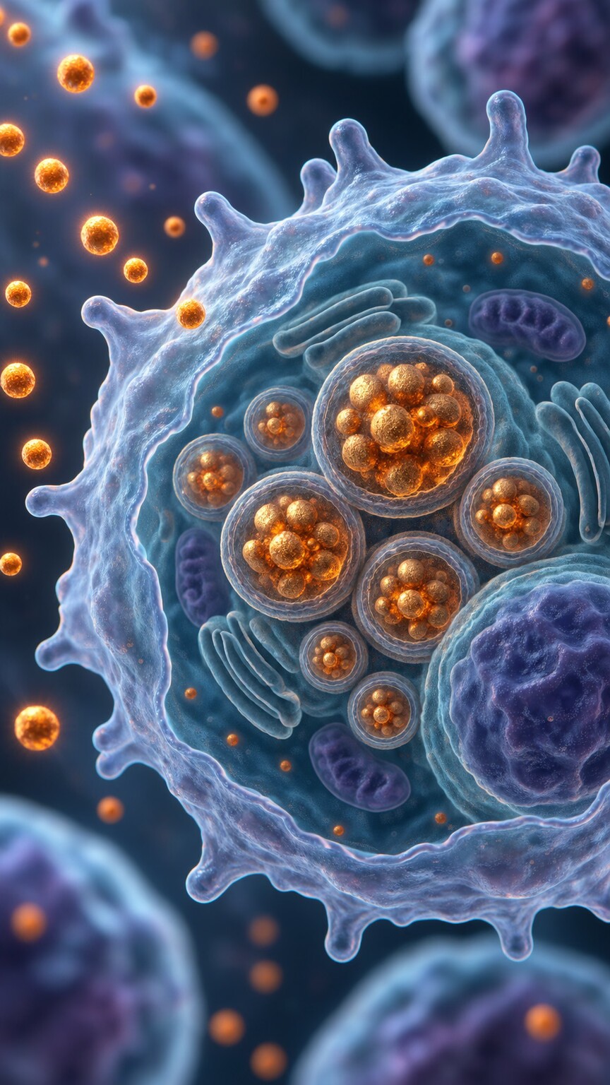
Figure 4. Chronic inflammation causes the body to trap and withhold available iron inside storage cells, making it inaccessible for red blood cell production even though total body iron stores may actually be normal or high.
Ongoing inflammatory conditions — rheumatoid arthritis, inflammatory bowel disease, chronic kidney disease, and certain long-standing infections among them — can produce a low MCH through an entirely different mechanism than true iron deficiency. Inflammation triggers the release of a hormone called hepcidin, which specifically restricts how much stored iron gets released for use in making new hemoglobin, effectively locking iron away inside storage cells even when the body's total iron reserves are perfectly adequate or even elevated.
This distinction — a genuine shortage of iron versus plenty of iron that simply isn't being released for use — is exactly why anemia of chronic disease requires a different diagnostic approach and different management than straightforward iron deficiency. Giving iron supplements to someone with anemia of chronic disease generally doesn't resolve the underlying problem, since the issue isn't a lack of iron but a lack of access to iron that's already present; the more effective approach usually involves treating the underlying inflammatory condition directly, which allows iron regulation to normalize once the inflammatory trigger settles down.
Anemia of chronic disease also tends to produce a somewhat different overall pattern than true iron deficiency, even though the MCH itself may look similarly low in both. In many cases of anemia of chronic disease, the MCV stays closer to the normal range even as the MCH dips slightly, producing what's sometimes described as a normocytic, mildly hypochromic picture rather than the more pronounced microcytic, hypochromic pattern typically seen with iron deficiency. The degree of anemia in chronic disease is also usually milder and progresses more slowly, tracking the severity and duration of the underlying inflammatory condition rather than an acute or rapidly worsening process.

Figure 5. Ferritin and RDW results, read together rather than in isolation, are usually what actually separates a genuine iron shortage from thalassemia trait or chronic inflammation as the explanation for a low MCH.
Distinguishing anemia of chronic disease from a combined problem — someone with both an underlying inflammatory condition and a genuine, separate iron deficiency at the same time — adds another layer of complexity that clinicians occasionally have to untangle. This overlap is common enough that ferritin alone can be misleading in this specific scenario, since inflammation itself tends to push ferritin upward independent of actual iron stores, potentially masking a true deficiency that would otherwise show up as a low ferritin. In these more ambiguous cases, additional markers such as transferrin saturation or soluble transferrin receptor levels are sometimes used to clarify the picture further.
Lead Poisoning and Other Rarer Causes
Lead directly interferes with several of the enzymatic steps involved in building heme, the iron-containing core of the hemoglobin molecule, meaning lead exposure can produce a low MCH through direct interference with hemoglobin assembly rather than through any shortage of raw materials. This cause is considerably less common than iron deficiency or thalassemia trait in most populations, but it's specifically worth considering in children with unexplained low MCH, particularly those living in older housing with a documented history of lead paint, given how much more sensitive a developing child's nervous system is to lead's other toxic effects beyond blood.
Adults can also develop lead-related changes to their red blood cells, most commonly through occupational exposure in settings like battery manufacturing, radiator repair, certain construction and renovation work involving older lead-based paint, or firing-range instruction without adequate ventilation and protective measures. Unlike iron deficiency or thalassemia trait, lead-related changes to MCH typically resolve once the source of exposure is identified and removed, though more significant or prolonged exposures sometimes require chelation therapy, a medical treatment that helps the body eliminate excess lead more quickly than it otherwise would on its own.
Sideroblastic anemia, a rarer group of conditions where the bone marrow has iron available but can't properly incorporate it into hemoglobin due to a defect somewhere in the heme-building pathway itself, represents another uncommon but important cause. Some forms are inherited, while others develop later in life from certain medications, alcohol use, or as part of a bone marrow disorder — distinguishing this cause typically requires specialized testing beyond a standard CBC, including direct examination of bone marrow cells under a microscope.
A few other, even less common contributors round out the full list. Copper deficiency, though rare, plays a role because copper is needed for the body to properly transport and utilize iron, and a deficiency can produce a low MCH pattern that closely resembles iron deficiency despite normal or even high iron stores — this is occasionally seen in people who have had extensive gastric bypass surgery or who take excessive zinc supplements over a long period, since high zinc intake interferes with copper absorption. Certain chronic infections, particularly long-standing ones affecting the gastrointestinal tract, can produce a low MCH through a combination of low-grade blood loss and the same inflammatory iron-restriction mechanism seen in anemia of chronic disease more broadly. And in rare cases, myelodysplastic syndrome, a group of bone marrow disorders more common in older adults, can produce a low MCH as one part of a broader pattern of abnormal blood cell production, usually alongside other CBC abnormalities that prompt more urgent specialist evaluation.
Symptoms That Sometimes Accompany a Low MCH
A low MCH by itself, especially a mild one, frequently produces no noticeable symptoms at all — it's often caught incidentally on routine bloodwork done for an unrelated reason. When symptoms do appear, they generally stem from the reduced oxygen-carrying capacity that a shortage of functional hemoglobin represents, rather than from the low MCH value as an isolated number: persistent fatigue that doesn't improve with rest, noticeable shortness of breath during activity that previously felt easy, unusual paleness of the skin or the inside of the lower eyelid, lightheadedness on standing, and a reduced tolerance for exercise are among the most commonly reported.

Figure 6. Fatigue, noticeable paleness, and reduced exercise tolerance are among the more common signs of reduced oxygen-carrying capacity, though many people with a mildly low MCH notice nothing at all.
Certain symptoms point more specifically toward one underlying cause than another, which can offer a useful clue even before lab results come back. Cravings for non-food substances such as ice, dirt, or starch — a phenomenon known as pica — are a distinctive, though not universal, sign specifically associated with iron deficiency rather than the other causes discussed here. Brittle, spoon-shaped nails and a sore, unusually smooth tongue are two other findings more specific to significant, longer-standing iron deficiency than to thalassemia trait or anemia of chronic disease. Because thalassemia trait so rarely causes any symptoms at all given how mild the associated red blood cell changes typically are, the presence of noticeable symptoms in someone eventually diagnosed with thalassemia trait should generally prompt a look for a second, coexisting explanation rather than being attributed to the trait itself.
Cardiovascular symptoms deserve particular mention because they're easy to overlook or attribute to something else entirely, especially in older adults. A heart working to compensate for reduced oxygen-carrying capacity may beat faster or harder than usual, sometimes producing a noticeable fluttering sensation or palpitations, and in more significant or long-standing cases, this added workload can occasionally unmask or worsen an underlying heart condition that had otherwise been stable. Headaches and difficulty concentrating are also reported with some frequency, reflecting the brain's high sensitivity to even modest reductions in oxygen delivery. None of these symptoms are specific to a low MCH on their own — they overlap with dozens of other conditions — which is exactly why a symptom pattern alone is never sufficient for diagnosis and why the actual lab values, interpreted together, remain the more reliable starting point.
It's also worth noting that the severity of symptoms doesn't always track neatly with how far below normal the MCH itself falls. Someone with a gradually developing, longstanding iron deficiency can sometimes adapt remarkably well and report surprisingly few symptoms despite fairly low values, since the body has had time to make compensatory adjustments to blood flow and oxygen delivery. Conversely, someone with a more acute drop, such as from sudden significant blood loss, can feel dramatically unwell even at values that might otherwise seem only moderately abnormal, simply because there's been no time for the body to adapt. This is one more reason clinicians weigh the full clinical context — how quickly a change developed, alongside other symptoms and findings — rather than reacting to a single isolated number.
How Doctors Actually Distinguish Between These Causes
Given how similarly several of these conditions can present on a basic CBC, working through a low MCH result typically starts with a ferritin test, which measures the body's stored iron reserves and is usually the single most useful next step for separating true iron deficiency from the other possibilities. A low ferritin strongly supports genuine iron deficiency, while a normal or high ferritin alongside a low MCH points the investigation away from iron deficiency and toward thalassemia trait, anemia of chronic disease, or one of the rarer causes instead.
Red cell distribution width, or RDW, adds another useful layer of information to this picture. Iron deficiency typically produces a high RDW, reflecting a genuinely mixed population of red blood cells at different stages of hemoglobin deprivation, while thalassemia trait more often shows a normal RDW despite the low MCH, since the cells, though uniformly small, tend to be more consistently sized with each other. When the ferritin and RDW results don't clearly point toward a single answer, hemoglobin electrophoresis — a test that separates and measures the different types of hemoglobin present in the blood — is typically the next step, since it can directly confirm or rule out thalassemia trait by identifying the specific pattern of hemoglobin chain production involved.
A handful of additional tests round out the picture when the initial results still leave uncertainty. A peripheral blood smear, in which a hematologist or laboratory technician directly examines a thin layer of blood under a microscope, can reveal characteristic cell shapes that point toward a specific cause — target cells and basophilic stippling are more typical of thalassemia, while pencil-shaped cells are a classic, though not perfectly specific, finding in significant iron deficiency. A reticulocyte count, which measures how many young, newly produced red blood cells are circulating, helps clarify whether the bone marrow is actively responding to a shortage by ramping up production, or whether production itself is impaired. In cases where lead exposure is a genuine concern, a blood lead level is drawn directly, and where sideroblastic anemia is suspected, a bone marrow biopsy with special staining for iron-containing precursor cells, known as ring sideroblasts, provides the definitive answer.
The overall sequence, in most routine practice, moves from least to most invasive: a basic CBC with red cell indices first, ferritin and RDW next since both come from the same blood draw and carry low cost and low risk, and only then hemoglobin electrophoresis, peripheral smear review, or bone marrow evaluation if the simpler tests haven't already provided a clear answer. Most cases of low MCH are fully explained well before reaching the more invasive end of that sequence.
Managing and Preventing Low MCH Going Forward
Once the underlying cause of a low MCH has been identified, the practical management differs considerably depending on which explanation actually applies, which is exactly why establishing the correct cause matters so much before starting any treatment. For confirmed iron deficiency, oral iron supplementation remains the standard first-line approach, typically taken on an empty stomach or with a source of vitamin C to improve absorption, and away from calcium-rich foods, coffee, or tea, all of which can meaningfully reduce how much iron the body actually absorbs from a given dose. Follow-up testing after a defined course of supplementation, usually somewhere around eight to twelve weeks, confirms whether the levels have responded as expected or whether an underlying source of ongoing blood loss still needs to be identified and addressed.
For thalassemia trait, no treatment is generally needed at all beyond documentation of the diagnosis for future reference, avoidance of unnecessary iron supplementation, and, when relevant, genetic counseling for anyone planning to have children with a partner who might also carry a thalassemia trait. For anemia of chronic disease, the most effective long-term strategy centers on managing the underlying inflammatory or chronic illness itself as effectively as possible, since the low MCH in this setting is a downstream reflection of that condition rather than an independent problem requiring its own separate treatment. Dietary strategies that support healthy iron status more broadly — regular intake of iron-rich foods such as red meat, poultry, fish, legumes, and iron-fortified grains, paired with vitamin-C-rich foods to enhance absorption from plant sources in particular — are reasonable for most people to incorporate regardless of the specific underlying cause, simply as a general measure supporting healthy red blood cell production over time.
Ongoing monitoring matters too, even after an initial cause has been identified and addressed. For someone who's had one episode of iron deficiency, particularly if a clear cause like heavy menstrual bleeding is still present, periodic rechecking of the CBC and ferritin — often annually, or sooner if symptoms return — helps catch a recurrence early, before it progresses to the point of causing noticeable symptoms again. For someone with thalassemia trait, no ongoing monitoring is typically needed at all beyond the one-time confirmation, since the underlying genetic finding doesn't change or progress over a person's lifetime.
A Worked Example: The Same Low MCH, Three Different Explanations
Consider a 28-year-old woman with heavy menstrual periods and new-onset fatigue, whose CBC shows a low MCH alongside a low MCV and a high RDW. Her ferritin comes back significantly low, confirming genuine iron deficiency, and her physician begins iron supplementation while also investigating the specific cause of her heavy bleeding, ultimately identifying and treating a benign uterine condition responsible for the excess blood loss.
Now consider a completely asymptomatic 24-year-old man who has an identical low MCH and low MCV discovered incidentally on a pre-employment physical, but with a normal RDW and a normal red blood cell count that's actually on the higher end of normal. His ferritin comes back entirely normal, and given this specific combination — normal iron stores, normal RDW, elevated-normal red cell count — hemoglobin electrophoresis is ordered and confirms alpha-thalassemia trait, a lifelong, harmless genetic finding requiring no treatment at all, simply documentation for his own future reference and for genetic counseling purposes if he plans to have children.
A third patient, a 61-year-old with long-standing rheumatoid arthritis, shows the same low MCH pattern, but her ferritin comes back elevated rather than low, consistent with anemia of chronic disease rather than either of the previous two explanations. Her treatment focuses on better controlling her underlying rheumatoid arthritis rather than iron supplementation, since her iron stores were never actually deficient to begin with. Three patients, an identical starting lab value, three genuinely different underlying stories, and three completely different appropriate responses.
A fourth scenario illustrates why the combination of markers matters more than any single number in isolation. A 45-year-old man with a decade-long history of Crohn's disease presents with a low MCH, a mildly low MCV, and a ferritin that comes back within the normal range — on its own, this ferritin result might seem reassuring, but given his active inflammatory bowel disease, his physician also orders a transferrin saturation, which comes back low, suggesting his true iron stores are lower than the ferritin alone implies, since inflammation is artificially propping the ferritin number up. His clinical picture ends up representing a genuine overlap: a degree of true iron deficiency, likely related to chronic low-grade blood loss from his intestinal disease, layered on top of the iron-restricting effect of ongoing inflammation. His management addresses both threads at once, combining cautious iron repletion with tighter control of his underlying Crohn's disease.
These four scenarios share the same starting point — a single abnormal number on a routine CBC — but diverge completely once the supporting evidence is examined, which is really the central lesson behind interpreting a low MCH responsibly: the number itself is a starting question, never a finished answer.
Frequently Asked Questions
Does low MCH always mean I have anemia?
Not necessarily on its own, though it often does correlate with it. MCH reflects hemoglobin content per cell, while anemia specifically refers to a low overall hemoglobin or red blood cell count — the two are related but measure different things.
Can iron supplements help if I have thalassemia trait instead of iron deficiency?
No. Thalassemia trait isn't caused by insufficient iron, so supplementation provides no real benefit and, if continued unnecessarily over a long period, could theoretically contribute to iron overload.
Why would my ferritin be normal or high if my MCH is low?
This pattern often points toward anemia of chronic disease, where iron is present in the body but trapped inside storage cells due to inflammation, or toward thalassemia trait, which isn't related to iron levels at all.
Is a low MCH ever a sign of something urgent?
Rarely urgent on its own, though the underlying cause sometimes needs prompt attention, such as unexplained gastrointestinal bleeding causing iron deficiency, or lead exposure in a child.
What test confirms thalassemia trait specifically?
Hemoglobin electrophoresis is the standard confirmatory test, since it directly identifies the specific pattern of hemoglobin chain production associated with this genetic condition.
How long does it take for MCH to return to normal after starting iron supplements?
Hemoglobin itself typically starts improving within two to three weeks of consistent supplementation, but MCH and MCV, which reflect the properties of red blood cells already in circulation, can take six to twelve weeks to normalize, since older, hemoglobin-deficient cells need to be gradually replaced by newly produced ones.
Can a low MCH result from something as simple as not eating enough iron-rich food?
Yes, dietary iron intake that consistently falls short of the body's needs is a genuine and common contributor, particularly in people following long-term vegetarian or vegan diets without deliberate attention to iron-rich plant sources or supplementation, though it's usually one contributing factor among several rather than the sole explanation.
Conclusion
A low MCH is a specific, well-defined signal that your red blood cells are carrying less hemoglobin than they should, on average — but the reason behind that signal varies considerably depending on what's actually happening upstream, whether that's a genuine shortage of iron, a lifelong genetic difference in how hemoglobin gets built, inflammation quietly locking iron away, or a rarer toxic or bone marrow-related cause. Sorting through these possibilities usually takes just a handful of straightforward follow-up tests, and getting the right answer matters enormously, since the correct next step differs completely depending on which explanation actually applies.
The most practical takeaway from all of this is that a single low MCH value, viewed in isolation, should prompt curiosity rather than alarm. Paired with the right handful of follow-up tests — most often a ferritin, an RDW, and occasionally a hemoglobin electrophoresis or peripheral smear — it becomes one of the more genuinely useful clues a routine blood test can offer, capable of pointing toward anything from a straightforward dietary fix to a lifelong genetic finding worth simply knowing about to an underlying condition that benefits from more active management.
Still Not Sure What Your Results Mean?
Upload your labs and get a complete, visual, plain-language interpretation of every biomarker — delivered to your inbox in under 15 minutes.
Get My ReportThis article is for educational purposes only and does not constitute medical advice. Always consult your healthcare provider regarding your specific lab results.