
Insulin and Cortisol Can Influence Each Other More Than Expected
Cortisol is usually described in casual conversation as "the stress hormone" and insulin as "the blood-sugar hormone," as if they belong to two entirely separate categories of biology handling two entirely separate jobs, filed under different chapters in a biology textbook and rarely discussed in the same breath. Research tells a more tangled story. Cortisol directly reduces how effectively your muscle cells respond to insulin, sometimes cutting insulin-stimulated glucose uptake by 40% to 60%, and insulin, in turn, appears to actively boost how much genuinely active cortisol your own fat tissue quietly generates internally on its own, creating a feedback relationship that runs in both directions rather than a simple one-way chain of command flowing from stress hormone down to blood-sugar hormone. Nearly 70% of people diagnosed with Cushing syndrome, a condition defined specifically by chronically excessive cortisol exposure, develop some measurable form of impaired glucose regulation as a direct consequence, and the same underlying mechanisms appear to operate at a smaller, more everyday scale in ordinary chronic stress. This article walks carefully through exactly how these two hormone systems affect each other, the specific cellular mechanisms involved — spanning muscle, liver, fat tissue, and the pancreas itself — and why chronic stress and everyday metabolic health turn out to be far more tightly and measurably connected than most people ever realize.
Two Hormones, Two Very Different Origins
It's worth pausing on just how unrelated these two systems appear to be when studied purely in the classroom, before looking at how they actually behave in a living body. Endocrinology curricula typically teach the HPA axis and cortisol as part of the stress response system, alongside adrenaline and the broader sympathetic nervous system, while insulin and glucose regulation are taught separately under pancreatic and metabolic endocrinology, often in an entirely different unit or semester. This particular educational separation isn't wrong exactly — the two systems really do have distinct primary triggers, distinct release patterns, and distinct primary jobs to perform — but it can leave the lasting impression that they operate in parallel, non-interacting lanes running side by side without ever crossing, which the actual cellular research simply doesn't support once you look closely enough at where each hormone's effects actually land. The overlap becomes obvious once you start looking specifically at shared tissue: muscle, liver, and fat cells all carry receptors for both insulin and cortisol, sitting side by side on the same cell surface and inside the same cell nucleus, meaning both hormones are constantly and continuously competing to influence the exact same underlying cellular machinery, often the exact same individual genes and the exact same transport proteins, rather than operating quietly on entirely separate, non-overlapping populations of cells scattered throughout different parts of the body.
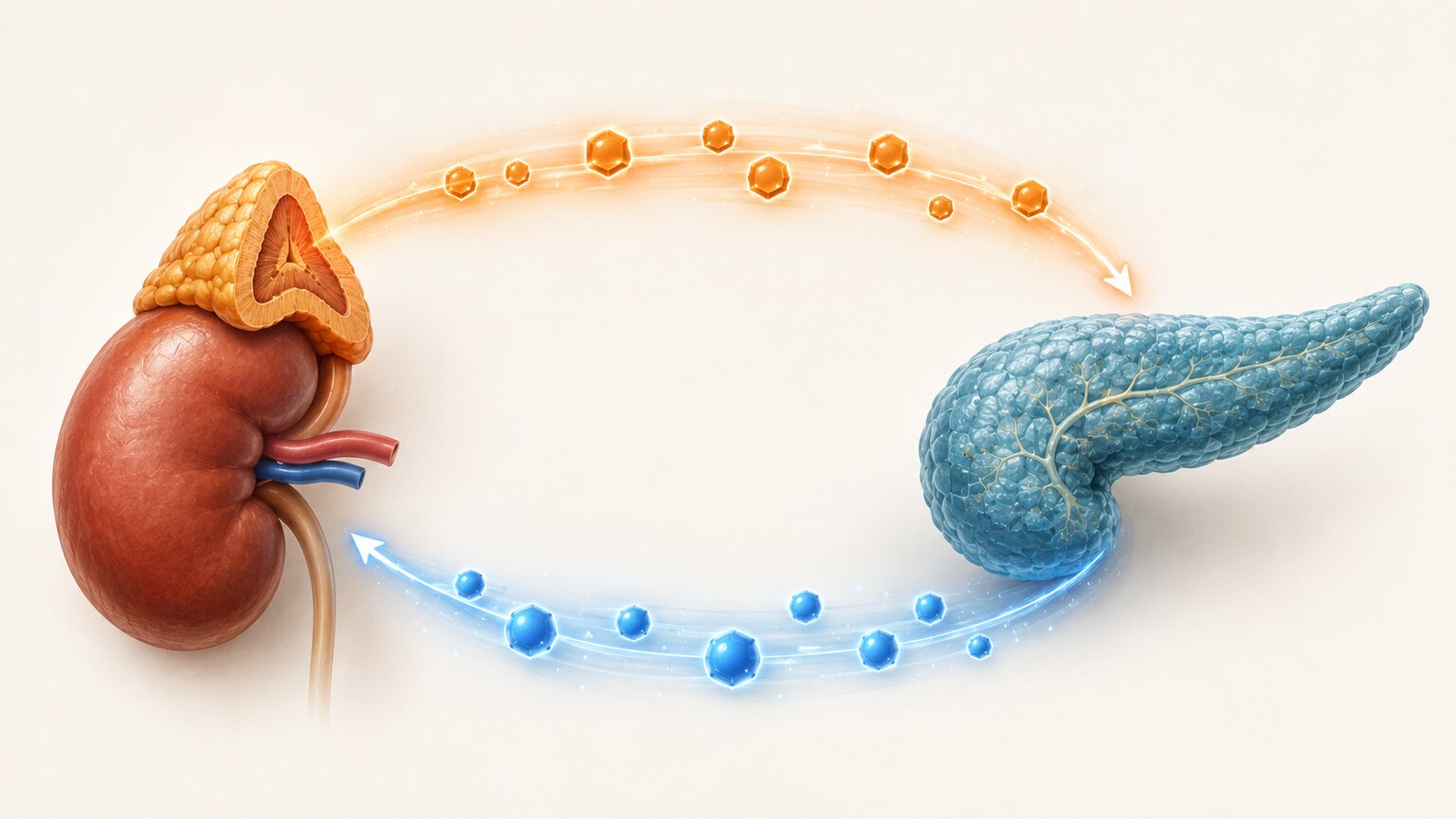
Before getting into how these hormones affect each other, it helps to understand where each one actually comes from and what job it was originally built to do. Cortisol is produced by the adrenal glands, two small structures sitting on top of each kidney, and its release is governed by the hypothalamic-pituitary-adrenal axis, commonly shortened to the HPA axis: the hypothalamus releases corticotropin-releasing hormone, which prompts the pituitary gland to release ACTH, which in turn signals the adrenal glands to synthesize cortisol and release it into general circulation. This entire hormonal cascade activates in response to either physical or psychological stress and follows a built-in negative feedback loop, where high cortisol levels signal back to the hypothalamus and pituitary to slow further release once enough has been produced, a self-limiting biological design meant to keep a normal stress response contained and genuinely temporary rather than open-ended and unchecked. Insulin, by clear contrast, is produced by specialized beta cells clustered together within the pancreas and released directly in response to rising blood glucose after a meal, with the straightforward job of telling cells throughout the entire body to pull glucose out of the bloodstream and either use it immediately for energy or store it away for later use. On paper, these two hormones represent completely separate regulatory systems, triggered by entirely different signals, governed by entirely different feedback loops, and historically studied by entirely different branches of endocrinology altogether. In actual practice, cortisol reaches directly into insulin's territory by interfering with how cells respond to insulin's signal, and insulin reaches back into cortisol's territory by influencing how much active cortisol certain tissues generate for themselves, making the two systems far more entangled at the cellular level than their separate textbook descriptions would suggest.
How Cortisol Directly Blocks Insulin's Signal
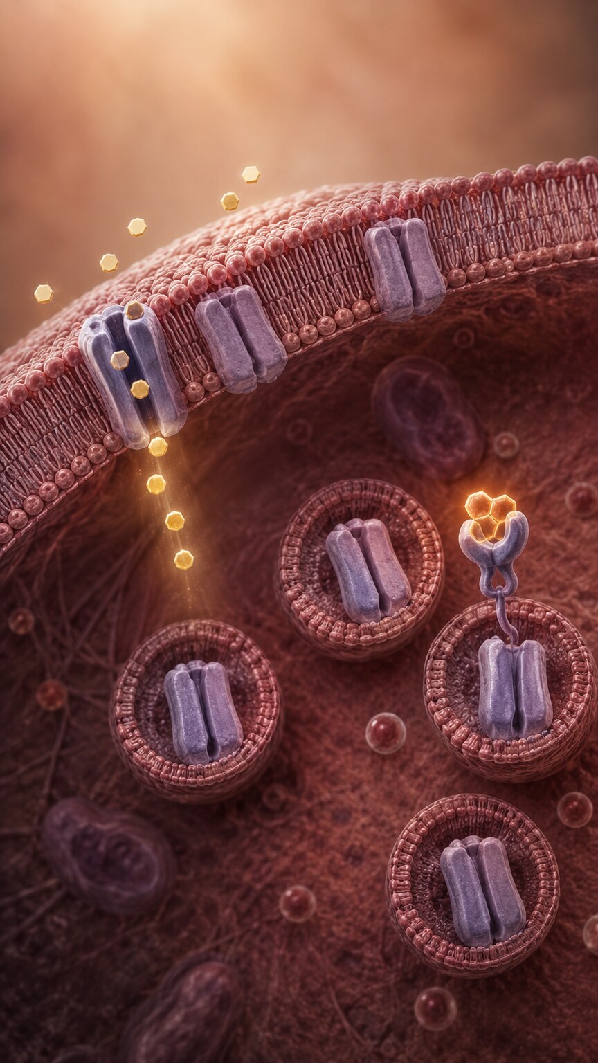
Cortisol's interference with insulin happens at a specific, well-documented cellular level rather than through some vague, general "stress effect" that's hard to pin down. Muscle cells specifically rely on a transporter protein called GLUT4 to physically pull glucose out of the bloodstream once insulin gives the signal, and this transporter normally sits in reserve inside small internal storage vesicles until insulin triggers its movement to the cell surface, where it can begin actively shuttling glucose inward. Cortisol disrupts this process directly: it reduces the translocation of GLUT4 to the cell membrane, meaning fewer of these transporter channels actually make it to the surface even when insulin is present and actively signaling exactly as it should. Research has found that this interference can reduce insulin-stimulated glucose uptake by roughly 40% to 60%, a substantial reduction that occurs even when insulin itself is being produced and released completely normally by the pancreas — the bottleneck sits downstream, at the muscle cell's response, rather than upstream at insulin's own supply. Cortisol compounds this effect by simultaneously downregulating the glycolytic enzymes cells use to actually process glucose once it manages to get inside, while at the same time stimulating an entirely opposite set of enzymes involved in gluconeogenesis, the liver's process of manufacturing brand-new glucose from other raw materials. In short, cortisol doesn't just make it harder for glucose to enter cells — it also pushes the liver to produce more glucose at the exact same time, working against insulin's basic job from two directions simultaneously rather than through a single isolated mechanism.
Curious how your own insulin and cortisol-related bloodwork lines up? Upload your labs and get a complete, plain-language breakdown in under 15 minutes.
Analyze My ResultsWhy the Liver Becomes a Battleground Between the Two Hormones
The liver is where cortisol and insulin's opposing agendas collide most directly, and understanding this collision helps explain why blood sugar problems can develop even when the pancreas itself is working perfectly well. Cortisol activates two key enzymes, glucose-6-phosphatase and phosphoenolpyruvate carboxykinase, that drive gluconeogenesis forward step by step, using free fatty acids released from fat breakdown and amino acids released from ongoing muscle protein breakdown as the raw material the liver needs to manufacture entirely new glucose molecules from scratch. Insulin's job under normal circumstances is to suppress exactly this process, signaling the liver clearly and directly to stop producing new glucose once blood sugar is already adequately supplied from a recent meal and doesn't need any additional supplementation. When cortisol is chronically elevated, it actively works against this suppression, effectively telling the liver to keep manufacturing glucose even while insulin is simultaneously, and just as forcefully, trying to tell it to stop — two competing hormonal instructions arriving at the same organ at the same time, pulling in opposite directions. This ongoing tug-of-war between the two hormones explains why chronically elevated cortisol reliably produces higher blood glucose levels even in people whose pancreas is producing entirely normal, healthy amounts of insulin on schedule — the problem isn't a lack of insulin at all in this scenario, it's cortisol actively counteracting the signal insulin is trying to send at the exact organ where that signal needs to land.
What Happens When This Goes to the Extreme: Cushing Syndrome
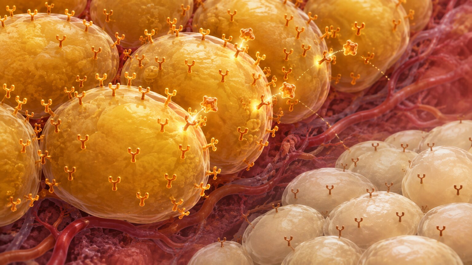
Cushing syndrome, a condition defined by prolonged exposure to abnormally high cortisol levels — whether from a tumor affecting the adrenal or pituitary gland, or from long-term use of corticosteroid medications at high doses — offers the clearest real-world demonstration of how far this relationship can go when taken to an extreme. The condition produces a recognizable, well-documented set of metabolic consequences that read almost like a checklist of everything described in this article happening simultaneously and continuously in one single person: central obesity concentrated specifically around the abdomen, muscle wasting from ongoing protein breakdown feeding gluconeogenesis, elevated blood pressure, thinning skin prone to easy bruising, and glucose intolerance. Research has found that impaired glucose tolerance affects roughly 10% to 30% of people with Cushing disease specifically, while the overall prevalence of some form of glucose metabolism impairment across the broader Cushing syndrome population reaches nearly 70%, making metabolic disruption genuinely one of the single most consistent, reliably observed features of the condition rather than merely an occasional complication seen in only a small subset of cases. Part of why the resulting fat distribution concentrates so specifically around the abdomen has to do with biology at the tissue level rather than simple caloric overflow: abdominal visceral fat contains an unusually high density of glucocorticoid receptors compared to fat elsewhere in the body, such as fat in the hips or limbs, making visceral fat specifically, and quite disproportionately, responsive to cortisol's ongoing signals to store additional fat in that exact anatomical location. This is why chronic cortisol excess, whether from Cushing syndrome itself or from sustained everyday psychological stress operating through the exact same hormonal pathway, tends to reshape body composition in this particular, recognizable pattern rather than distributing fat evenly throughout the body as ordinary weight gain typically does.
How Insulin Reaches Back Into Cortisol's Territory
The relationship isn't a one-way street where only cortisol affects insulin — insulin appears to actively influence how much active cortisol certain tissues generate for themselves, through a genuinely fascinating enzymatic mechanism that most people have never heard of despite how directly it connects everyday eating to local hormone activity. Your body produces an inactive precursor form of cortisol called cortisone, essentially a dormant version of the hormone circulating harmlessly until it's needed, and a specific enzyme, 11-beta hydroxysteroid dehydrogenase type 1 (11β-HSD1), converts that inactive cortisone into fully active cortisol directly within certain tissues, essentially manufacturing local cortisol on-site rather than relying solely on what the adrenal glands release into general circulation from further away. Research has found that a single mixed meal triggers a measurable rise in this local cortisol regeneration throughout the body, and this effect appears to be specifically and directly mediated by the resulting rise in insulin that naturally follows that particular meal, rather than by any other component of digestion occurring at the same time. In other words, eating triggers an insulin response, and that insulin response appears to directly activate more local cortisol production at the tissue level, essentially converting a routine, everyday metabolic event into a small, localized hormonal one as well — a genuinely two-way relationship where each hormone actively shapes the other's effective activity, not just a case of cortisol acting on insulin while insulin passively sits on the receiving end without any influence flowing back the other way.
Why This Matters Especially in Fat Tissue
Fat tissue isn't just a passive, inert storage depot for excess dietary energy — it functions as a genuinely active endocrine organ in its own right, capable of locally manufacturing and responding to hormones in ways that don't necessarily show up when measuring circulating levels in the bloodstream from a standard venous blood draw. This local, tissue-level hormonal activity is exactly what makes the insulin-driven 11β-HSD1 pathway described earlier so genuinely significant: it means a person's fat tissue can effectively be exposed to meaningfully more active cortisol than what a routine blood test would ever reveal, since the conversion from inactive cortisone to fully active cortisol is happening locally, quietly, inside the fat cells themselves, rather than out in general circulation where a standard venous blood draw would actually be able to detect and quantify it.
This insulin-driven cortisol activation appears to be particularly relevant in adipose tissue specifically, and it helps explain a pattern that clinicians and researchers have long noticed but struggled to fully account for using circulating hormone levels alone: obesity and elevated cortisol activity so often travel together in the same individuals, even in people without any diagnosed adrenal or pituitary condition and without circulating cortisol levels in the general bloodstream that would look obviously abnormal on a standard blood test taken at a routine visit. and research has found that 11β-HSD1 activity is measurably increased in both visceral and subcutaneous fat tissue in people who are obese compared to people at a healthy weight, with overexpression of this enzyme in visceral fat specifically associated with hyperinsulinemia in patients with severe obesity. This creates the conditions for a genuinely self-reinforcing cycle, one that can be difficult to notice from the outside since no single lab value obviously points to it: excess insulin appears to drive greater local cortisol activation within fat tissue, and that locally elevated cortisol, through the exact same GLUT4-blocking and gluconeogenesis-promoting mechanisms described earlier in this article, further worsens insulin resistance throughout the body, which in turn can drive circulating insulin levels even higher still as the pancreas compensates for reduced cellular sensitivity by producing more. Once fully established over time, a cycle like this can become genuinely difficult to interrupt at just one single point in the loop, since each hormone is actively reinforcing conditions that favor more of the other's unwanted downstream effects, rather than either hormone simply returning quietly to its own independent baseline once the immediate original trigger has passed and conditions have otherwise stabilized.
Cortisol Also Acts Directly on the Cells That Produce Insulin
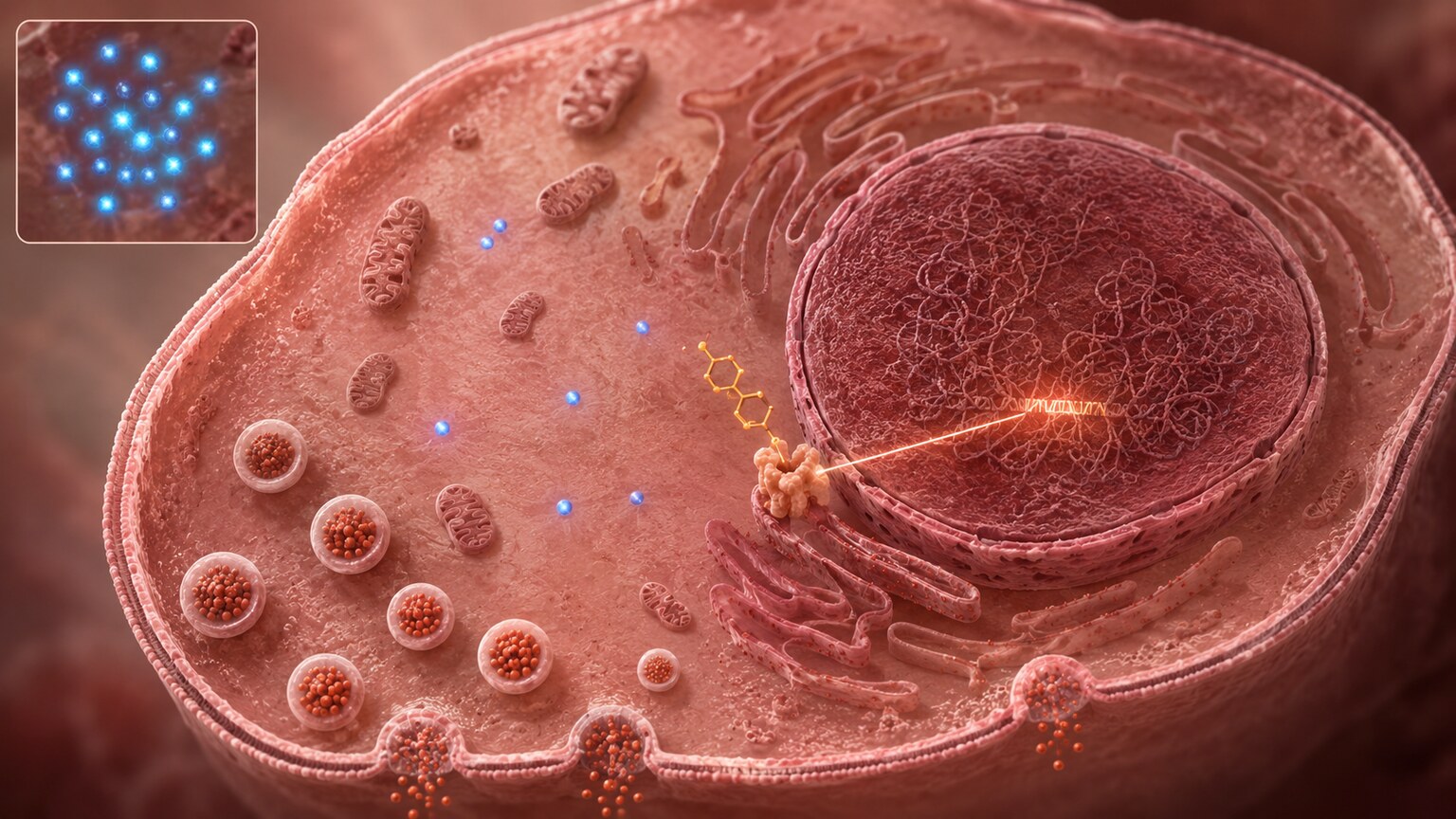
Everything described so far involves cortisol interfering with how tissues respond to insulin after it's already been released into circulation — but research has found that cortisol also acts directly on the pancreatic beta cells responsible for producing insulin in the first place, meaning the interference doesn't stop conveniently at the receiving end of the signal. Laboratory studies have found that glucocorticoids directly inhibit insulin secretion from beta cells in a dose-dependent manner, meaning higher cortisol exposure produces a correspondingly larger suppression of insulin release, an effect that comes on slowly over time and reverses only slowly once cortisol levels eventually drop back down, rather than switching on and off quickly alongside momentary, short-lived cortisol fluctuations the way some other hormonal effects do. Several distinct molecular mechanisms appear to be involved, each acting through a different piece of the cell's internal machinery: cortisol induces a protein called NECAB1 inside beta cells, which acts as a negative regulator and reduces the intracellular calcium signaling that insulin release directly and critically depends on; separately, cortisol has been shown to down-regulate Pdx-1, a transcription factor essential for normal insulin gene expression inside the cell nucleus, while simultaneously inducing a different competing factor that further suppresses that same gene expression from the opposite direction. In effect, chronically elevated cortisol doesn't just make tissues throughout the body less responsive to whatever insulin happens to be circulating at any given moment — it can also directly blunt the pancreas's own underlying capacity to produce adequate insulin in the first place, adding a genuine third mechanism on top of the liver and muscle effects already described earlier, all three pushing blood sugar regulation in the exact same unfavorable direction simultaneously and from three separate angles.
Chronic Stress, Not Just Acute Stress, Is the Real Concern
A brief, occasional cortisol spike from a genuinely stressful event isn't the primary concern here — that kind of short-term rise is a normal, entirely adaptive response that resolves on its own once the stressor passes and the HPA axis's own built-in negative feedback loop brings cortisol back down to baseline within a matter of hours. The real metabolic concern is chronic, sustained activation of the HPA axis, the kind produced by ongoing psychological stress, chronic poor sleep, unmanaged chronic illness, or persistent overtraining without adequate recovery, all of which keep cortisol elevated for extended periods of days, weeks, or months rather than allowing it to spike briefly and then fully recover back to a resting level. Research specifically describes chronic hypercortisolism, meaning sustained activation of the HPA axis over an extended stretch of time rather than a single brief episode, as producing hyperglycemia, unfavorable lipid changes, insulin resistance, and low-grade inflammation — a cluster of findings that closely mirrors, almost feature for feature, what's typically seen in people who have already been diagnosed with type 2 diabetes for some time. This is part of why chronic stress management is increasingly discussed in clinical and research settings as a genuine, evidence-based component of metabolic health, rather than a purely psychological or vague quality-of-life consideration entirely disconnected from measurable blood sugar and insulin outcomes on an actual lab report.
Why Cortisol's Daily Rhythm Matters for Interpreting a Test
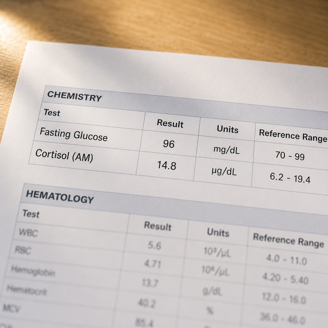
Unlike insulin, which rises and falls sharply within a matter of minutes depending on when you last ate, cortisol follows a distinct daily rhythm called the diurnal cortisol pattern, naturally peaking shortly after waking in the morning, holding relatively high through the early part of the day, and then gradually declining throughout the afternoon and evening to its lowest point around midnight before beginning to climb again quietly ahead of the next morning's wake time. This built-in rhythm is precisely why a single, isolated cortisol measurement means very little on its own without knowing exactly when during the day it was actually drawn — a level considered perfectly normal at eight in the morning would look distinctly abnormal if that exact same number showed up instead on a test drawn at ten at night, since the expected reference range itself shifts substantially across the full 24-hour cycle rather than staying fixed. Chronic stress and certain HPA axis disorders can flatten this normal, otherwise predictable rhythm, keeping cortisol elevated later into the day than it should be or blunting the size of the expected morning peak entirely, which is why some testing approaches specifically measure cortisol at multiple points across a single day, or measure it in saliva collected at bedtime specifically, precisely when it should be at its lowest, rather than relying on a single random blood draw taken at whatever hour happened to be convenient for scheduling. Because insulin's release pattern is tied primarily to meal timing rather than to a fixed time of day in the same rhythmic, clock-driven sense that cortisol follows, comparing the two hormones meaningfully side by side on paper requires understanding that they're being measured against genuinely different expected baselines depending on exactly when each individual sample was actually collected relative to both the clock and the person's last meal.
What This Means If You Have Elevated Levels of Either Hormone
Given how tightly these two systems interact at the cellular level, unexplained findings in one often genuinely warrant a closer look at the other rather than being evaluated in isolation. Someone with new insulin resistance and no obvious cause like diet, activity level, or recent weight change may benefit from investigating whether chronic stress or an underlying cortisol abnormality is quietly contributing to the picture, particularly if other Cushing-syndrome-adjacent signs like central weight gain concentrated around the midsection, muscle weakness, unusually thin or easily bruised skin, or persistently elevated blood pressure are also present alongside the insulin resistance itself. Someone being evaluated for possible cortisol dysregulation, whether prompted by physical symptoms or an initial abnormal cortisol result, may similarly benefit from having glucose and insulin checked alongside cortisol testing, since metabolic findings can sometimes provide useful supporting evidence toward a cortisol-related diagnosis that isn't yet fully confirmed by cortisol testing alone, especially in earlier or more borderline cases where the underlying cortisol abnormality itself is still relatively subtle and hasn't yet produced clearly obvious physical signs. Neither hormone should really be interpreted in complete isolation from the other, given how directly and genuinely bidirectionally they've been shown to interact at the cellular level across muscle, liver, fat tissue, and the pancreas alike, each organ contributing its own piece to the overall picture.
Frequently Asked Questions
Does cortisol actually cause insulin resistance?
Yes, through a well-documented mechanism: cortisol reduces GLUT4 transporter movement to the cell surface, cutting insulin-stimulated glucose uptake by roughly 40% to 60%, while simultaneously promoting glucose production in the liver.
Does insulin affect cortisol, or does it only work the other way around?
It works both ways. Rising insulin after a meal appears to activate an enzyme called 11β-HSD1, which converts inactive cortisone into active cortisol directly within certain tissues, particularly fat tissue.
How common is impaired glucose regulation in Cushing syndrome?
Very common. Research has found impaired glucose tolerance in roughly 10% to 30% of people with Cushing disease specifically, with overall glucose metabolism impairment reaching nearly 70% across the broader Cushing syndrome population.
Why does cortisol-related weight gain concentrate around the abdomen specifically?
Abdominal visceral fat contains an unusually high density of glucocorticoid receptors compared to fat elsewhere in the body, making it especially responsive to cortisol's signals to store fat in that specific location.
Is a brief stressful moment enough to meaningfully affect insulin sensitivity?
Not typically. The metabolic concern centers on chronic, sustained cortisol elevation rather than short-term spikes, since a brief rise usually resolves through the body's own negative feedback loop before it produces lasting effects.
Does cortisol affect the pancreas directly, not just how tissues respond to insulin?
Yes. Research shows cortisol directly inhibits insulin secretion from pancreatic beta cells by reducing calcium signaling and suppressing key genes needed for normal insulin production.
Why does the timing of a cortisol test matter so much?
Cortisol follows a natural daily rhythm, peaking in the morning and declining through the day, so a single result can only be interpreted correctly when compared against the reference range expected for that specific time of day.
Conclusion
Insulin and cortisol are often taught as if they belong to two unrelated chapters of biology, one filed under stress physiology and the other under metabolic endocrinology, but the evidence tells a genuinely interconnected story that spans nearly every major organ involved in blood sugar regulation. Cortisol directly interferes with insulin's ability to move glucose into muscle cells while simultaneously pushing the liver to produce more glucose of its own, and it also acts directly on the pancreas itself, blunting insulin secretion at the source rather than only interfering downstream. Insulin, in turn, appears to actively drive local cortisol activation within tissue like fat, particularly in the hours following a meal, through the 11β-HSD1 enzyme pathway, creating a feedback relationship that can reinforce itself once established rather than staying neatly contained to a single direction. Conditions like Cushing syndrome show just how far this relationship can go when cortisol is chronically excessive, producing a recognizable and consistent cluster of metabolic consequences in a large majority of affected people, and the same underlying mechanisms appear to operate, at a considerably smaller scale, in the kind of ordinary chronic stress many people experience for months or years without ever connecting it to their blood sugar or insulin results. Understanding this bidirectional relationship, along with practical details like cortisol's own daily rhythm, turns two seemingly separate hormone panels into a single, more complete and more accurately interpreted picture of overall metabolic health.
Still Not Sure What Your Results Mean?
Upload your labs and get a complete, visual, plain-language interpretation of every biomarker — delivered to your inbox in under 15 minutes.
Get My ReportThis article is for educational purposes only and does not constitute medical advice. Always consult your healthcare provider regarding your specific lab results.