
What's the Link Between Eczema and Elevated IgE Levels?
Eczema — the itchy, red, often cracked skin condition doctors call atopic dermatitis — and elevated IgE (immunoglobulin E, one of the antibodies your immune system produces) are linked, but not in the simple cause-and-effect way most people assume. Roughly 70 to 80 percent of people with moderate to severe eczema have higher-than-normal IgE levels in their blood, and the two conditions share the same underlying biology: a skin barrier that lets things in that shouldn't get in, and an immune system that overreacts once they do. But eczema isn't caused by high IgE the way a fever is caused by an infection — it's more accurate to say both are downstream symptoms of the same broken process. Some people have textbook eczema with completely normal IgE, and some people have sky-high IgE with no skin symptoms at all. Understanding why requires looking at what actually happens at the surface of the skin, and why that skin surface talks to your immune system far more than most people realize. By the end of this article, an IgE number on a lab report should read less like a mysterious code and more like one chapter in a story that starts at the skin's own physical defenses.
What Eczema Actually Is, and Why It Involves the Immune System at All
It's tempting to think of eczema as a skin problem, full stop — dry patches, some redness, maybe a cream from the pharmacy. But underneath the visible rash, eczema is fundamentally a problem with your skin's outermost layer, the stratum corneum, which normally functions like a brick wall: skin cells are the bricks, and a mixture of fats called lipids is the mortar holding them tightly together. In healthy skin, this wall keeps water in and keeps everything else — allergens, bacteria, irritating chemicals — out. In eczema, that mortar is deficient. The wall develops gaps. Water escapes more easily, which is why eczema-prone skin is chronically dry no matter how much lotion goes on it, and outside substances get in more easily, which is the piece most people never hear about.
Once allergens, microbes, and irritants can slip past a compromised barrier and reach the living tissue underneath, they run straight into your immune system's surveillance cells, which sit just beneath the skin's surface waiting for exactly this kind of intrusion. That contact is the spark that begins the eczema-IgE connection — not a separate allergic event happening somewhere else in the body, but a direct consequence of the skin's own physical failure to keep its gates closed. This is the central idea to hold onto through everything that follows: the skin barrier problem comes first, and the immune and IgE changes are largely a reaction to it, not an independent process running in parallel.
The specific surveillance cells doing this work are called Langerhans cells, a type of dendritic cell that lives permanently within the epidermis, positioned like a network of sentries with long, branching arms that constantly sample whatever touches the skin's living layers. When a Langerhans cell picks up a piece of an allergen that has crossed the broken barrier, it doesn't just sound a local alarm — it physically migrates to the nearest lymph node, carrying a fragment of that allergen with it, and presents it to a type of immune cell called a T-helper cell. In eczema-prone skin, that presentation tends to activate a specific subtype of T-helper cell known as Th2, and Th2 cells are the ones responsible for instructing B cells — the antibody factories of the immune system — to start manufacturing IgE specifically against whatever allergen was just presented to them. This entire relay, from a gap in the skin barrier to a fully formed IgE antibody circulating in the blood, typically takes days to weeks the first time it happens, which is one reason sensitization to a new allergen isn't something a person notices happening in real time.
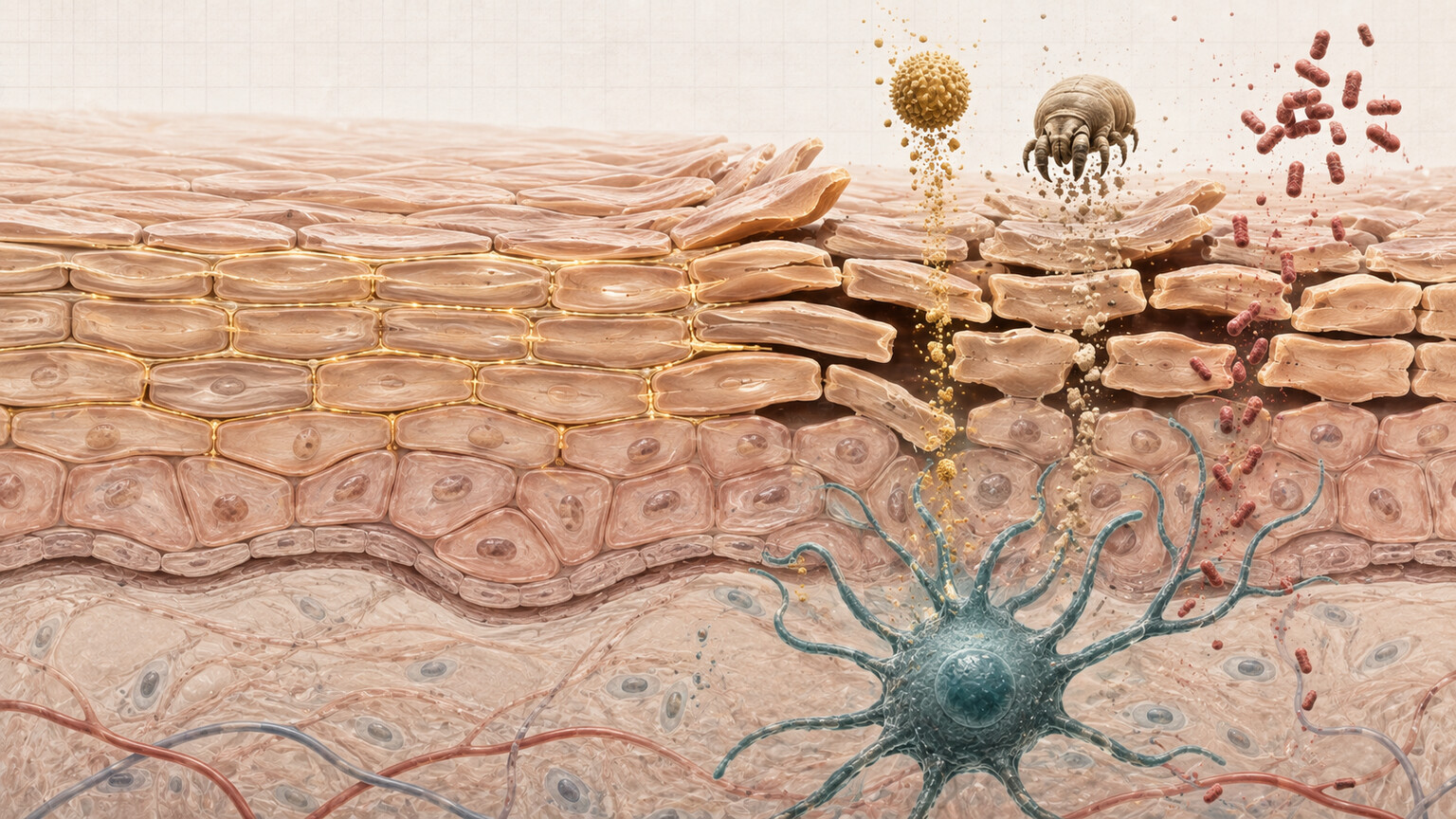
Figure 1. A cross-section of the skin's outer barrier, where gaps in the lipid layer allow allergens and microbes to reach immune cells beneath the surface.
What IgE Actually Is, and Why the Body Makes It in the First Place
IgE is one of five classes of antibody your immune system can produce, and by volume it's the rarest — most of the antibody circulating in your blood at any given moment is a different type called IgG, which handles routine defense against bacteria and viruses. IgE exists in vanishingly small amounts by comparison, and evolutionarily, it appears to have developed as a defense against parasitic worms, which is still its dominant job in parts of the world where parasitic infection is common. In industrialized countries, where parasitic infections are rare, that same IgE machinery gets redirected toward substances that were never actually dangerous — pollen, dust mite droppings, pet dander, certain foods, and, relevant here, whatever manages to cross a broken skin barrier.
Here's the part that surprises most people: IgE doesn't float around causing symptoms on its own. It works by attaching itself to the surface of specific immune cells — mainly mast cells and basophils — using a receptor built specifically to hold it in place, almost like a hook waiting for a very particular kind of bait. Once enough IgE molecules are sitting on a mast cell's surface, that cell becomes primed. It's now armed and waiting for the exact substance that IgE was built to recognize. The next time that substance shows up — even in a tiny amount — the mast cell reacts explosively, and that reaction is what actually produces the redness, swelling, and itching people associate with an allergic response. IgE is the trigger mechanism; the mast cell is the loaded gun.
The signals that push a B cell to start manufacturing IgE instead of one of the other antibody types come primarily from two proteins released by activated Th2 cells: interleukin-4 (IL-4) and interleukin-13 (IL-13). Think of these as a specific instruction handed to the B cell — essentially telling it "switch your production line to IgE." This detail matters well beyond biochemistry trivia, because it's the exact instruction that some of the newest eczema medications are designed to interrupt, a connection this article returns to later. It also explains why eczema, food allergy, asthma, and allergic rhinitis so often travel together in the same person or the same family: they're all, at their core, powered by this same IL-4/IL-13-driven, Th2-skewed immune pattern, just expressed in different tissues — skin in eczema, airways in asthma, nasal passages in allergic rhinitis.
Why a Broken Skin Barrier Is the Real Starting Point

Figure 2. Skin already inflamed from eczema gives food proteins encountered around the mouth an easier route to immune cells than intact skin would.
One of the most consistent findings in modern eczema and allergy research is that a specific gene, called filaggrin (short for "filament aggregating protein"), plays an outsized role in how well the skin barrier holds together. Filaggrin helps bind skin cells tightly and contributes to the skin's natural moisturizing factor, the mix of molecules that keeps the outer layer hydrated. Somewhere between 20 and 50 percent of people with moderate to severe eczema carry a mutation in the filaggrin gene that reduces how much of this protein they can make, and those mutations are one of the strongest known genetic risk factors for developing eczema at all.
What matters for the IgE story is what a filaggrin mutation does beyond dry skin: it makes the barrier measurably more permeable to things it should be blocking. Researchers have shown that when skin barrier function is experimentally disrupted — even in people without eczema — allergen exposure through that compromised skin is far more likely to trigger sensitization, meaning the immune system starts producing IgE against that specific allergen going forward. This is sometimes called the "outside-in" hypothesis of eczema and allergy development, and it has increasingly replaced an older idea that food and airborne allergies came first and caused skin symptoms as a side effect. The current understanding runs the other direction: allergen exposure through inflamed, barrier-broken skin is what teaches the immune system to start making IgE against foods and environmental substances in the first place.
This is part of why infants with early, severe eczema are considered at meaningfully higher risk of developing food allergies later — not because the eczema and the allergy are unrelated coincidences, but because inflamed skin around the mouth and cheeks, encountering food residue during early feeding, may be functioning as an unintended point of entry for sensitization, well before any food is deliberately eaten.
Curious what your own IgE or allergy panel results are actually telling you? Upload your results and get a complete, plain-language breakdown in under 15 minutes.
Analyze My ResultsHow IgE Actually Triggers the Redness, Itching, and Swelling of Eczema
Once IgE specific to a particular allergen is circulating and attached to mast cells sitting throughout the skin, the mechanism that produces an eczema flare becomes fairly mechanical. When that allergen — say, a dust mite protein or a food particle — crosses paths with two IgE molecules sitting close together on the same mast cell, it bridges them, a process immunologists call cross-linking. That bridging event is the actual trigger. It causes the mast cell to rapidly dump the contents of internal granules into the surrounding tissue, releasing histamine and a cascade of other inflammatory chemicals within minutes.
Histamine widens nearby blood vessels, which is why the skin turns red and can feel warm to the touch, and it makes those vessels temporarily leakier, which produces the swelling. It also directly stimulates nerve endings in the skin responsible for the sensation of itch — which is why eczema is so intensely itchy rather than just sore or tender. Scratching in response then physically damages the already-weakened skin barrier further, letting in more allergens, which can trigger more mast cells, which produces more itching. This is the itch-scratch cycle that makes eczema so difficult to break without addressing the underlying barrier and immune activity directly, rather than just treating the itch symptom in isolation.
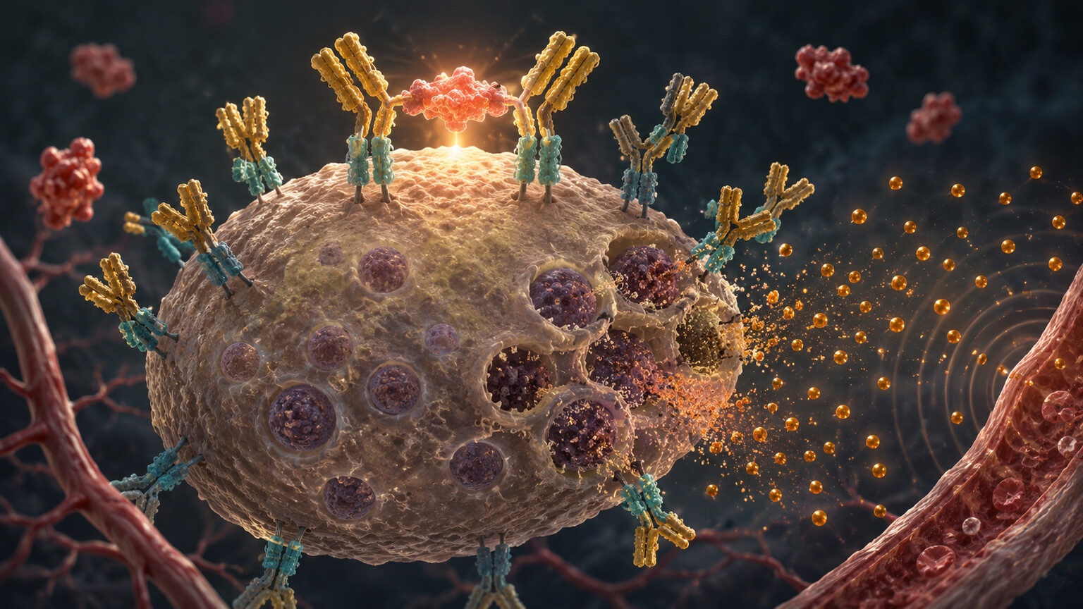
Figure 3. An allergen bridging two IgE antibodies bound to a mast cell's surface receptors, the exact molecular event that triggers histamine release.
Not All Eczema Raises IgE — Extrinsic Versus Intrinsic Atopic Dermatitis
If IgE and eczema were simply the same thing wearing two names, every person with eczema would have elevated IgE, and that isn't the case. Dermatologists and allergists divide eczema into two broad patterns based on exactly this distinction. The far more common form, sometimes called extrinsic or allergic atopic dermatitis, does involve elevated total IgE and detectable IgE against specific allergens — foods, dust mites, pet dander — and this pattern accounts for roughly 70 to 80 percent of eczema cases, more in children than adults.
The other pattern, called intrinsic (or non-allergic) atopic dermatitis, produces skin that looks clinically identical under a dermatologist's exam — the same redness, the same distribution across elbow creases and behind the knees, the same intense itch — but without elevated IgE and without positive allergy testing to explain it. This form is less common, tends to appear more often in adult-onset eczema and in women, and is thought to involve a different, still not fully understood combination of skin barrier dysfunction and immune activity that doesn't route primarily through the IgE pathway. The practical takeaway is that a normal IgE result does not rule out eczema, and clinicians diagnose eczema by examining the skin and taking a history, not by ordering an IgE test — IgE testing is used to investigate potential allergic triggers once eczema is already suspected or confirmed, not to diagnose the eczema itself.
The "Atopic March" — Why Eczema Often Shows Up Before Other Allergic Conditions
Pediatricians and allergists use the term "atopic march" to describe a pattern seen in a meaningful subset of children: eczema tends to appear first, often in the first year of life, followed over subsequent years by food allergies, then allergic rhinitis (hay fever), and then asthma, roughly in that order, though not every child who develops eczema goes on to develop the later conditions. Longitudinal studies following large groups of infants with eczema have found that a substantial proportion go on to develop at least one additional allergic condition within several years, and the severity of early eczema — along with how early it appears and how high the associated IgE levels run — has been linked to a higher likelihood of progressing along this path.
The current leading explanation for the atopic march loops directly back to the skin barrier story: if allergen exposure through broken skin is what teaches the immune system to produce allergen-specific IgE in the first place, then a child whose skin has been chronically compromised by eczema has effectively had more opportunities for that sensitization process to happen, across more allergens, over a longer stretch of early development. This is also why some pediatric research has explored whether aggressively treating eczema and repairing the skin barrier early — through consistent moisturizing and anti-inflammatory treatment — might reduce the likelihood of a child going on to develop food allergies or asthma later, an idea sometimes referred to as the "skin barrier hypothesis" of allergic disease prevention.
Environmental and Lifestyle Factors That Influence This Whole Relationship
Genetics and skin barrier biology set the stage, but they don't act in a vacuum — a number of everyday environmental factors measurably influence how much allergen exposure a compromised skin barrier actually experiences, and by extension, how much IgE-driven activity results. Hard water, which contains higher concentrations of calcium and magnesium, has been studied specifically in relation to eczema, with research suggesting it can interact with soap to leave a residue that's more irritating to already-compromised skin, and some studies have found associations between hard water exposure in early childhood and higher eczema risk. Harsh soaps, fragranced detergents, and hot bathing water all strip away more of the skin's natural lipid layer, widening the same gaps in the barrier that allergens use to get through — which is why dermatologists consistently recommend lukewarm (not hot) showers, fragrance-free cleansers, and moisturizing within minutes of bathing, while the skin is still damp, as basic eczema management.
Climate and season play a role too. Low humidity — common in winter, in heated indoor air, and in dry climates — increases water loss straight through the skin barrier, which is why eczema often visibly worsens in colder months even without any new allergen exposure. Indoor allergen load matters independently of climate: dust mites thrive in warm, humid bedding and carpeting, and homes with higher dust mite burden have been linked to higher rates of dust-mite-specific IgE sensitization in eczema-prone children. None of these factors cause eczema on their own the way a filaggrin mutation predisposes someone to it, but each one changes how much allergen exposure reaches a barrier that's already compromised — which is exactly the variable that determines how much IgE-driven inflammation follows. This is also why two people with a similar genetic eczema risk can have noticeably different disease courses depending on where they live, what products they use, and how humid their home environment tends to be.
There's also a broader idea worth mentioning, sometimes called the hygiene hypothesis: the observation that allergic conditions, including eczema, have become dramatically more common in industrialized countries over the past several decades, alongside reduced early-life exposure to a diverse range of microbes, larger family sizes, and rural farm environments. The leading explanation is that early, varied microbial exposure helps calibrate the developing immune system away from the Th2-skewed, IgE-prone pattern described earlier in this article, and that a more sanitized, lower-microbial-diversity early environment leaves that calibration incomplete in some children. This remains an active area of research rather than settled fact, but it fits within the same overall picture: eczema and elevated IgE are shaped by a mix of inherited skin barrier vulnerability and a lifetime of environmental exposures layered on top of it, not by a single fixed cause.
How Doctors Actually Use an IgE Result Alongside a Skin Exam
In practice, an allergist or dermatologist rarely looks at an IgE number in isolation. The typical workflow starts with a physical exam and a detailed history — where on the body the eczema shows up, how long it's been present, whether flares seem to follow any pattern, and whether there's a personal or family history of asthma, hay fever, or food allergy, since atopic conditions cluster in families. IgE testing, when ordered, is used to build on that picture rather than replace it: a clinician might order a panel of specific IgE tests targeted at the allergens that history already made suspicious, rather than a broad, untargeted screen for dozens of substances with no connection to the patient's actual symptoms.
This targeted approach matters because of a statistical reality of allergy testing: the more allergens tested at once without a specific reason to suspect each one, the higher the chance of encountering a false positive — a positive specific IgE result for something that isn't actually causing any real-world reaction. Ordering IgE tests broadly and without a clinical rationale tends to generate confusing, sometimes contradictory results that can lead to unnecessary dietary restrictions or anxiety, which is part of why professional allergy society guidelines specifically recommend history-directed testing over blanket panels. When a result does come back positive for something the history didn't already suggest, the standard next step isn't automatically eliminating that substance — it's discussing whether the result is clinically meaningful at all, sometimes confirmed later with a supervised food challenge for food allergens specifically.
For eczema management specifically, IgE testing tends to be most useful in a few concrete scenarios: when eczema is severe and not responding to standard topical treatment, raising the question of whether an unaddressed allergen is a contributing factor; when there's a suspicious pattern linking specific eczema flares to specific food exposures; or when a child with eczema is being evaluated for risk of the broader atopic march described earlier, to help guide monitoring and, in some cases, timing around introducing common allergenic foods. Outside of these situations, routine IgE testing for every person with eczema isn't generally recommended, since it doesn't reliably change day-to-day management for milder, well-controlled cases.
Total IgE Versus Specific IgE — What Your Test Results Are Actually Measuring
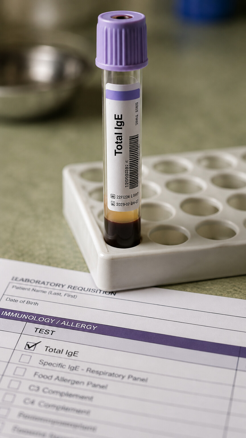
Figure 4. A total IgE blood test measures overall antibody levels but doesn't identify which specific substances the immune system is reacting to.
When a doctor orders IgE testing for someone with eczema, there are two meaningfully different tests being discussed, and confusing them is one of the most common sources of misunderstanding about lab results. A total IgE test measures the overall amount of IgE antibody circulating in the blood, regardless of what it's targeted against. It's a useful general marker — a high total IgE supports the idea that an allergic, IgE-driven process is contributing to someone's eczema — but on its own, it says nothing about which specific allergen, if any, is actually responsible.
Specific IgE testing is a different, more targeted approach: it measures antibody levels against one particular substance at a time — peanut, egg, dust mite, a specific pollen — and modern lab panels can screen for dozens of individual allergens from a single blood draw using a technique called a multiplex assay. This is the test that actually helps identify a trigger worth addressing, whereas total IgE mainly helps establish the broader pattern. It's entirely possible, and not unusual, to have a high total IgE with no single specific IgE result standing out significantly, which usually points toward a more generalized allergic tendency (sometimes described as an atopic predisposition) rather than one identifiable culprit driving the eczema.
Reference ranges for total IgE also shift meaningfully with age — children's normal ranges are lower than adults', and ranges climb through childhood before leveling off — so a result has to be interpreted against an age-appropriate range, not a single fixed adult cutoff, to mean anything at all.
Why a High IgE Number Doesn't Automatically Mean "Cause Found"
It's worth being direct about a limitation that trips up a lot of people reviewing their own lab results: a positive specific IgE test means the immune system has produced antibodies against that substance, but it does not, by itself, prove that substance is actually causing symptoms when encountered. This gap between "sensitized" and "clinically allergic" is well documented in allergy research — a meaningful percentage of positive specific IgE results, particularly for foods, don't correspond to any real-world reaction when that food is actually eaten under medical supervision.
This is exactly why allergists caution against using an IgE panel alone to justify eliminating multiple foods from someone's diet, especially a child's, without a clinical history that actually supports it or, when appropriate, a supervised oral food challenge to confirm the connection. An elevated specific IgE result is a genuine, useful clue — it tells a doctor where to look — but it functions as one piece of a larger diagnostic picture that also includes symptom timing, skin exam findings, and sometimes a trial elimination and reintroduction, rather than as a standalone verdict.
The Role of Skin Infections and Staph Colonization in Spiking IgE
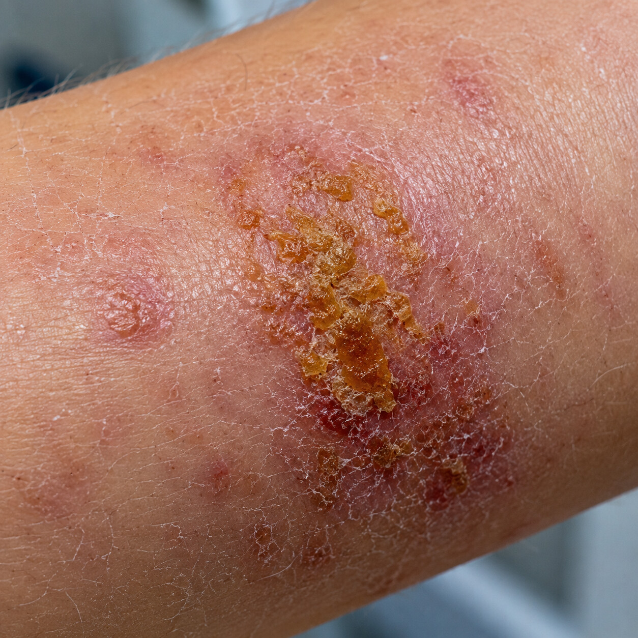
Figure 5. The honey-colored crusting seen here is characteristic of Staphylococcus aureus colonizing eczema-affected skin, a common driver of IgE spikes.
One factor that surprises a lot of people is how much of a role bacteria — specifically a species called Staphylococcus aureus — plays in this entire process. Research using skin swabs has repeatedly found that people with eczema carry far higher populations of Staph aureus on their skin than people without eczema, including on skin that looks clinically uninvolved and not actively flaring. Staph aureus doesn't just sit passively on eczema-prone skin; it produces proteins called superantigens that are unusually effective at provoking a strong, broad immune response, including further IgE production, essentially adding fuel directly onto the fire that's already burning.
This creates a self-reinforcing loop that's important to understand if eczema flares seem to keep coming back despite treatment: a broken skin barrier lets in allergens and lets Staph aureus overgrow at the same time; the resulting inflammation further damages the barrier; the damaged barrier lets in more allergens and supports even more bacterial growth; and the ongoing Staph exposure keeps driving IgE production upward. This is one of the reasons dermatologists sometimes address suspected bacterial overgrowth directly — through dilute bleach baths, topical antiseptics, or, when there's a clear infection, antibiotics — as part of managing eczema that isn't responding well to moisturizers and topical steroids alone, rather than treating the skin barrier and the bacterial factor as unrelated issues.
Does a Higher IgE Level Mean More Severe Eczema?
There is a real, measurable correlation between IgE levels and eczema severity in population-level research — on average, people with more extensive, more severe eczema tend to have higher total IgE than people with mild, limited disease, and IgE has been studied as one potential biomarker among several for tracking disease activity over time. But it's a loose correlation, not a precise dial. IgE levels can vary by a wide margin between two people with clinically similar-looking eczema, and IgE doesn't reliably predict how a specific person will respond to a specific treatment the way some other inflammatory markers do in other diseases.
Because of that variability, IgE is generally treated by clinicians as one contextual data point rather than the primary measure of how someone's eczema is doing. Physical exam findings — how much of the body is affected, how intense the redness and thickening of the skin are, how disrupted sleep is from itching — remain the main way eczema severity gets assessed and tracked, with IgE serving more as supporting evidence about the allergic component underlying the picture. Standardized scoring tools used in dermatology clinics and clinical trials, such as the SCORAD or EASI index, are built almost entirely around what can be seen and felt on the skin itself, precisely because these visual and symptom-based measures have proven more reliable than any single blood marker at tracking how a person's eczema is actually behaving from one visit to the next.
How This Connection Changes Treatment — From Moisturizers to Targeted Biologics
Understanding the skin-barrier-to-IgE pathway isn't just academic; it directly explains why eczema treatment is layered the way it is. The foundation of nearly every eczema treatment plan is barrier repair — thick, fragrance-free moisturizers applied generously and frequently — because reinforcing that physical wall reduces how much allergen and bacteria get through in the first place, addressing the process at its actual starting point rather than only managing the downstream inflammation. Topical corticosteroids and topical calcineurin inhibitors work further downstream, calming the immune activity already happening in the skin once barrier repair alone isn't enough.
For moderate to severe eczema that doesn't respond adequately to topical treatment, a newer category of injectable biologic medications has become an important option, and their mechanism connects directly back to everything discussed above. Dupilumab, the most widely used of these, works by blocking the receptor for two signaling proteins — interleukin-4 and interleukin-13 — that are central to instructing immune cells to produce IgE in the first place. By interrupting that instruction, these medications reduce the ongoing drive toward new IgE production, and clinical trials have shown that total IgE levels measurably decline over months of treatment in many patients, alongside improvement in skin symptoms. This is a useful real-world illustration of the eczema-IgE relationship: a treatment that blocks the immune signal upstream of IgE production improves both the antibody level and the visible skin disease together, because they were always connected through the same pathway to begin with.
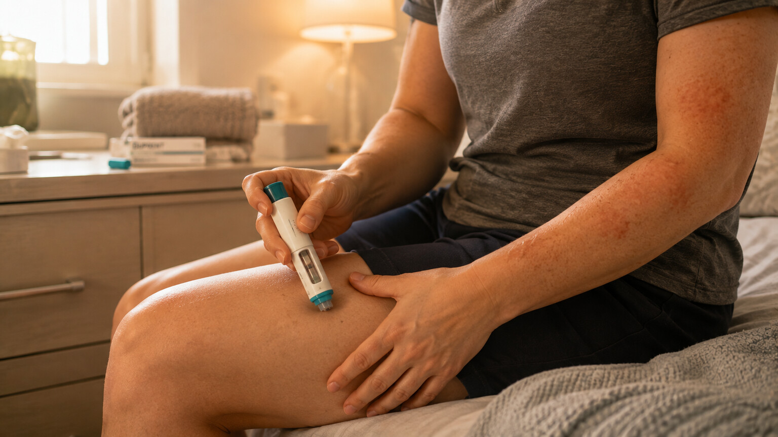
Figure 6. Biologic medications like dupilumab block the IL-4 and IL-13 signals that drive IgE production, improving skin symptoms and IgE levels together.
Frequently Asked Questions
Can I have eczema with a completely normal IgE level?
Yes. Around 20 to 30 percent of people with atopic dermatitis have the intrinsic, non-allergic pattern, where IgE and specific allergy testing come back normal despite clinically obvious eczema. A normal IgE result does not rule out eczema, and diagnosis is based on the appearance and pattern of the skin, not on antibody levels.
If my IgE is high, does that mean a specific food is causing my eczema?
Not necessarily. A high total IgE reflects general allergic tendency, and even a positive specific IgE test for a food means the immune system has made antibodies against it — not that eating it is guaranteed to trigger a reaction. Confirming a true food trigger usually requires correlating test results with symptom history, and sometimes a supervised food challenge.
Will treating my eczema lower my IgE level?
It can, particularly with treatments that target the immune pathway driving IgE production, such as dupilumab, which has been shown in clinical studies to reduce total IgE over months of use alongside skin improvement. Topical treatment and barrier repair alone have a smaller, slower effect on circulating IgE, since they primarily calm inflammation already present rather than blocking new antibody production upstream.
Does having eczema as a baby mean my child will definitely develop allergies or asthma?
No. The "atopic march" describes a pattern seen in a meaningful subset of children with eczema, not a guaranteed outcome. Earlier onset, greater severity, and higher associated IgE levels are linked to higher likelihood of progressing to food allergy, hay fever, or asthma, but many children with eczema never develop these additional conditions.
Why does my eczema keep flaring even though I moisturize every day?
Persistent flares despite consistent moisturizing can point toward ongoing Staphylococcus aureus overgrowth on the skin, an unaddressed specific allergen trigger, or eczema severe enough to need a prescription anti-inflammatory or biologic treatment in addition to barrier repair. It's worth discussing with a dermatologist rather than assuming moisturizer alone should be sufficient for every case.
Is it worth testing IgE levels again after starting eczema treatment?
Sometimes, particularly with biologic treatment, where a falling IgE level over several months can serve as one piece of supporting evidence that the underlying immune activity is responding, alongside the skin itself looking and feeling better. It isn't usually necessary for topical-only treatment plans, since IgE tends to move more slowly than visible skin symptoms do.
Conclusion
Eczema and elevated IgE are connected through a shared root cause rather than a simple one-causes-the-other relationship: a compromised skin barrier lets allergens and bacteria reach immune cells that were never meant to encounter them there, and the resulting IgE production, mast cell activation, and inflammation are what produce both the visible rash and the elevated antibody levels seen on a blood test. That's true for most people with eczema, but not all of them — a meaningful minority have eczema running through a different, non-IgE pathway entirely, which is exactly why doctors diagnose eczema by examining the skin rather than by ordering an antibody test. Understanding your own IgE results, whether they're markedly elevated, only mildly so, or squarely normal, is far more useful in the context of your actual skin symptoms and history than as a number viewed in isolation.
None of this requires memorizing immunology to act on. In practical terms, it means three things are worth prioritizing together rather than separately: consistent barrier repair through moisturizing, since that's the physical starting point of the whole cascade; a conversation with a clinician about targeted, history-guided allergy testing if flares seem tied to a specific exposure; and, for eczema that stays severe despite the basics, an honest discussion about escalating treatment rather than persisting indefinitely with a plan that isn't working. Each of those steps addresses a different link in the same chain, and together they tend to move both the visible skin disease and the underlying IgE-driven biology in the same direction.
Still Not Sure What Your Results Mean?
Upload your labs and get a complete, visual, plain-language interpretation of every biomarker — delivered to your inbox in under 15 minutes.
Get My ReportThis article is for educational purposes only and does not constitute medical advice. Always consult your healthcare provider regarding your specific lab results.