
Understanding the Difference Between IgG, IgM, and IgA
If your lab report lists three unfamiliar codes — IgG, IgM, and IgA — sitting next to numbers that seem to mean nothing on their own, you're looking at your immune system's paper trail. These three letters stand for three different classes of antibodies, the Y-shaped proteins your immune system builds to recognize and neutralize anything it identifies as foreign: a virus, a bacterium, an allergen, even a vaccine. IgG, IgM, and IgA aren't three versions of the same thing measured three different ways — they're three separate tools your body deploys for different jobs, at different moments, in different locations. IgM shows up first and fades fast. IgG shows up later and can stick around for decades, sometimes for life. IgA barely circulates in your bloodstream at all, spending most of its career standing guard at the surfaces where the outside world touches your inside world — your gut lining, your airways, your tears, your saliva. Understanding which antibody is doing what, and why your doctor ordered this specific combination, turns three cryptic abbreviations into a genuinely readable story about your immune system's history and its current state.
What Antibodies Actually Are, in Plain English
Before the differences between IgG, IgM, and IgA mean anything, it helps to understand what an antibody is doing in the first place. Every antibody — also called an immunoglobulin, which is just the formal name for this entire family of proteins ("Ig" is short for immunoglobulin) — is built by a type of white blood cell called a B cell. When a B cell first encounters something your body doesn't recognize, called an antigen (any molecule, from a fragment of a virus's outer shell to a protein in peanuts, that your immune system can specifically detect and remember), it can mature into a plasma cell, which functions like a small, dedicated biological factory. That factory's entire job is to manufacture one very specific antibody, shaped to fit that one antigen the way a key is cut to fit a single lock.
Structurally, every antibody looks roughly like the letter Y. The two upper arms of the Y are the business end of the molecule — they're what physically grabs onto the antigen — and the exact shape of those arms is what makes one antibody bind, say, a flu virus, while a completely different antibody binds a peanut protein instead. The stem of the Y is what tells the rest of the immune system what to do once the antibody has latched on: whether to summon cells that destroy what's been tagged, trigger a cascade of blood proteins called complement that punch holes directly through a bacterium's outer membrane, or simply flag the invader so a scavenger cell can engulf and digest it.
Here's the part that trips most people up: your body doesn't build one universal antibody and call it done. It builds several different classes of antibodies — five, technically, though only three of them (IgG, IgM, and IgA) show up on a routine panel, with the other two (IgD and IgE) reserved for more specialized roles, including allergic reactions in the case of IgE. Each class keeps the same Y-shaped, antigen-grabbing arms, but changes the stem of the Y — and that single structural difference is what determines where in the body that class of antibody works best, how long it survives once it's made, and what kind of alarm it's able to sound once it's attached to something foreign.
Meet the Three Classes: IgG, IgM, and IgA at a Glance
Think of IgM, IgG, and IgA less as three flavors of the same tool and more as three members of a response team with genuinely different jobs and genuinely different personalities.
IgM is the first responder. It's the largest of the three by far — assembled as a cluster of five Y-shaped units joined together at their stems in a starburst arrangement called a pentamer — and it's the antibody class your body throws together fastest when it meets something new, often within the first several days of exposure. Because it's built in such a hurry, an individual IgM antibody isn't especially precise or strongly binding on its own, but its five-armed shape more than compensates by letting it grab onto several copies of an antigen simultaneously, and it happens to be exceptionally good at triggering that complement cascade mentioned above.
IgG is the record-keeper. It shows up later — typically one to three weeks after IgM, once your immune system has had time to refine and strengthen its aim through a process called affinity maturation — and it exists as a single Y-shaped unit, smaller and more nimble than IgM, which lets it slip out of the bloodstream and into surrounding tissues where IgM generally can't follow. IgG makes up roughly 70 to 75% of all the antibody protein circulating in a healthy adult's blood, and unlike IgM, it can persist for years, sometimes for a lifetime, which is exactly what allows a single childhood chickenpox infection, or a single course of a vaccine, to protect a person for decades afterward.
IgA is the border guard. Only a modest fraction of the body's total IgA actually circulates in the bloodstream at any given time; the vast majority of it lives at what immunologists call mucosal surfaces — the moist linings of the gut, the airways and sinuses, the tear ducts, and breast milk — where it intercepts antigens before they ever get the chance to cross into the bloodstream at all. IgA typically travels as a pair of Y-shaped units linked together, called a dimer, with an extra protective piece called a secretory component attached to it, which helps the whole structure survive the harsh, enzyme-rich environment of, for example, the digestive tract, where a less reinforced protein would simply be torn apart.
A routine blood test almost never measures all five immunoglobulin classes individually. What's typically ordered — a "quantitative immunoglobulins" panel — measures only the total amount of IgG, IgM, and IgA circulating in the blood, giving a broad snapshot of whether each arm of this system is present in a normal amount. It does not tell your doctor which specific antigen any of that antibody is actually aimed at; that requires a separate, targeted test.
IgM: Your Immune System's First Responder
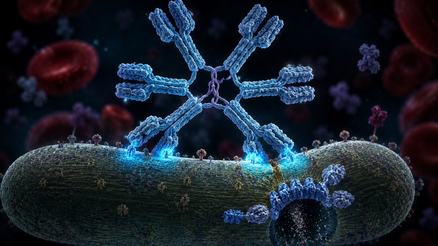
Figure 1. A pentameric IgM molecule, assembled from five antibody units joined by a J chain, binds multiple antigen sites on a bacterial surface and triggers the complement cascade.
When your immune system meets a genuinely new threat — a virus it has never dealt with, a bacterium it has no memory of — IgM is almost always the first antibody class to appear in measurable amounts in your blood, often within three to five days. This timing is exactly why an IgM result is so useful diagnostically: a positive IgM against a specific pathogen is one of the strongest available signals that an infection is recent or currently active, rather than something that happened years ago. That's the logic behind tests like hepatitis A IgM, or the IgM component of an Epstein-Barr virus (the virus behind mono) panel — a positive IgM points toward "this is happening now," while a negative IgM in the same person often points toward "this either isn't the cause, or it happened a long time ago."
IgM's five-armed pentamer shape isn't just a structural curiosity; it's the reason IgM is so effective despite being built quickly and somewhat imprecisely. Because each pentamer offers up to ten antigen-binding arms at once, it can latch onto multiple copies of a repeating surface pattern — the kind bacteria and viruses commonly display — far more efficiently than a single Y-shaped antibody ever could. That same multi-armed structure also makes IgM extraordinarily efficient at activating complement, the cascade of blood proteins that can physically punch holes through a bacterium's outer membrane, causing it to rupture. In effect, IgM trades precision for raw, immediate effectiveness, buying the immune system time while it builds a more refined, longer-lasting IgG response behind the scenes.
IgM levels typically peak within the first one to two weeks of a new infection and then decline over the following weeks to months, usually becoming undetectable again once the acute illness has resolved — which is part of why IgM is rarely useful for confirming immunity from something that happened long ago. There's one other place IgM plays an outsized role that has nothing to do with infection at all: it's the antibody class responsible for the "natural" antibodies against A and B blood group antigens, which is a large part of why blood typing and cross-matching before a transfusion is taken so seriously.
IgG: The Immune System's Long-Term Memory
If IgM is the immune system's rapid, blunt-force first draft, IgG is the polished, edited final version — and it's the antibody class most people mean when they casually talk about "having immunity" to something. After the initial IgM surge, certain B cells go through a deliberate refinement process, gradually swapping the class of antibody they produce from IgM to IgG while simultaneously improving how tightly that antibody binds its target, a process called affinity maturation. The result, appearing roughly one to three weeks after the original exposure, is an antibody that binds its target more precisely than the original IgM ever did, and that survives in the body dramatically longer.
That longevity is IgG's defining feature. A subset of the B cells that produced it don't disappear once the infection clears; some become long-lived memory B cells and long-lived plasma cells that can persist for years, quietly maintaining a baseline level of IgG production even with no active infection present. This is the entire biological basis of vaccination: a vaccine deliberately exposes the immune system to a harmless piece or weakened version of a pathogen specifically so it builds this IgG memory response, without requiring the person to go through the actual illness first. It's also why a positive IgG result against, say, measles or hepatitis B usually means "you have immunity, likely from past infection or vaccination," rather than "you are currently sick" — the opposite interpretation from a positive IgM.
IgG is technically further divided into four subclasses (IgG1 through IgG4), each slightly biased toward different types of targets — IgG1 and IgG3 tend to dominate responses against viruses and protein antigens, for instance, while IgG2 leans toward responses against the sugar-based coatings found on certain bacteria. Subclass testing exists but is reserved for more specialized situations, such as investigating certain recurrent bacterial infections; a standard immunoglobulin panel reports total IgG as a single combined number, not broken out by subclass.
Because IgG is built as a single, relatively compact Y-shaped unit rather than IgM's bulky five-unit cluster, it's able to leave the bloodstream and diffuse into surrounding tissues far more readily, reaching sites of infection or inflammation throughout the body. That same compact size gives IgG one more remarkable ability that no other antibody class has: it's small enough, and specifically recognized by a transport receptor, to cross the placenta from a pregnant person into a developing fetus — a mechanism covered in more depth further down this article.
Curious what your own lab results are really telling you? Upload them and get a complete, plain-language breakdown in under 15 minutes.
Analyze My ResultsIgA: The Antibody Guarding Your Body's Entrances

Figure 2. Secretory IgA, reinforced with a protective secretory component, patrols the mucosal lining of the gut, intercepting antigens before they cross into the bloodstream.
IgG and IgM both do most of their work inside the bloodstream and the tissues that blood reaches. IgA operates on an entirely different battlefield: the roughly 400 square meters of mucosal surface — the moist linings of the gut, the respiratory tract, the sinuses, and the eyes — where the outside world is in constant, direct physical contact with the inside of your body. This form, called secretory IgA, is built specifically to survive being dumped into an environment full of digestive enzymes, stomach acid, and bacteria without falling apart, thanks to that extra secretory component piece riveted onto the antibody as it's transported across the mucosal cell layer and out into the mucus itself.
Rather than destroying invaders the way IgG and IgM often do by triggering complement or recruiting immune cells, secretory IgA's primary job is closer to a bouncer at a door: it coats bacteria, viruses, and toxins and physically prevents them from attaching to and penetrating the cells lining the gut or airway, a process immunologists call immune exclusion. This is a fundamentally quieter, less inflammatory form of defense than what IgG and IgM do inside the bloodstream — which makes sense, given that the gut lining is in constant contact with trillions of harmless resident bacteria that the immune system needs to tolerate, not attack, every single day.
IgA has one more remarkable role that's easy to overlook: it's the dominant antibody in breast milk, particularly in the first days of milk called colostrum. A newborn's own immune system is still immature and produces very little antibody of its own for the first several months of life, so the secretory IgA delivered through breastfeeding provides the infant's gut with a ready-made layer of mucosal protection at exactly the moment it's needed most, without that antibody ever being absorbed into the baby's own bloodstream — it works entirely locally, right there in the infant's digestive tract.
IgA also comes up in a specific, commonly misunderstood diagnostic context: celiac disease, an autoimmune reaction to gluten, is typically screened for using a blood test called tissue transglutaminase IgA (tTG-IgA). Because that test relies specifically on the IgA class, a person who happens to have selective IgA deficiency — discussed later in this article — can produce a falsely normal-looking tTG-IgA result even if they truly have celiac disease, simply because they don't make enough IgA of any kind to generate a meaningful antibody response. This is exactly why celiac screening panels are usually paired with a total IgA level: if total IgA comes back very low, the tTG-IgA result can't be trusted on its own, and doctors switch to an IgG-based version of the same test instead.
Why Doctors Order an Immunoglobulin Class Panel

Figure 3. A quantitative immunoglobulin panel measures total circulating IgG, IgM, and IgA from a single blood draw, most often ordered to investigate recurrent infections.
A quantitative immunoglobulins test isn't part of routine annual bloodwork the way a basic metabolic panel or a complete blood count often is — it's typically ordered when something specific has raised a question about how well the antibody-producing side of the immune system is functioning. The single most common reason is a pattern of recurrent infections: repeated bouts of sinusitis, bronchitis, ear infections, or pneumonia, especially in a pattern that seems more frequent or more severe than what would be considered ordinary. Because IgG, IgM, and IgA each defend different territory, measuring all three at once helps a doctor see whether one specific arm of that defense is unusually low, rather than treating "the immune system" as a single undifferentiated unit.
The test also gets ordered in the opposite direction, when a doctor is concerned about an immunoglobulin level being too high rather than too low. A broad, proportional rise across all three classes — called a polyclonal elevation — is a fairly common and often unremarkable finding in chronic inflammatory conditions, chronic infections, or liver disease, since a persistently activated immune system simply produces more antibody protein overall. A very different, more concerning pattern is a sharp rise in just one class, especially IgG or IgM, accompanied by a drop in the other two — a pattern called a monoclonal spike, which can be a sign that a single clone of antibody-producing cells has begun growing out of control, as seen in conditions like multiple myeloma or Waldenström macroglobulinemia. Distinguishing a reassuring polyclonal pattern from a concerning monoclonal one is one of the main reasons this test gets ordered in older adults with unexplained fatigue, bone pain, or anemia.
Beyond infection frequency and monoclonal screening, this panel is also used to monitor people already known to have an immune deficiency, to check immunoglobulin levels before and during certain immunosuppressive therapies, and as part of the workup for unexplained chronic diarrhea or malabsorption, since gut conditions that cause significant protein loss can drag immunoglobulin levels down as a side effect, independent of how the immune system itself is functioning.
How to Read Your Own Immunoglobulin Results
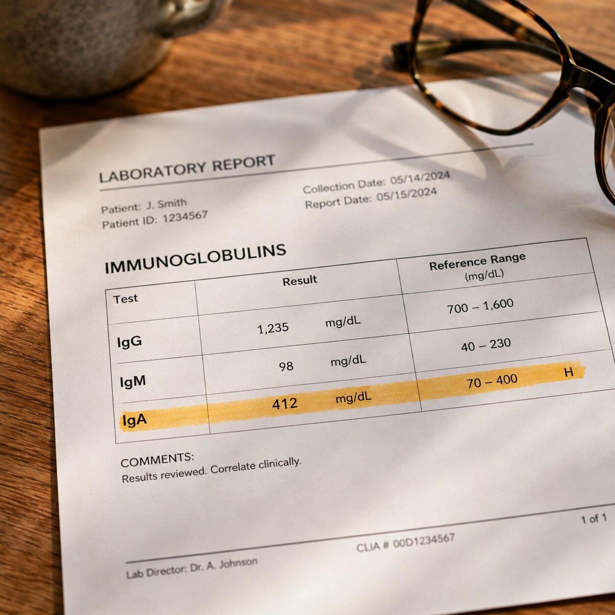
Figure 4. Reference ranges for IgG, IgM, and IgA vary by laboratory and by age, and a result outside those ranges is interpreted alongside symptoms and history, not in isolation.
Immunoglobulin results are reported in milligrams per deciliter (mg/dL), a measure of how much of that antibody protein is present in a given volume of blood. As a rough adult reference point — and it's genuinely important to treat this as a rough point of orientation rather than a strict cutoff, since every laboratory sets its own reference range based on its own equipment and population — total IgG typically falls somewhere between about 700 and 1,600 mg/dL, total IgM between roughly 40 and 230 mg/dL, and total IgA between about 70 and 400 mg/dL. Your own lab report will list the specific range used by the laboratory that processed your sample directly next to your result, and that's the range that actually applies to your number, not any figure quoted in an article.
A single class running mildly outside its range, especially IgA, is common and is often not clinically significant on its own. Selective IgA deficiency, where IgA is very low or absent while IgG and IgM remain normal, is thought to affect somewhere in the range of 1 in 500 to 1 in 700 people of European descent, and the majority of people with it are entirely healthy and never find out unless they happen to have bloodwork done for an unrelated reason, or unless it's picked up incidentally through something like that celiac testing caveat described earlier. A pattern that carries more weight is when more than one class is low simultaneously, particularly IgG together with either IgM or IgA — that combination is what starts to raise the question of a broader antibody deficiency rather than an isolated, usually harmless quirk in a single class.
On the high side, a modestly elevated IgG or IgA in someone with a chronic infection, ongoing inflammation, or a chronic liver condition like cirrhosis is a fairly expected, proportionate response and is usually interpreted in that clinical context rather than treated as an alarming finding by itself. What tends to prompt further workup is a result that's dramatically elevated, elevated in only one class while the others stay flat or drop, or elevated alongside other red-flag findings like unexplained anemia, kidney dysfunction, or bone pain — at that point, additional testing such as protein electrophoresis is typically used to look more closely at exactly what kind of antibody is being overproduced, and by how many different cell clones.
It's also worth knowing that immunoglobulin levels aren't fixed for life. They shift with age — newborns start with almost none of their own and gradually build up their own IgG, IgM, and IgA production over the first several years of childhood — and they can be temporarily suppressed by certain medications, most notably long-term corticosteroids and other immunosuppressive drugs, or by conditions that cause the body to lose protein faster than it can replace it, such as nephrotic syndrome in the kidneys or protein-losing enteropathy in the gut. A low result in the setting of one of these known causes is generally interpreted very differently than the same low number appearing with no clear explanation at all.
Newborns and Passive Immunity from Mom

Figure 5. IgG is the only immunoglobulin class transported across the placenta, giving a newborn temporary passive immunity that fades over the first several months of life.
The way these three antibody classes behave around pregnancy and birth is one of the clearest illustrations of how differently IgG, IgM, and IgA are built and deployed. Starting around the second trimester and increasing sharply in the third, a specialized transport receptor in the placenta actively pulls IgG — and only IgG, because of its small, single-unit size and the specific way its stem is shaped — out of the mother's bloodstream and delivers it into the fetal circulation. By the time a full-term baby is born, its blood may actually contain a slightly higher concentration of IgG than its mother's does, almost none of which the baby made itself; it's essentially a temporary loan of the mother's own accumulated immune memory, protecting the newborn against the specific pathogens she's encountered or been vaccinated against over her own lifetime.
IgM, by contrast, is far too large and structurally different to cross the placenta at all under normal circumstances. This turns out to be diagnostically important: because a healthy fetus is normally shielded from the outside world, any IgM detected in a newborn's blood at birth must have been made by the baby itself, since it couldn't have come from the mother. That fact is the basis of TORCH testing, a panel used when a newborn is suspected of having acquired an infection before birth (from organisms like toxoplasma, rubella, cytomegalovirus, or herpes simplex virus) — finding pathogen-specific IgM in the newborn's own blood is strong evidence that the infection happened in utero, rather than being passively transferred maternal antibody.
IgA follows yet a third pattern. It doesn't cross the placenta before birth, and a newborn's own gut isn't yet capable of producing meaningful amounts of it — which is exactly the gap that breastfeeding fills, delivering secretory IgA directly into the infant's digestive tract through colostrum and, later, ongoing breast milk, protecting the gut lining locally without that antibody ever needing to enter the baby's bloodstream at all.
There's a well-documented and entirely normal dip that follows all of this: the IgG a baby receives from its mother naturally degrades over the first several months of life, typically bottoming out somewhere around three to six months of age, before the infant's own antibody production has fully ramped up to compensate. Pediatric immunologists refer to this window as transient hypogammaglobulinemia of infancy, and in most babies it resolves on its own as their own immune system matures — it's one of the reasons very young infants are more vulnerable to certain infections during this specific stretch of early life, and it's a normal developmental stage rather than a diagnosis on its own.
When Low Levels Signal Something Bigger: Understanding Immunodeficiency

Figure 6. Recurrent sinus and respiratory infections are a hallmark clinical sign that prompts testing for primary antibody deficiencies such as selective IgA deficiency or CVID.
As mentioned above, selective IgA deficiency is the most common of the primary immunodeficiencies — conditions someone is essentially born with, as opposed to acquiring later from a medication or an illness — and the majority of people who have it live entirely normal lives without ever knowing. A smaller subset does experience recurrent sinus and respiratory infections, more frequent gastrointestinal infections, or a higher rate of certain autoimmune conditions, though researchers still don't fully understand why the same underlying deficiency produces no noticeable effect in some people and a real clinical pattern in others.
A more consequential condition sits further along the same spectrum: Common Variable Immunodeficiency, or CVID, which involves low IgG together with a low IgA and/or IgM, and — critically — an impaired ability to mount a normal antibody response even when vaccinated or exposed to an infection. CVID is usually diagnosed in adulthood, often only after years of recurrent sinus infections, bronchitis, or pneumonia that seem disproportionate to what would be considered ordinary, sometimes alongside chronic gastrointestinal symptoms or an increased rate of certain autoimmune conditions. Unlike selective IgA deficiency, CVID typically does require active treatment, most often regular infusions of immunoglobulin replacement therapy, which supplies the antibody protection the person's own plasma cells aren't reliably producing on their own.
It's worth clearly separating these primary, inborn causes of low immunoglobulins from secondary causes, which are far more common in day-to-day medical practice. Certain blood cancers, particularly chronic lymphocytic leukemia and multiple myeloma, can suppress normal antibody production even while one abnormal clone of cells produces too much of a single type. Long-term use of corticosteroids or other immunosuppressive medications, conditions that cause significant loss of protein through the kidneys or gut, and severe malnutrition can all lower immunoglobulin levels as a downstream effect, without reflecting a fundamental problem with the immune system's underlying architecture. Because the treatment and the outlook for these secondary causes are completely different from a primary immunodeficiency, distinguishing between the two is usually the first branch point once a low immunoglobulin result has been confirmed.
The practical takeaway for anyone looking at their own low result is that context does almost all of the interpretive work. A single mildly low IgA in someone with no history of frequent infections is a very different finding from the same number in someone who has had four sinus infections this year and a lifelong pattern of getting sicker, for longer, than people around them. If that second pattern sounds familiar, it's a reasonable and worthwhile thing to raise directly with a primary care doctor, who can decide whether a referral to an immunologist for further evaluation makes sense.
Using IgM and IgG Together to Time an Infection
One of the most practically useful things about having both an IgM and an IgG result for the same pathogen is that, read together, they can roughly place an infection in time — something neither number can do reliably on its own. This combined approach comes up constantly in testing for infections like toxoplasmosis, rubella, cytomegalovirus, and Epstein-Barr virus, and it follows a fairly consistent logic across all of them.
IgM positive, IgG negative generally points to a genuinely new, recent infection — the fast-but-imprecise first responder has shown up, but the immune system hasn't yet had time to build its longer-lasting IgG response. IgM negative, IgG positive is the pattern of past infection or established immunity: the acute phase has long since resolved, but the durable IgG memory response remains, which is exactly the pattern doctors look for when confirming that someone is immune to something like rubella before a pregnancy, or checking whether a childhood vaccine series against hepatitis B or varicella actually produced lasting protection. Both IgM and IgG positive together is the trickiest pattern to interpret in isolation, since it can represent a recent infection that's already transitioning into the IgG memory phase, a reinfection, or, in some tests, a false-positive IgM — which is part of why a positive IgM result is often followed up with a second blood draw two to three weeks later to see whether the IgG level is rising, confirming a genuinely active, evolving immune response rather than a one-off lab artifact.
For situations where timing precision matters even more, some laboratories offer IgG avidity testing, which measures how tightly the IgG antibodies present are binding to their target. Because affinity maturation is a gradual process, IgG produced very early in an infection binds relatively loosely (low avidity), while IgG from an infection that happened months or years ago binds tightly (high avidity) — giving doctors a second, independent way to distinguish a genuinely recent infection from an old one, which becomes especially important in situations like suspected infection during pregnancy, where the exact timing can meaningfully change the recommended next steps.
This same IgG-based logic is also the foundation of vaccine titer testing, sometimes ordered for healthcare workers, students, or anyone who needs documented proof of immunity to something like measles, mumps, rubella, or hepatitis B. In that context, a lab isn't checking for IgM at all — it's specifically measuring IgG, because IgG is the class that reflects durable, established immune memory, which is the entire point of a titer test in the first place.
Frequently Asked Questions
Can I have low IgA and never know it?
Yes, and it's actually the most common outcome. Selective IgA deficiency affects roughly 1 in 500 to 1 in 700 people of European descent, and the majority never experience any noticeable symptoms or find out unless it's picked up incidentally on unrelated bloodwork.
Does a positive IgM always mean an active infection?
Not always, but it's the strongest single signal available on a routine antibody test that something is recent rather than old. Because IgM can occasionally give a false-positive result, doctors sometimes confirm a positive IgM with a repeat test a few weeks later to check whether IgG is also rising, which supports a genuinely active infection.
Why did my celiac test include a total IgA level?
Because the standard celiac antibody test (tTG-IgA) only works reliably if you make normal amounts of IgA in the first place. If total IgA comes back very low, that test can give a falsely reassuring result even if celiac disease is present, so labs check total IgA alongside it and switch to an IgG-based version of the test if IgA is deficient.
Is it normal for a newborn's immunoglobulin levels to be low?
Yes. A baby is born with a supply of maternal IgG that naturally declines over the first several months, often dipping lowest around three to six months of age before the infant's own antibody production catches up. This normal dip is called transient hypogammaglobulinemia of infancy.
What's the difference between low IgA alone and Common Variable Immunodeficiency?
Selective IgA deficiency involves only IgA running low while IgG and IgM stay normal, and it's usually harmless. CVID involves low IgG together with low IgA and/or IgM, along with a poor antibody response to vaccines or infections, and it typically causes recurrent infections that require ongoing treatment.
Conclusion
IgG, IgM, and IgA were never meant to be read as three interchangeable versions of "your antibodies" — they're three specialized tools built for three different moments and three different locations in the body. IgM arrives first and fast, a blunt but effective early warning system. IgG arrives later, refined and built to last, carrying the immune system's long-term memory of everything it has ever successfully fought off or been vaccinated against. IgA works quietly at the body's mucosal borders, intercepting threats before they ever reach the bloodstream at all. Seen this way, a quantitative immunoglobulin panel isn't three redundant numbers — it's a snapshot of three distinct defense systems, and understanding what each one is actually doing is what turns a page of unfamiliar abbreviations into a genuinely useful piece of information about your own health.
Still Not Sure What Your Results Mean?
Upload your labs and get a complete, visual, plain-language interpretation of every biomarker — delivered to your inbox in under 15 minutes.
Get My ReportThis article is for educational purposes only and does not constitute medical advice. Always consult your healthcare provider regarding your specific lab results.