
What Does a Rising Antibody Titer Between Tests Suggest?
If you've had your blood drawn twice for the same antibody test — once when you first got sick, and again a couple of weeks later — and the second number came back noticeably higher than the first, that pattern has a very specific meaning in medicine. It isn't just "your immune system is doing more." A rising antibody titer between two paired blood draws is one of the oldest and most reliable ways doctors confirm that a recent infection actually happened, distinguishing it from old immunity, a lab fluke, or a false alarm. But the word "rising" is doing a lot of work in that sentence, and not every increase counts. There's a specific, validated threshold — a fourfold rise — that separates a meaningful signal from ordinary lab-to-lab wobble, and there's a specific reason doctors insist on drawing the second sample two to four weeks after the first rather than the next day. This article walks through what a titer actually is, why doctors compare two of them instead of trusting one, exactly how big a rise has to be before it counts, and what a genuine rise usually tells your care team about what's happening — and what happened recently — inside your immune system.

Figure 1. A titer is measured through serial dilution: each well contains half the antibody concentration of the one before it, and the last well still showing a positive reaction sets the number.
Curious what the rest of your bloodwork is telling you? Upload your results and get a complete, plain-language breakdown in under 15 minutes.
Analyze My ResultsWhat a "Titer" Number Actually Is
Before any of this makes sense, it helps to understand what a titer is measuring, because the number itself looks strange the first time you see it — something like "1:320" instead of a normal lab value with a unit attached. A titer is the answer to a very specific laboratory question: how many times can we dilute your blood serum and still detect the antibody we're looking for? To find that answer, a lab technician takes your serum — the clear, cell-free liquid part of your blood — and mixes it with a testing solution in a small well. Then they take some of that mixture, mix it with an equal amount of plain saline to cut the concentration in half, and test that. Then they cut it in half again. And again. This is called serial dilution, and it continues through a whole row of wells, each one twice as dilute as the last: 1:10, 1:20, 1:40, 1:80, 1:160, 1:320, and so on. The titer is reported as the highest dilution — the most watered-down version of your blood — that still produces a positive, detectable reaction. A titer of 1:320 means your serum still had enough antibody to react even after being diluted 320-fold; a titer of 1:20 means the antibody ran out much sooner. Higher numbers after the colon mean more antibody was present, not less — a common point of confusion, since intuitively a "bigger" fraction like 1:320 can look smaller than 1:20 if you're reading it as a plain fraction. It isn't a fraction in that sense; it's a dilution ratio, and a bigger second number always means a stronger, more concentrated antibody response.
How the Lab Actually Detects a "Positive" Well
The serial dilution process explains how the number is generated, but it skips over an equally important question: what does the lab actually watch for to decide a given well is still "positive"? The answer depends on which of several detection methods the lab uses, and understanding the basic idea behind them makes the whole process feel less like a mysterious black box. One of the oldest techniques, hemagglutination inhibition, relies on the fact that certain viruses can clump red blood cells together into a visible mat at the bottom of a well — a reaction called hemagglutination. If your serum contains antibody against that virus, the antibody coats the virus particles and blocks them from clumping the red cells, so instead of a mat, the red cells simply settle into a small, compact dot at the bottom of the well, like a marble rolling to the center of a bowl. The technician reads across the row of increasingly diluted wells and finds the last one where that compact dot still appears — the last well where your antibody was still concentrated enough to stop the clumping. Complement fixation, another classic method, works on a related principle: it measures whether your antibody, combined with a separate blood-testing protein complex called complement, can destroy red blood cells added to the mixture, and again the titer is read as the last dilution where that destruction still happens. Newer, more common methods like enzyme-linked immunosorbent assay, usually shortened to ELISA, use a color-changing chemical reaction instead: a well containing your antibody binding correctly to its target changes color, often to a visible blue or yellow, and modern lab instruments measure exactly how strong that color change is at each dilution using a light-based sensor, translating a visual color change into the same kind of titer number a technician would once have read by eye. Whichever method is used, the core logic stays identical: keep cutting the sample's concentration in half until whatever signals a positive reaction — a settled dot of cells, a burst of red cell destruction, a color change — finally disappears, and report the last dilution where it was still there.
Why Doctors Draw Two Blood Samples, Weeks Apart
A single titer, taken once, tells you that antibody is present and roughly how much of it there is at that moment — but it can't tell you when your immune system made it. Antibodies from an infection you had five years ago can still show up on a test today, sometimes at a respectable level, simply because your immune system built long-lasting memory cells that keep producing a background level of protection. This is exactly why, when a doctor needs to confirm that an infection is happening right now rather than sometime in the past, a single number isn't enough evidence on its own. The solution is to draw two samples at two different points in the timeline of the illness. The first, called the acute-phase sample, is drawn as early as possible after symptoms start, ideally within the first week. The second, called the convalescent-phase sample, is drawn two to four weeks later, once the body has had time to mount a full antibody response if the infection is real and recent. Comparing the two numbers side by side turns a single snapshot into something much more useful: a measurement of direction and speed, not just a static level.
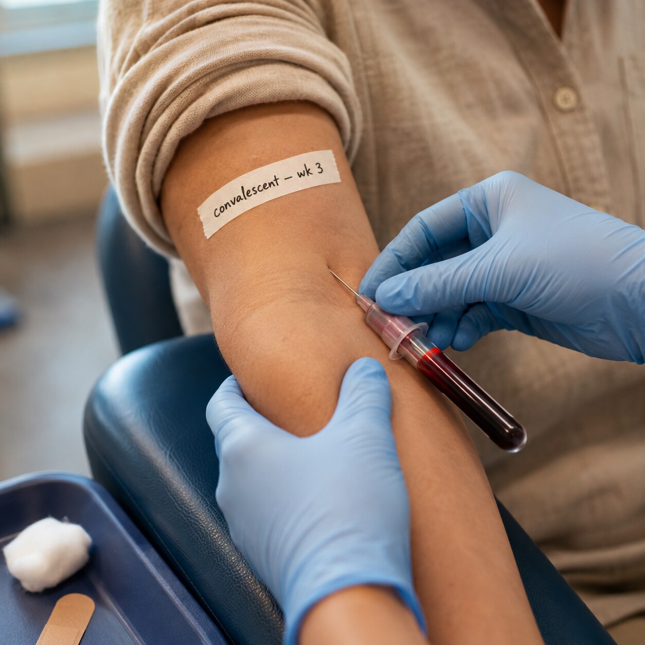
Figure 2. The second, convalescent-phase draw is timed two to four weeks after the first so the body has had a full window to mount a measurable antibody response.
This two-sample approach is one of the oldest confirmatory tools in infectious disease diagnosis, predating modern molecular testing by decades, and it's still used today precisely because it answers a question that a single test — or even a genetic test looking for the pathogen itself — sometimes can't. A PCR test, for instance, only detects a pathogen if it's still actively present in your body at the moment of the swab or draw; by the time some illnesses are recognized and investigated, the organism itself may already be gone, cleared by the very immune response the paired titers are measuring. In those situations, watching the antibody response rise between two time points becomes the most reliable remaining evidence that the infection actually occurred, even after the pathogen itself is no longer detectable.
The Fourfold Rule — How Big a Rise Has to Be Before It Counts
Because titers are measured in doubling steps — 1:10, then 1:20, then 1:40 — small, single-step differences between two tests aren't treated as meaningful on their own. Lab-to-lab and even run-to-run variability in how these dilution assays are read means a titer can bounce by one dilution step in either direction without anything real having changed in your body. To filter out that noise, the long-established clinical and public health standard is what's called a fourfold rise: the convalescent titer has to be at least four times higher than the acute titer — equivalent to two full doubling steps — before it's accepted as genuine, seroconversion-confirming evidence of a recent infection. A move from 1:10 to 1:20 is only a twofold change and, by itself, isn't considered diagnostic. A move from 1:10 to 1:40, or from 1:80 to 1:320, clears the fourfold bar and is read as strong evidence that your immune system mounted a real, new response to that specific pathogen during the window between the two draws. This threshold isn't an arbitrary round number — it was chosen specifically because it reliably separates true immunologic change from the ordinary measurement variability built into how these dilution-based assays work, and it remains the standard definition used in serologic case confirmation for illnesses ranging from measles and mumps to a range of tick-borne and respiratory infections.

Figure 3. Each antibody-producing plasma cell releases Y-shaped IgG molecules that bind a specific viral antigen; a rising titer reflects a several-fold increase in the density of these molecules in circulation.
It helps to picture what's physically happening behind that number. Each Y-shaped antibody molecule circulating in your blood is capable of binding to a specific target — an antigen — on the surface of the exact virus or bacterium your immune system is responding to. When an infection first begins, only a small number of antibody-producing cells have been activated, so the antibody molecules floating in your serum are relatively sparse; dilute the serum a little, and you quickly run out of enough antibody to produce a detectable reaction, which is why the acute titer often reads low. Over the following two to three weeks, if the infection is real and ongoing, your immune system dramatically ramps up production, and the number of these binding-ready antibody molecules in every drop of serum multiplies many times over. That's the biological reality a fourfold titer rise is capturing on paper: not an abstract statistic, but a measurable, several-fold increase in the density of pathogen-specific antibody molecules physically present in your bloodstream between the two draws.
IgM vs. IgG — Why Timing Changes What a Rising Titer Means
Not all antibodies behave the same way over the course of an infection, and understanding the difference between the two main classes involved in a typical immune response — IgM and IgG — explains a lot about why the timing of these two blood draws is so deliberate. IgM is the immune system's first responder: it's the first antibody type your body manufactures after encountering a new pathogen, often becoming detectable within the first week of illness, and it tends to fade relatively quickly, usually becoming undetectable within a few weeks to a couple of months. IgG is the second wave — slower to appear, typically becoming detectable one to two weeks after IgM, but far more durable, often persisting for years and sometimes for life, forming the backbone of your long-term immune memory. Because of this staggered timeline, a rising IgG titer between an acute and convalescent sample specifically confirms that a genuine, active immune response occurred during that window — the immune system was still actively ramping up production during the very days between your two blood draws. A high IgM alone, without a paired rising IgG, can sometimes be trickier to interpret, since IgM assays are more prone to certain types of cross-reaction and false positives than the fourfold IgG rise, which is why the paired-titer, doubling-dilution approach remains the more definitive confirmatory tool in ambiguous cases.
When a Rising Titer Signals Reinfection or Reactivation, Not a First-Time Illness
A rising titer doesn't only happen the first time you're ever exposed to a pathogen — it can also happen when someone with pre-existing immunity encounters the same organism again, or when a dormant infection reactivates. This is explained by something immunologists call the anamnestic response, from a Greek root meaning "not forgetting." Certain white blood cells called memory B cells persist for years after an initial infection or vaccination, quietly standing by without actively producing much antibody. When they encounter the same antigen a second time, they don't have to start from scratch the way a naive immune system does on first exposure — they recognize the threat immediately and differentiate into antibody-producing plasma cells far faster and in far greater numbers than the first time around. The result is a titer that was already measurable at baseline — reflecting old immunity — but that climbs sharply and quickly on a repeat test, sometimes rising even faster and higher than it did during the original infection. This pattern is clinically important because it tells a very different story than a first-time infection: it suggests your immune system already knew this pathogen and mounted an accelerated, experienced response to a fresh encounter, rather than building protection completely from the ground up.

Figure 4. In the anamnestic response, memory B cells from a prior exposure recognize the same antigen instantly and multiply into antibody-producing plasma cells far faster than on a first encounter.
This same mechanism explains why titers are also used to check whether a vaccine "took." After a vaccination against a disease like measles, mumps, rubella, or hepatitis B, doctors sometimes order a titer weeks later specifically to confirm that the immune system responded and built a protective level of antibody. If a follow-up titer is drawn after a booster dose in someone who was already immune, a fourfold or greater rise from their pre-booster baseline is a good sign — it means the booster successfully re-engaged existing memory cells and pushed the antibody level meaningfully higher, exactly the anamnestic pattern described above, confirming the booster did what it was supposed to do.
What Can Make a Titer Look Like It's Rising When It Isn't
Because so much rides on comparing two numbers correctly, a handful of technical factors can create a misleading appearance of change even when nothing meaningful shifted in your immune system. The single most important one is testing consistency: paired acute and convalescent samples should ideally be run side by side, in the same laboratory, using the same assay method and the same reagent lot, and tested at the same time rather than weeks apart on different equipment. Different labs — or even the same lab using a different testing platform between visits — can produce titers that aren't directly comparable, since different assay techniques (traditional methods like complement fixation or hemagglutination inhibition versus newer enzyme immunoassays) don't always report numbers on an identical scale. A shift from a low titer at one lab to a higher titer at a different lab isn't automatically a real fourfold rise; it may simply reflect two different measuring sticks. This is one of the most common, and most avoidable, sources of confusion for patients comparing two report printouts from different visits, especially if care was transferred between providers or health systems partway through an illness.
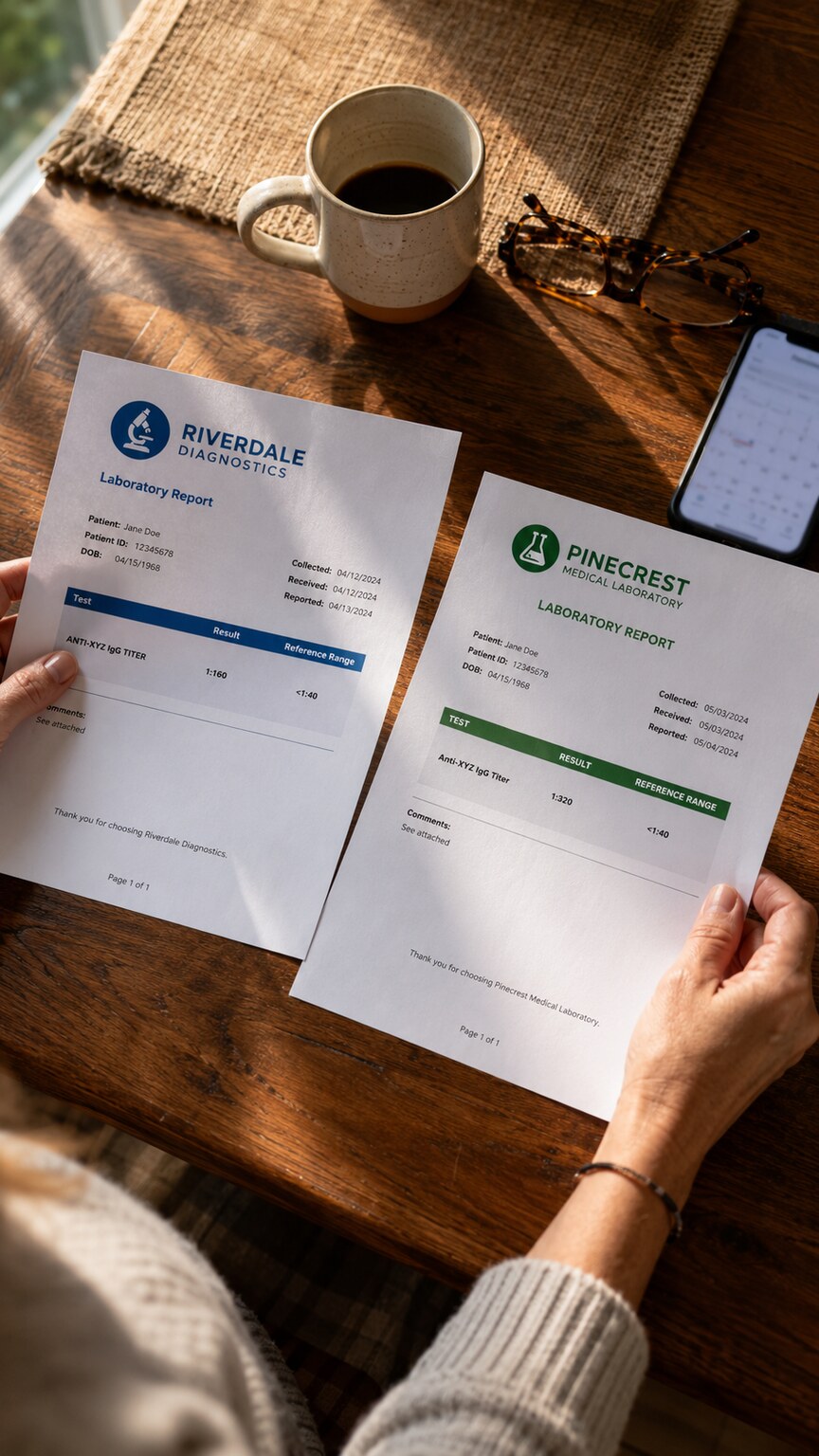
Figure 5. Comparing paired titer results requires checking that both samples were drawn on the dates and tested at the laboratory recorded on each report, since differing methods between visits can distort the comparison.
The dilution step size itself also limits precision in a way that's worth understanding: because titers only move in doubling increments, there's no such thing as a titer of 1:15 or 1:25 — the true underlying antibody level gets rounded into the nearest dilution bracket the assay tested. This means the reported number is always a slight approximation of what's really circulating in your blood, and a result sitting right at the edge of a dilution step can occasionally shift by one step purely due to the normal, small variability inherent in pipetting and reading these plates by eye or by instrument — which is exactly the kind of single-step wobble the fourfold rule exists to filter out. Finally, the timing of the draws matters as much as the lab running them: a convalescent sample drawn too early, before the antibody response has had time to fully develop, can understate the true rise and lead to a falsely reassuring — or falsely negative — comparison, which is part of why the standard two-to-four-week window between draws isn't arbitrary and shouldn't be shortened without a specific clinical reason.
A Concrete Walk-Through: Following One Result From Draw to Diagnosis
It can help to trace the whole process through a single hypothetical example from start to finish. Imagine someone develops a fever, joint aches, and a faint rash about a week after spending a weekend hiking, and their doctor suspects a tick-borne illness. An acute-phase blood sample is drawn that same visit, and the titer comes back at 1:40 — technically positive, but low, and not yet conclusive on its own, since it's hard to know from a single number whether this reflects a brand-new infection just getting started, a much older exposure from years ago, or even a borderline, nonspecific reaction unrelated to the current illness. Rather than treating that single number as an answer, the doctor schedules a second draw for three weeks later and, in the meantime, treats the symptoms based on the overall clinical picture. When the second sample comes back, the titer now reads 1:320. Dividing 320 by 40 gives 8 — an eightfold increase, comfortably above the fourfold threshold — and because both samples were run at the same laboratory using the same testing method, the comparison is considered reliable rather than an artifact of switching labs. Taken together with the timeline of symptoms and the recent outdoor exposure, this paired result becomes strong, specific evidence that a real, active infection was unfolding in the weeks between those two blood draws, converting an ambiguous single number into a confident, actionable answer. This same basic sequence — an early sample, a deliberate waiting period, a second sample, and a side-by-side comparison against the fourfold threshold — plays out in essentially the same shape regardless of which specific pathogen is being investigated, which is part of what makes paired titer testing such a durable, broadly applicable tool across so many different areas of medicine.
Immunity Screening — When a Titer Confirms Protection Rather Than Infection
Not every titer conversation is about diagnosing an active illness. Healthcare workers, for example, are frequently required to have their immunity to diseases like varicella (chickenpox), measles, mumps, and rubella confirmed through titer testing before starting a new job, specifically because they may come into contact with vulnerable patients who can't be vaccinated or who are at higher risk of severe illness. In this context, there's no "acute" sample to compare against — the goal is simply to check whether an existing titer sits above the level considered protective. If it doesn't, or if the result is borderline, the standard next step is a booster dose of the relevant vaccine followed by a repeat titer several weeks later, and that follow-up titer is read using the same fourfold, anamnestic-response logic already described: a clear rise confirms the booster successfully re-engaged the immune system's memory and pushed protection back up to a reliable level. This is a good example of how the exact same laboratory technique and the exact same underlying biology — memory B cells, plasma cells, doubling dilutions — gets reused across genuinely different clinical questions, from confirming a brand-new infection to confirming that decades-old childhood immunity is still holding up.
How Doctors Put a Rising Titer Into Context
A confirmed fourfold rise is rarely interpreted in isolation. Doctors weigh it alongside the timeline of your symptoms, whether you had any known exposure to a specific illness, and — when available — more direct tests like PCR or culture that can detect the pathogen itself rather than just your immune response to it. A rising titer that lines up with a recent, matching illness is treated as strong confirmatory evidence; a rising titer with no corresponding symptoms at all is more likely to prompt questions about whether a recent, mild, or even asymptomatic exposure occurred without anyone realizing it at the time. For certain conditions, the specific pathogen the titer is checking also changes how the result gets used clinically — a rising titer against a respiratory virus might simply close the loop on a diagnosis that's already fairly clear from symptoms, while a rising titer in the context of suspected heart or nervous system involvement can meaningfully change a treatment plan. In every case, the two numbers on their own — 1:40 and 1:320, for example — become clinically meaningful only once they're read together, as a pair, against the calendar of when each sample was actually drawn.
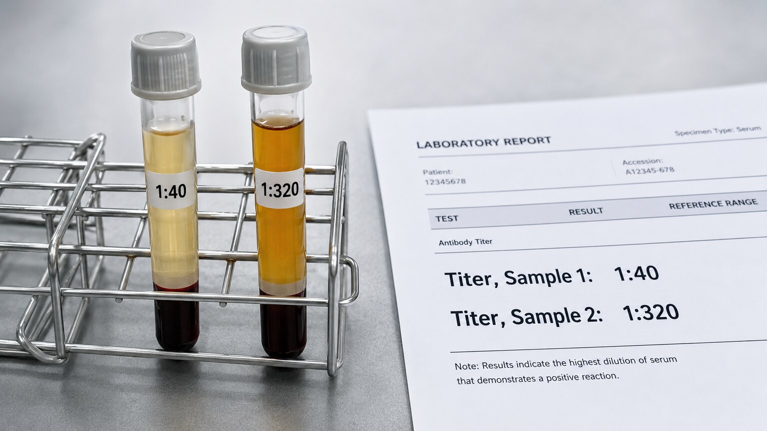
Figure 6. A rise from 1:40 to 1:320 — an eightfold increase — comfortably clears the fourfold threshold used to confirm a recent, genuine immune response.
What a Titer That Doesn't Rise Actually Tells You
It's worth spending a moment on the opposite scenario, because it's just as informative as a genuine rise and comes up just as often in real practice. If your acute and convalescent titers come back essentially the same — say, 1:160 on both draws — that stability is itself a meaningful result, not a non-result. It generally means one of two things. The first possibility is that you were never actively fighting this particular pathogen during the window between the two draws; whatever symptoms prompted the testing had a different cause, or the antibody detected was left over from an old, unrelated exposure or vaccination rather than reflecting anything currently happening. The second possibility, more reassuring in a different way, is that your immune system had already finished its work before the first sample was ever drawn — the antibody response peaked and plateaued earlier than expected, so both draws simply caught the same, already-established plateau rather than catching it mid-rise. This is part of why the timing of the first, acute-phase draw matters just as much as the second: a first sample drawn too late in the illness, after the antibody level has already climbed and leveled off, can make a genuine recent infection look like old, stable immunity, since there's no earlier, lower baseline left to compare against. A flat pair of titers doesn't rule out a past infection with this organism — it specifically rules out an active, currently unfolding immune response during the exact days separating your two blood draws.
Real-World Situations Where Paired Titers Are Actually Used
This comparison isn't a rare, obscure lab maneuver — it shows up across a wide range of everyday clinical situations, each with its own twist on how the rising-titer pattern gets applied. In suspected Lyme disease, paired testing is built directly into the standard two-tier protocol: an initial screening test is followed, when positive or ambiguous, by a more specific confirmatory test, and in cases where the first sample is drawn very early in the illness — sometimes before the body has had time to produce a detectable antibody response at all — a second sample drawn two to four weeks later can catch a seroconversion that the first sample entirely missed, turning an initially negative result into a confirmed diagnosis once the paired comparison is complete. In suspected Epstein-Barr virus infection, the virus responsible for mononucleosis, doctors sometimes track specific antibody types against different viral proteins over time to distinguish a brand-new infection from a past one that's simply flaring up detectably, since EBV establishes lifelong latency and different antibody patterns mark different stages of that relationship with the virus. Pregnancy care relies on a related but distinct use of titers: a rubella immunity titer drawn early in pregnancy confirms whether a person already has lasting protective antibody from childhood vaccination, and if it's borderline or unclear, it may be rechecked to make sure the reading is stable and reliable, since active rubella infection during pregnancy carries serious risks to fetal development. And after a hepatitis B vaccine series, a follow-up antibody titer — sometimes repeated after a booster dose in someone whose first check came back low — is used specifically to confirm that vaccination successfully built a protective level of antibody, applying the same anamnestic-response logic already described earlier in this article.
A Brief History of Why the Fourfold Threshold Became the Standard
The fourfold rule isn't a modern invention — it traces back to some of the earliest formal methods in diagnostic virology, developed well before molecular testing existed, when serology was essentially the only tool available for confirming that a specific infection had actually occurred. Early researchers working with serial dilution assays needed a way to separate genuine biological change from the ordinary, small-scale variability baked into manually diluting and reading a row of test wells, and repeated observation showed that single-step shifts happened often enough by chance alone that they couldn't be trusted as evidence of anything. A two-step, fourfold change, by contrast, occurred far less often through chance variability and lined up consistently with cases where a real infection was independently confirmed through other means. That empirical finding became formalized into the case-confirmation criteria still used today by public health and reference laboratories worldwide, spanning everything from classic childhood illnesses like measles and mumps to newer respiratory and vector-borne diseases, and it remains one of the more durable pieces of twentieth-century diagnostic methodology still in active daily use, essentially unchanged, in twenty-first-century laboratories.
Frequently Asked Questions
Does any increase between two titers count as significant?
No. Because titers are measured in doubling dilution steps, a single-step increase is considered within normal lab variability. The accepted threshold for a clinically meaningful change is a fourfold rise — the convalescent titer needs to be at least four times the acute titer — which corresponds to two full doubling steps, such as a move from 1:20 to 1:80 or higher.
Why does my doctor want a second blood draw weeks later instead of retesting right away?
The immune system needs time to build a full antibody response. Convalescent samples are typically drawn two to four weeks after the first, acute-phase sample specifically so the comparison reflects the immune system's complete response rather than an incomplete, still-developing one.
Can a rising titer happen even if I already had this infection before?
Yes. This is called an anamnestic response. Memory cells from a past infection or vaccination can react to a new exposure faster and more strongly than a first-time immune response, producing a sharp titer rise even in someone who already had detectable, pre-existing antibody.
Can my titer look like it rose just because two different labs ran the tests?
Yes, and this is a common source of confusion. Different laboratories and different assay methods aren't always directly comparable, so an apparent rise between results from two different labs may reflect a difference in measuring technique rather than a real change in your immune response. Paired samples are ideally tested together, at the same lab, using the same method.
If my two titers stayed the same, does that mean I never had the infection?
Not necessarily. A flat, unchanged titer usually means either that this particular infection wasn't active during the window between your two draws, or that your immune response had already peaked and plateaued before the first sample was even drawn. It rules out an active response happening between the two tests, but it doesn't rule out a past infection or vaccination that already ran its full course earlier.
Is titer testing only used for infections, or also for things like autoimmune conditions?
Paired titer testing is most established for confirming infections and checking vaccine response, since both situations involve a specific antigen the immune system is reacting to. Some autoimmune antibody tests are also reported as titers and can be tracked over time, but they're generally interpreted differently, since autoimmune antibody levels don't follow the same acute-versus-convalescent infection timeline this article describes.
Conclusion
A rising antibody titer between two paired blood draws is one of the more elegant tools in diagnostic medicine — a way of turning a single, ambiguous snapshot into a clear signal of direction and timing. The number itself, expressed as a dilution ratio like 1:320, can look unfamiliar at first, but the underlying idea is straightforward: it measures how much antibody your immune system had built by a specific point in time. What actually matters clinically isn't whether the second number is a little higher than the first — it's whether it climbed by at least fourfold, drawn two to four weeks apart, ideally at the same lab using the same method. When it does, that rise is strong, time-stamped evidence that your immune system was actively mounting a genuine response to a specific pathogen during that exact window, whether that's a brand-new infection, a fast, experienced reaction to something your body has met before, or confirmation that a vaccine successfully did its job. Just as importantly, a titer that doesn't rise is informative too — it tells your care team that nothing new was actively unfolding during that particular stretch of time, even if it can't rule out something that happened earlier. If you're looking at two titer results and trying to make sense of what changed between them, bringing both reports — and the exact dates and labs they came from — to your healthcare provider is the most useful next step, since the comparison, not either number alone, is where the real answer lives. Two numbers on two separate pieces of paper can feel disconnected from each other at first glance, but read together, in order, with the right amount of time between them, they tell a coherent story about exactly what your immune system was doing during a specific stretch of your life — which is a remarkable amount of insight to get from something as simple as diluting a few drops of blood, over and over, until a reaction finally stops.
Still Not Sure What Your Results Mean?
Upload your labs and get a complete, visual, plain-language interpretation of every biomarker — delivered to your inbox in under 15 minutes.
Get My ReportThis article is for educational purposes only and does not constitute medical advice. Always consult your healthcare provider regarding your specific lab results.