
Can a Viral Infection Cause a Temporary Positive Autoimmune Test?
Yes — a viral infection can genuinely cause a temporary positive autoimmune antibody test, and this isn't a rare fluke or lab error. It's a well-documented, mechanistically understood phenomenon, and certain viruses are notorious enough for triggering it that clinicians specifically ask about recent illness when a young, otherwise healthy person shows up with an unexpected positive ANA (antinuclear antibody) test. The immune response your body mounts against a virus and the immune response involved in true autoimmune disease actually share overlapping machinery — B cells, antibody production, and inflammatory signaling — and a sufficiently intense viral infection can, through several distinct biological pathways, temporarily produce antibodies that also happen to react against your own tissue. Epstein-Barr virus (the cause of mononucleosis), parvovirus B19, cytomegalovirus, hepatitis C, and more recently SARS-CoV-2 are the most extensively documented examples. This article walks through the specific mechanisms behind why this happens, which viruses are most associated with it, the important nuance that a virus can sometimes be a genuine trigger for lasting autoimmune disease rather than just a temporary mimic, and how doctors actually distinguish a real, ongoing autoimmune condition from a passing viral echo.
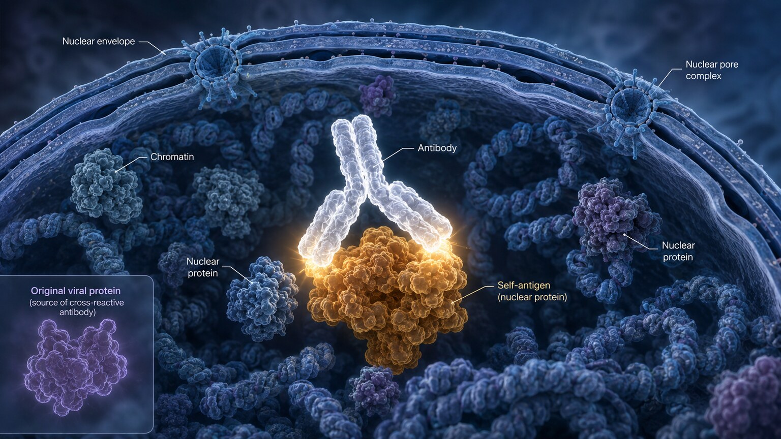
Figure 1. Molecular mimicry occurs when a viral protein's structure closely resembles a human self-antigen closely enough that antibodies made against the virus also bind to the body's own tissue.
What Your Immune System Is Actually Doing When It Fights a Virus
To understand why this happens, it helps to remember what antibody production is fundamentally built to do: identify a very specific molecular shape (called an antigen) and manufacture a matching antibody that binds to it, marking it for destruction. When you catch a virus, your immune system's B cells generate a wide range of antibodies aimed at the virus's various surface and internal proteins. Under normal circumstances, this process is remarkably precise — but it isn't perfect, and the sheer scale and intensity of the immune activity involved in fighting off a significant viral infection creates several distinct openings for antibodies or immune cells that also happen to react against your own body's proteins to slip through, at least temporarily, before the immune system settles back into its normal, tightly regulated state once the infection clears.
This isn't unique or freakish biology — your immune system already contains a background population of low-level autoreactive B cells and T cells even when you're completely healthy, normally kept in check by multiple layers of immune tolerance. A viral infection can transiently disrupt that tolerance through several specific, well-studied mechanisms, covered one at a time below.
It's worth being precise about what an autoimmune antibody test is actually built to detect, since it clarifies why a viral infection can produce a positive result without a person having true autoimmune disease. A test like ANA doesn't detect "autoimmune disease" directly — no lab test does. It detects the presence of antibodies that bind to specific components of the human cell nucleus, full stop, regardless of why those antibodies happen to be there. In someone with genuine lupus or a related condition, those antibodies are being produced because of a sustained, dysregulated autoimmune process. In someone recovering from a significant viral illness, the antibodies detected by the exact same test can be present for one of the entirely different, self-limited reasons described in this article. The test itself can't distinguish between these two very different underlying stories — that distinction requires everything covered in the sections that follow.
Curious what the rest of your lab results are actually telling you? Upload your results and get a complete, plain-language breakdown in under 15 minutes.
Analyze My ResultsMechanism 1: Molecular Mimicry — When a Viral Protein Looks Like You
Molecular mimicry is the best-known of these mechanisms and the one shown in the illustration above. Some viral proteins happen to share a strong structural or sequence resemblance to specific human self-proteins, purely by evolutionary coincidence, or in some cases because certain viruses have evolved surface structures that help them evade immune detection by resembling host tissue. When your immune system builds antibodies specifically targeted at the viral protein, those same antibodies can, due to the shared shape, also bind to the structurally similar human protein — cross-reacting with your own tissue as an unintended side effect of an otherwise appropriate antiviral response.
Epstein-Barr virus offers one of the best-studied examples: a specific EBV protein called EBNA-1 (Epstein-Barr nuclear antigen 1) shares meaningful structural similarity with certain human nuclear self-proteins, including some targeted by antibodies seen in lupus. Antibodies generated against EBNA-1 during an active or recent EBV infection can cross-react with these human proteins, contributing to a positive ANA or other autoantibody result. This same mimicry mechanism is part of why EBV in particular occupies a more complicated role than a simple "temporary false alarm," discussed further below.
It's worth understanding why this kind of coincidental resemblance happens at all rather than being vanishingly rare. Proteins are built from a limited alphabet of twenty amino acids, and functional protein structures — particularly small, exposed surface regions that antibodies actually latch onto, called epitopes — recur across otherwise unrelated organisms simply because there are only so many chemically stable ways to fold a short stretch of amino acids into a shape capable of doing a given job. Viruses, having evolved over millions of years alongside their human hosts, have in some cases converged on protein shapes that happen to overlap with human self-proteins performing an unrelated function, not because the virus is deliberately disguising itself, but simply as a byproduct of the limited structural vocabulary evolution has to work with. Antibodies bind based on shape and chemical fit, not on knowing anything about an antigen's origin — so a close enough structural match is, from the antibody's perspective, functionally indistinguishable regardless of whether it came from a virus or from the person's own body.
Mechanism 2: Bystander Activation — Breaking Tolerance Through Inflammation Alone
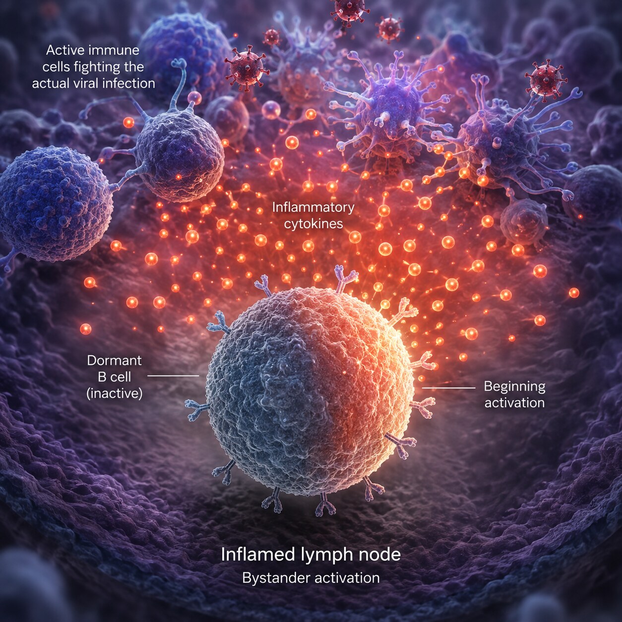
Figure 2. Bystander activation occurs when the intense cytokine storm surrounding a viral infection nonspecifically stimulates dormant self-reactive B cells that had nothing to do with the virus itself.
Unlike molecular mimicry, bystander activation doesn't require any structural resemblance between the virus and your own tissue at all. A significant viral infection floods the surrounding tissue and lymph nodes with inflammatory signaling molecules — cytokines like type I interferon, interleukin-6, and tumor necrosis factor-alpha — as part of the normal antiviral response. This intense local inflammatory environment can nonspecifically activate B cells sitting nearby that were never targeting the virus at all, including some of the dormant, low-level autoreactive B cells that exist in the background of a healthy immune system under normal tolerance controls. Essentially, the inflammatory "noise" surrounding the infection can be loud enough to wake up immune cells that have nothing to do with the actual viral fight, prompting them to start producing antibody, including autoantibody, purely as a side effect of being caught in the inflammatory crossfire.
This mechanism helps explain why the degree and severity of a viral illness often correlates with how likely a temporary autoantibody positivity is to appear — a more severe, more inflammatory infection creates a stronger bystander activation signal, which is part of why this phenomenon is documented more consistently with infections known for producing an intense systemic inflammatory response, such as significant EBV mononucleosis or severe COVID-19, than with milder, more localized viral illnesses.
A useful way to picture bystander activation is a crowded room where a fire alarm suddenly goes off. Most people in the room react appropriately to the actual emergency — heading for the exits in response to genuine danger. But a fire alarm is loud enough, and disruptive enough, that it can also startle people who weren't paying attention to anything related to the emergency at all, prompting reactions that have nothing to do with the fire itself. Bystander activation works similarly: the cytokine "alarm" produced by a genuine antiviral response is loud enough, immunologically speaking, that some nearby autoreactive B cells react simply because the alarm went off nearby, not because they detected anything related to the virus specifically.
Mechanism 3: Polyclonal B Cell Activation — When a Virus Directly Hijacks B Cells
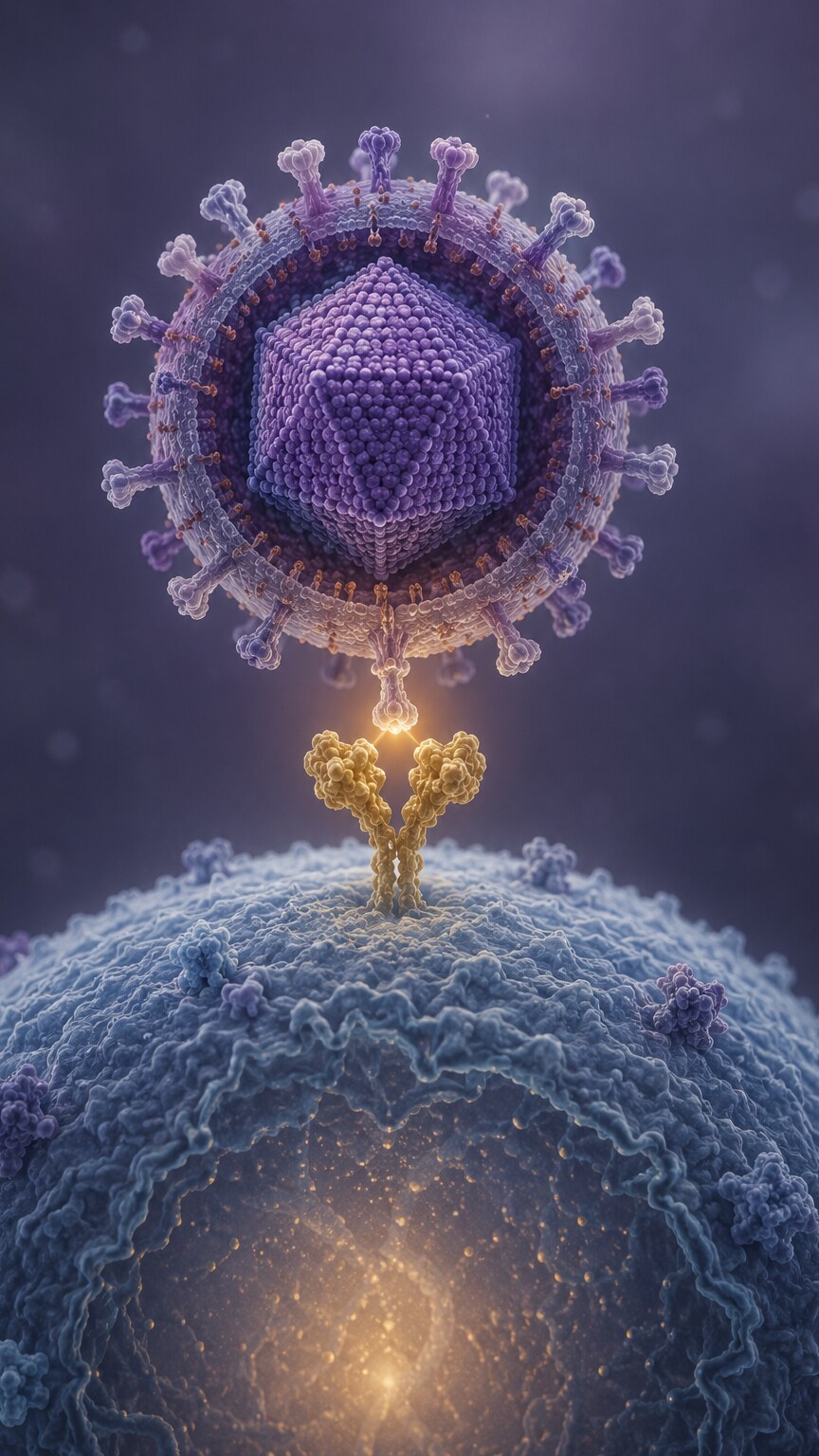
Figure 3. Epstein-Barr virus infects B cells directly by binding the CD21 receptor on their surface, driving the widespread, nonspecific antibody production characteristic of infectious mononucleosis.
A third mechanism is specific to a small number of viruses capable of directly infecting the very B cells responsible for making antibodies in the first place — and Epstein-Barr virus is, once again, the textbook example. EBV specifically targets and infects B lymphocytes by binding to the CD21 receptor on their surface, the same receptor these cells normally use as part of a different immune signaling pathway. Once inside, EBV doesn't just hide there quietly; it drives infected B cells into a state of vigorous, largely nonspecific proliferation and antibody production, a phenomenon called polyclonal B cell activation. Rather than a narrowly targeted response aimed precisely at EBV's own proteins, this creates a broad, somewhat indiscriminate wave of antibody output from many different B cell lineages simultaneously, some of which inevitably produce autoreactive antibody purely as a statistical byproduct of so many B cell clones being switched on at once.
This mechanism is directly responsible for one of the oldest diagnostic quirks in medicine: the heterophile antibody, the nonspecific antibody detected by the classic "monospot" test used to help diagnose mononucleosis, is itself a byproduct of this same polyclonal B cell activation — a helpful diagnostic tool built directly on the very same mechanism that also happens to produce incidental autoantibody positivity in a meaningful share of people during acute EBV infection.
Estimates of exactly how common transient ANA positivity is during acute infectious mononucleosis vary across different studies, partly because of differences in which antibody assay was used and how long after symptom onset testing was performed, but multiple published case series have documented positivity rates high enough that clinicians experienced with EBV are rarely surprised by it. This is a large part of why a thorough history — specifically asking about recent sore throat, significant fatigue, swollen lymph nodes, or a known mono diagnosis in the weeks before a positive ANA was drawn — is considered a routine, expected part of working up a new positive result in a young patient, rather than an obscure detail only relevant in unusual cases.
Mechanism 4: Cell Death and the Sudden Exposure of Hidden Self-Antigens
A fourth mechanism centers on what happens to the cells a virus actually infects and destroys. Many of the proteins your immune system later learns to recognize in true autoimmune disease — components of the cell nucleus, DNA-binding proteins, RNA-processing machinery — normally sit safely tucked away inside intact, living cells, essentially invisible to the immune system under ordinary circumstances. When a virus infects and kills large numbers of cells, whether through direct viral damage or through the immune system's own effort to eliminate infected cells, these normally hidden intracellular contents spill out into the surrounding tissue and bloodstream in unusually large quantities, all at once, in the middle of an already highly inflamed environment.
This sudden, large-scale exposure of self-antigens that the immune system rarely encounters in this form or quantity, combined with the surrounding inflammatory "danger signals" from the infection, can be enough to transiently prompt an antibody response against those self-antigens — essentially catching the immune system's tolerance mechanisms off guard by presenting them with material they weren't prepared to encounter at that scale. Once the infection resolves and cell death slows back to its normal, low-level baseline, this source of antigen exposure fades, and the transient autoantibody response it triggered typically fades along with it.
Researchers distinguish between two broad patterns of cell death that matter for this mechanism: ordinary, quiet apoptosis, in which a dying cell's contents are neatly packaged and cleared away by nearby immune cells without triggering much alarm, versus more disorderly necrosis or a specific inflammatory form of cell death associated with viral infection, in which cellular contents spill out more chaotically alongside additional "danger signal" molecules that actively recruit and activate immune attention. It's this second, messier pattern of cell death — common during an active, aggressive viral infection — that carries the greater risk of triggering a transient autoantibody response, since it combines the exposure of normally hidden self-antigen with an inflammatory context that specifically encourages the immune system to react to whatever it finds nearby.
Mechanism 5: The Type I Interferon Connection
The final mechanism worth understanding ties directly back to the actual biology of genuine autoimmune disease, and it's part of why this topic is more nuanced than a simple "false alarm" framing suggests. Type I interferons (interferon-alpha and interferon-beta) are among the very first signaling molecules your body releases in response to a viral infection, and they're extremely effective at limiting viral replication. But type I interferon signaling isn't just an antiviral tool — it's also one of the most well-established drivers of autoantibody production in genuine autoimmune conditions like lupus, so consistently that researchers refer to an elevated "interferon signature" as a hallmark feature of active lupus itself, independent of any viral trigger at all.
This creates a genuinely interesting overlap: because a significant viral infection triggers a strong type I interferon surge as a completely normal, appropriate antiviral defense, that same interferon surge can transiently push the immune system's antibody-producing machinery in a direction that mimics part of what's happening biologically in real, chronic autoimmune disease — without the person actually having lupus or another autoimmune condition. This shared interferon pathway is one of the more actively researched links between infection and autoimmunity, and it partly explains why viral-triggered autoantibody positivity isn't simply a coincidental lab artifact, but a genuine, mechanistically grounded overlap between two immune processes that share more underlying biology than their outcomes would suggest.
Interferon's role here also helps explain an observation that puzzled researchers for years: why lupus itself sometimes appears to flare, or occasionally to first present, in the weeks following an unrelated viral illness in people who already carry an underlying genetic predisposition to autoimmune disease. If interferon signaling is already primed to run hot in someone genetically inclined toward lupus, an added viral trigger supplying its own interferon surge on top of that existing predisposition may be enough to tip a borderline, subclinical process into a more clearly active one — another example of how the line between "temporary viral mimicry" and "a real disease process becoming apparent" isn't always a clean, easily drawn boundary.
Which Viruses Are Most Documented to Cause This

Figure 4. Epstein-Barr virus, the cause of infectious mononucleosis, is the single most extensively documented trigger of temporary autoantibody positivity among common viral infections.
While the mechanisms above can, in principle, apply to many viral infections, research has consistently documented this phenomenon most strongly with a specific handful of viruses. Epstein-Barr virus leads the list by a wide margin, given its unique ability to directly infect B cells and its well-characterized molecular mimicry with lupus-associated self-antigens; transient ANA and rheumatoid factor positivity during acute mononucleosis is common enough that it's a standard consideration in clinical evaluation of a young person presenting with a new positive ANA.
Parvovirus B19 is another well-documented example, notable because the acute infection it causes in adults commonly produces joint pain and swelling that can itself clinically resemble early rheumatoid arthritis or lupus, compounding the diagnostic confusion when it's accompanied by a positive autoantibody test — this combination of arthritis-like symptoms and positive serology makes parvovirus B19 one of the more frequent genuine diagnostic mimics of autoimmune joint disease in clinical practice. Unlike the childhood presentation of parvovirus B19 (the classic "slapped cheek" facial rash), the adult presentation often skips the rash entirely and centers almost exclusively on this joint involvement, which is part of why it's specifically an adult rheumatology consideration rather than a pediatric one in this context.
Cytomegalovirus, a relative of EBV in the herpesvirus family, has similarly been documented to produce transient autoantibody positivity through overlapping mechanisms, particularly in the context of a more significant, symptomatic CMV illness rather than the often silent, asymptomatic CMV infections many adults carry without ever knowing it. Hepatitis C virus, particularly with chronic infection, is well known for inducing a range of autoantibodies including ANA, rheumatoid factor, and anti-smooth muscle antibody, occasionally complicating the distinction from autoimmune liver disease — a distinction that matters enormously in practice, since the treatment for chronic viral hepatitis and the treatment for autoimmune hepatitis are entirely different, making accurate differentiation a genuine clinical priority rather than an academic curiosity. More recently, a substantial body of research generated during the COVID-19 pandemic documented transient ANA and other autoantibody positivity in a meaningful proportion of people during and after acute SARS-CoV-2 infection, with some researchers proposing this autoantibody activity may contribute to certain lingering post-COVID symptoms, an area of continued active research.
The Important Nuance: Sometimes the Virus Is a Real Trigger, Not Just a Mimic
It would be an oversimplification to treat every case of viral-associated autoantibody positivity as a clean, harmless coincidence that always resolves without consequence. For a genetically susceptible subset of people, the same viral mechanisms described throughout this article — particularly EBV's molecular mimicry and its direct effect on B cells — appear to function not as a temporary mimic but as an actual initiating trigger for lasting, genuine autoimmune disease. The evidence connecting EBV specifically to multiple sclerosis has become particularly strong in recent years, with large longitudinal studies finding that the overwhelming majority of multiple sclerosis cases are preceded by EBV infection, to the point that many researchers now consider EBV infection a necessary, if not sufficient, step in the chain of events leading to MS in susceptible individuals. EBV has also long been epidemiologically linked to an increased risk of developing lupus later in life.
This distinction matters enormously for how a positive result should actually be understood: it's not always a simple binary between "temporary viral mimicry that means nothing" and "an unrelated, coincidental autoimmune disease that happened to show up around the same time." In some people, the virus genuinely is the spark that, in combination with an underlying genetic susceptibility, sets a real and lasting autoimmune process into motion — which is exactly why a positive result after a viral illness, particularly one that doesn't fully resolve on repeat testing, deserves a proper, careful evaluation rather than being automatically dismissed as "just the virus."
Researchers describe this using a "multi-hit" framework: a single risk factor alone — carrying a susceptibility gene, or catching a particular virus — is rarely sufficient on its own to produce autoimmune disease in most people, since a large share of the population is exposed to EBV at some point (estimates suggest the substantial majority of adults worldwide have been infected at some point, often without ever developing noticeable symptoms) without ever developing lupus or multiple sclerosis. It's the combination of the right genetic background, the right environmental trigger, and often additional contributing factors like hormonal shifts or other environmental exposures, arriving together, that appears necessary to tip a susceptible person from "exposed" to "affected." This is precisely why most people who test positive after a viral illness never go on to develop lasting autoimmune disease, even though the underlying viral mechanisms capable of contributing to that outcome are common and well understood.
How Doctors Actually Tell the Difference
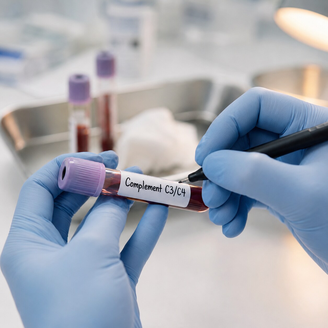
Figure 5. Unlike active autoimmune disease, which typically consumes and lowers complement proteins C3 and C4, viral-triggered autoantibody positivity usually leaves complement levels normal, making it a useful distinguishing test.
The single most useful tool for telling a temporary, viral-associated positive apart from a genuine autoimmune disease is simply time: repeating the antibody test roughly three to six months after the acute illness has fully resolved. A viral-triggered positive typically falls significantly or returns to negative once the infection and its accompanying inflammation have cleared, while a true underlying autoimmune disease tends to persist or, in some cases, climb further as time passes. This single piece of information — the trajectory over time, not the initial number — is often more diagnostically valuable than the original result itself.
Why does simple observation over time work so well here, when a snapshot in the moment often can't? Because the underlying biology of a temporary versus a persistent process is fundamentally different in a way that only reveals itself with time. A viral-triggered response is, by its nature, tied to an active, self-limited event — once the virus is cleared and inflammation subsides, the biological "fuel" driving the antibody production runs out, and production naturally winds down. A genuine autoimmune disease, by contrast, is sustained by an ongoing, self-perpetuating process that doesn't have a natural endpoint the way an infection does, so it keeps producing antibody indefinitely rather than tapering off. No single blood draw can distinguish "fuel that's about to run out" from "a process that will keep going" — but two blood draws, spaced appropriately apart, usually can.
Beyond simple retesting, several other tools help sharpen the distinction. Complement proteins C3 and C4, part of a separate branch of the immune system, are typically consumed and measurably lowered during active, genuine autoimmune inflammation (particularly in active lupus), but usually remain normal in viral-associated autoantibody positivity, making a complement panel a genuinely useful add-on test. A more detailed extractable nuclear antigen (ENA) panel and specific, more targeted anti-dsDNA testing (using an assay like the Crithidia luciliae method, which is considered more specific for genuine lupus-associated antibody than some standard assays) can further refine the picture, since viral-associated antibodies often don't show the same specific antibody subtype pattern typically seen in confirmed autoimmune disease. Finally, and perhaps most importantly, the actual clinical picture matters more than any single lab value: the presence or absence of hallmark autoimmune symptoms — a persistent, non-migrating joint swelling lasting many weeks, a photosensitive facial rash, unexplained kidney involvement, or organ-specific inflammation — carries substantial diagnostic weight alongside the lab trajectory.
The pattern seen on the ANA test itself, called the immunofluorescence staining pattern, can also offer a modest additional clue, though it's far from definitive on its own. Certain patterns, like a homogeneous or speckled appearance, are seen across a broad range of situations including viral-associated positivity and carry less specific diagnostic weight, while other, less common patterns are more tightly associated with particular genuine autoimmune conditions. Because staining pattern alone isn't reliable enough to confirm or rule out disease by itself, it's generally treated as one additional data point layered alongside titer, complement levels, symptom history, and the trajectory over time, rather than a standalone answer.
What This Means If You Just Had a Viral Illness and a Positive Result

Figure 6. A follow-up conversation weighing the repeat test trajectory alongside any ongoing symptoms is typically what actually resolves the uncertainty left by an initial positive result, more so than the first number alone.
If you've recently recovered from a significant viral illness and received an unexpected positive ANA or other autoantibody test, the most useful immediate step is simply making sure whoever ordered or is interpreting the test knows about the recent infection, since that single piece of context substantially changes how the result should be read. From there, the practical path forward usually involves a period of watchful waiting rather than immediate aggressive workup, particularly if you have no symptoms suggestive of genuine autoimmune disease — with a repeat test planned for a few months down the line to see whether the antibody trajectory falls, as expected with a viral-triggered positive, or persists, which would prompt a more thorough autoimmune evaluation.
Keeping a simple written timeline for yourself in the meantime — the date the viral illness started, when symptoms resolved, when the positive test was drawn, and any new symptoms that appear in the following months — can make that eventual follow-up conversation considerably more useful than trying to reconstruct the sequence of events from memory weeks or months later. This kind of concrete timeline is often exactly the detail that helps a clinician confidently place a given result into the "resolving as expected" category rather than needing to pursue a more extensive, and more anxiety-inducing, workup out of an abundance of caution alone.
It's worth remembering that a single positive ANA, especially at a low titer, is common in the general population even without any autoimmune disease or recent viral illness at all — some studies estimate a meaningful percentage of healthy people test positive at low titers without ever developing any related condition. A positive result following a documented viral illness, interpreted in that context and given the specific mechanisms covered in this article, is genuinely one of the more reassuring scenarios in which an unexpected positive autoimmune antibody test can appear.
It's also worth keeping the underlying purpose of the test in mind while waiting through this process. Autoimmune antibody testing exists to catch and characterize genuine disease as early as possible, when treatment tends to be most effective — not to generate anxiety over every incidental positive that crosses a threshold. A single result, taken in isolation and without the surrounding context this article has walked through, was never designed to be the final word on its own. The trajectory over time, combined with your actual symptoms and history, is what the test was always meant to be interpreted alongside, and giving that fuller picture time to reveal itself is a normal, expected part of how this kind of testing is meant to work.
Frequently Asked Questions
How long after a viral infection can a temporary positive autoimmune test last?
It varies by virus and by individual, but viral-associated autoantibody positivity typically declines over the weeks to a few months following the acute illness, tracking the resolution of the underlying inflammation. This is why doctors generally recommend retesting roughly three to six months after recovery rather than immediately, giving enough time for a genuinely temporary response to fall.
Which specific test result would suggest my positive is from a virus rather than a real autoimmune disease?
No single test proves this definitively, but a combination of clues is reassuring: normal complement (C3/C4) levels, a low antibody titer, a negative extractable nuclear antigen panel, absence of hallmark autoimmune symptoms, and — most importantly — a falling or negative result on repeat testing months later, all point toward a viral-associated cause rather than genuine autoimmune disease.
Does this mean EBV or other viruses actually cause autoimmune diseases like lupus?
The relationship is more nuanced than simple causation. In most people, a viral infection produces only a temporary antibody blip that resolves completely. In a genetically susceptible subset, however, research increasingly supports that certain viruses, particularly EBV, can act as a genuine triggering event that helps set lasting autoimmune disease into motion — which is why persistent, rather than resolving, positivity after a viral illness deserves proper follow-up.
Should I avoid getting tested for autoimmune antibodies right after being sick with a virus?
It's not that testing should be avoided, but timing and interpretation matter. If possible, and if symptoms allow it, waiting until you've fully recovered from an acute viral illness before autoimmune antibody testing can reduce the chance of a confusing, temporary positive result — though if there's a genuine clinical concern for autoimmune disease, your doctor may still test promptly and simply plan to interpret the result with the recent infection in mind.
Can a viral infection affect other autoimmune-related tests besides ANA, like rheumatoid factor?
Yes. The same mechanisms described in this article — molecular mimicry, bystander activation, polyclonal B cell activation — aren't specific to ANA alone. Rheumatoid factor is another commonly affected antibody test, particularly with EBV and parvovirus B19 infections, and can show a similar pattern of temporary positivity that resolves as the underlying viral illness clears.
Conclusion
A temporary positive autoimmune test after a viral infection is a real, biologically well-understood phenomenon, driven by molecular mimicry, bystander activation, direct viral effects on B cells, sudden self-antigen exposure from cell death, and shared interferon signaling — not a lab mistake or a meaningless coincidence. Epstein-Barr virus, parvovirus B19, cytomegalovirus, hepatitis C, and SARS-CoV-2 are the most well-documented examples, and for most people, this kind of result fades on its own as the infection resolves. The one genuine complication is that, in a smaller number of genetically susceptible people, certain viruses — EBV most notably — can act as an authentic trigger for lasting autoimmune disease rather than a passing mimic, which is exactly why a positive result that doesn't resolve on repeat testing, or that comes with real autoimmune symptoms, still deserves a careful, proper evaluation rather than automatic reassurance.
The underlying takeaway is less about memorizing five specific mechanisms and more about the broader relationship they collectively reveal: the same immune machinery that protects you from infection is not a separate system from the one implicated in autoimmune disease — it's the same system, capable of the same tools, occasionally pointed, briefly and usually harmlessly, in a direction its own tolerance mechanisms are normally built to prevent. Understanding that overlap is what turns an unexpected positive result from a source of open-ended worry into a specific, answerable question with a clear, evidence-based path toward resolving it.
Still Not Sure What Your Results Mean?
Upload your labs and get a complete, visual, plain-language interpretation of every biomarker — delivered to your inbox in under 15 minutes.
Get My ReportThis article is for educational purposes only and does not constitute medical advice. Always consult your healthcare provider regarding your specific lab results.