
Understanding the Different ANA Staining Patterns
An ANA staining pattern describes exactly where, physically, inside a cell's nucleus your own antibodies are actually attaching, and that specific, observable location serves as a genuine clue pointing toward which antibodies are actually present and, by direct extension, which autoimmune conditions are actually worth investigating further. The test itself works by mixing a diluted sample of your blood with a slide of human cells, then using a fluorescent tag to make any bound antibodies glow green under a microscope — and different antibodies bind to different structures inside the nucleus, producing visually distinct glowing patterns that a trained technologist can recognize and classify by name. This article walks through exactly what each major pattern looks like, the specific antibodies and structures responsible for producing it, and which autoimmune conditions each pattern is most strongly associated with — plus why the pattern alone is a starting clue, never a finished diagnosis.
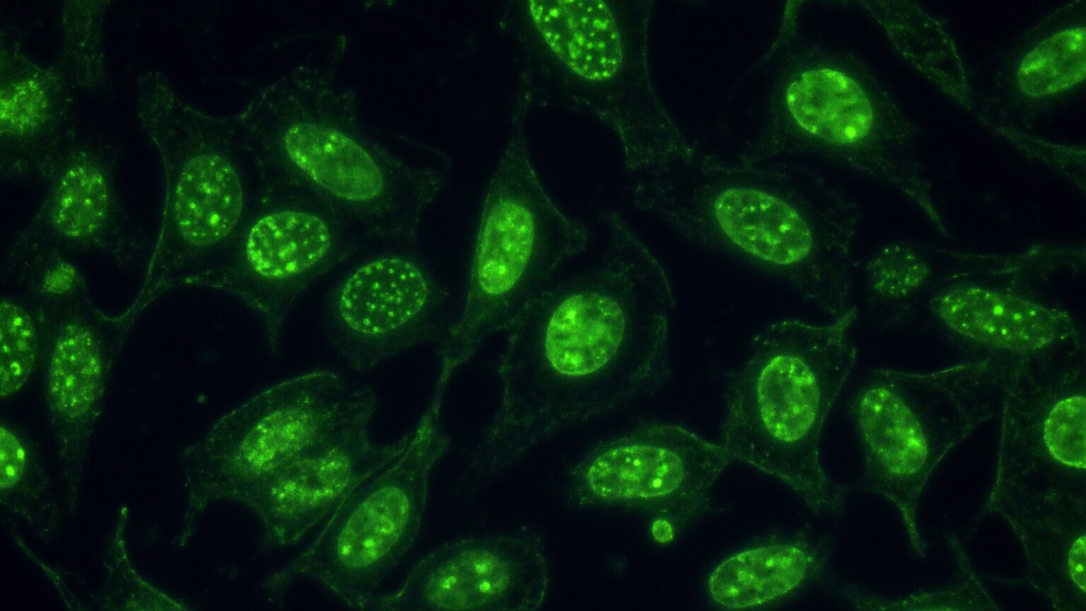
Figure 1. The ANA test exposes a slide of human cells to a patient's blood sample, then uses a fluorescent tag to make any bound antibodies glow visibly under a specialized microscope.
How the Test Actually Produces a Visible Pattern
The standard ANA test uses a technique called indirect immunofluorescence, performed on a specific line of laboratory-grown human cells called HEp-2 cells, chosen specifically because they're large, easy to examine, and actively dividing, which reveals certain structures — like the machinery involved in cell division — that wouldn't be visible in a resting cell. A diluted sample of the patient's blood is applied to a slide of these cells; if antibodies targeting any component of the cell nucleus are present, they bind directly to that specific structure and stay attached once the slide is rinsed.
The choice of HEp-2 cells specifically, rather than any other available cell type, reflects decades of refinement in autoimmune laboratory testing. Earlier versions of this test used tissue sections cut directly from rodent organs, which worked reasonably well but lacked the large, clearly visible nuclei and the reliably active cell division that HEp-2 cells provide, meaning several of the patterns covered later in this article, particularly the centromere pattern, were considerably harder to identify confidently using those older methods. The switch to HEp-2 cells, a human-derived cell line originally established from a laryngeal cancer sample decades ago, meaningfully improved both the sensitivity and the pattern-recognition capability of this test across the board, which is part of why it remains the standard substrate used in laboratories worldwide today.
A second reagent, a fluorescently tagged antibody designed to attach specifically to human antibodies, is then applied, effectively lighting up wherever the original patient antibody bound. Under a fluorescence microscope, this produces a glowing green pattern that traces the exact shape and location of whatever structure the antibody was actually targeting — and because different autoimmune-related antibodies target genuinely different structures inside the nucleus, the resulting visual pattern is itself a real, physical clue about which specific antibody is present.
This technique, developed decades ago, remains the recognized reference standard for ANA testing today, even as newer, faster, automated methods have become increasingly available in many labs. Automated solid-phase assays can screen for ANA positivity more quickly and with less reliance on a human technologist's subjective visual interpretation, but they generally can't report a specific staining pattern the way traditional immunofluorescence can, since they're designed to detect overall antibody binding rather than visually map exactly where that binding is occurring within the cell. This is precisely why a sample flagged as positive by one of these faster screening methods is often still sent on for traditional immunofluorescence testing specifically when the ordering physician wants pattern information to help guide the next diagnostic steps.
Reading these patterns accurately requires real training and experience, since subtle differences between similar-looking patterns can carry genuinely different clinical meaning, and a single sample can sometimes show more than one pattern layered together. Clinical laboratory scientists specializing in this kind of testing typically develop this pattern-recognition skill over years of examining thousands of slides, comparing their interpretations against confirmed reference samples, and participating in ongoing proficiency testing specifically designed to ensure consistent, reliable pattern classification across different laboratories and different technologists.
An international effort called the International Consensus on Antinuclear Antibody Patterns, often abbreviated ICAP, was specifically established to help standardize this classification process across laboratories worldwide, assigning each recognized pattern a standardized reference code and a detailed set of criteria for identifying it correctly. This standardization matters enormously in practice, since a patient's sample might be processed at one laboratory and their follow-up testing at a completely different one, and consistent terminology across that transition prevents confusion or, worse, a missed connection between a pattern reported under one name at one lab and the same underlying pattern reported under a slightly different name somewhere else.
This same standardization effort has also worked to formally distinguish patterns that were once informally used interchangeably despite representing genuinely different underlying biology, refining the field's overall precision over time. As research continues to link specific staining patterns to increasingly specific molecular targets, this classification system continues to evolve, occasionally adding newly recognized patterns or refining the criteria for distinguishing between two patterns that were previously grouped together under one broader, less precise category.
Homogeneous Pattern: A Uniformly Glowing Nucleus
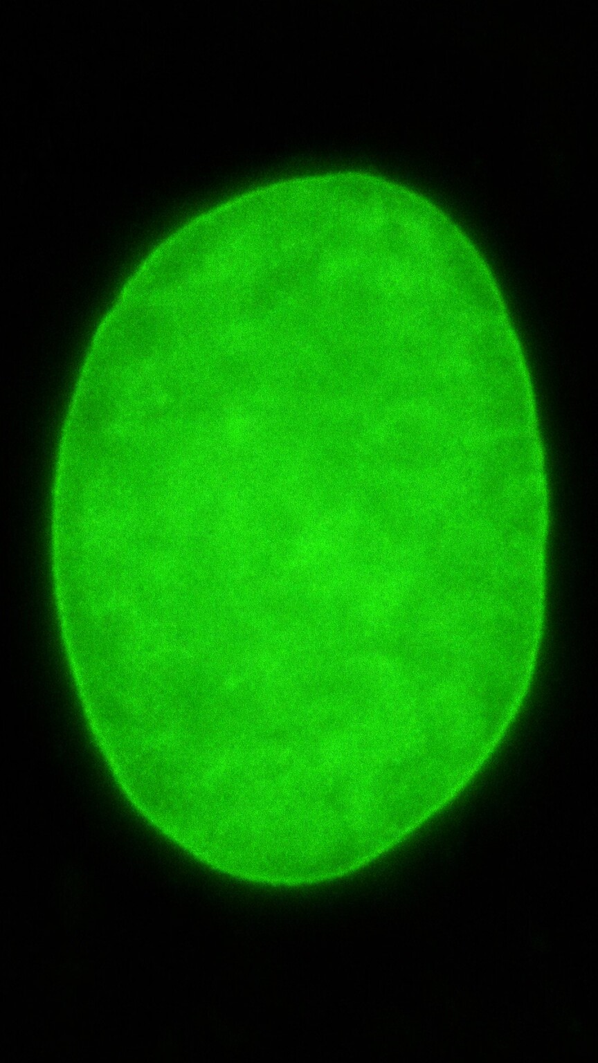
Figure 2. The homogeneous pattern shows an evenly, uniformly glowing nucleus, produced when antibodies bind broadly across chromatin, and is most classically associated with lupus.
The homogeneous pattern, also frequently referred to as the diffuse pattern, shows the entire visible nucleus glowing evenly and consistently uniform throughout, with no distinct dots, speckles, or clearly empty regions breaking up that continuous glow anywhere within it. This pattern is produced when antibodies bind broadly to chromatin — the combined structure of DNA wound around histone proteins that makes up the bulk of the nucleus's contents — rather than targeting any single, isolated structure within it.
The homogeneous pattern is the classic signature most strongly associated with systemic lupus erythematosus, historically linked specifically to anti-double-stranded DNA (anti-dsDNA) and anti-histone antibodies, both of which bind broadly across chromatin material in exactly the way that produces this uniform glow. Anti-histone antibodies specifically are also strongly associated with drug-induced lupus, a temporary, medication-triggered form of lupus-like illness that typically resolves once the responsible medication is stopped, making this pattern a genuinely useful early clue in that specific clinical scenario.
It's worth understanding why this particular pattern earned its "homogeneous" name specifically, since the terminology itself describes exactly what a technologist sees under the microscope. Chromatin, the DNA-and-histone complex making up the bulk of the nucleus, is distributed relatively evenly throughout the entire nuclear space during most of a cell's life cycle, rather than being clustered into a small number of discrete regions the way some other nuclear structures are. When an antibody binds broadly across this evenly distributed material, the resulting fluorescent signal naturally traces that same even distribution, producing the smooth, uniform glow that gives this pattern its descriptive name.
Clinically, the strength of this pattern's association with anti-dsDNA specifically matters beyond just diagnosis, since anti-dsDNA levels are also one of the more useful laboratory markers for tracking lupus disease activity over time once a diagnosis has already been established. Rising anti-dsDNA levels in someone with confirmed lupus often correlate with an approaching disease flare, particularly kidney involvement, giving this specific antibody genuine ongoing monitoring value well beyond the moment of initial diagnosis — a level of longitudinal usefulness that not every antibody associated with a different staining pattern shares to the same degree.
Curious what your own ANA result or the rest of your autoimmune panel is really telling you? Upload your results and get a complete, plain-language breakdown in under 15 minutes.
Analyze My ResultsSpeckled Pattern: The Broadest, Most Diverse Category
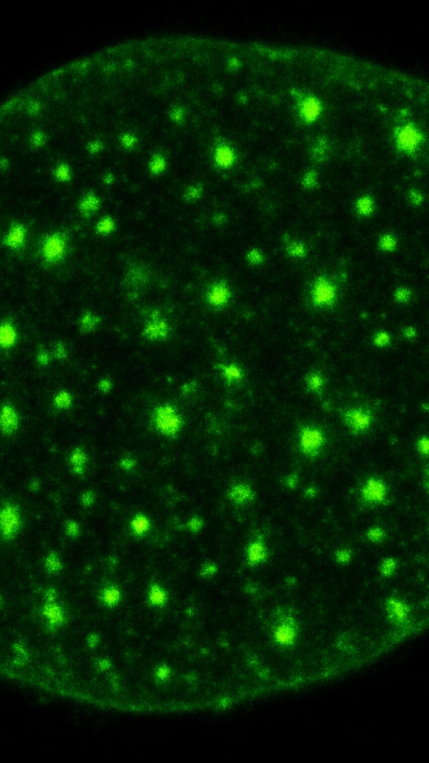
Figure 3. The speckled pattern shows numerous small, scattered glowing dots throughout the nucleus, produced by antibodies targeting a variety of distinct, non-chromatin proteins scattered within it.
The speckled pattern shows the entire nucleus filled with numerous small, distinct glowing dots scattered evenly throughout its interior, rather than one single uniform glow, and it's genuinely, by a wide margin, the broadest and most heterogeneous category on this entire list, since it can be produced by antibodies targeting several completely different, unrelated nuclear proteins that all happen to be scattered in small clusters rather than spread evenly across chromatin.
Anti-Sm and anti-RNP antibodies, which target small nuclear ribonucleoprotein complexes involved in processing genetic material, both produce a speckled pattern and are associated respectively with lupus specifically and with mixed connective tissue disease, a condition that shares overlapping features of lupus, scleroderma, and inflammatory muscle disease. Anti-SSA (Ro) and anti-SSB (La) antibodies, targeting different ribonucleoprotein complexes, also produce a speckled pattern and are most strongly associated with Sjögren's syndrome, a condition causing dry eyes and dry mouth from immune-mediated damage to moisture-producing glands, though they also appear in a meaningful subset of lupus patients as well.
Because so many genuinely different antibodies can all produce a visually similar speckled appearance, this particular pattern carries the least specific diagnostic weight on its own compared to the others covered in this article — it reliably tells a clinician that one of several possible antibodies is present, but confirming which one requires a separate, more specific confirmatory test targeting each individual antibody by name.
Laboratories have developed a useful sub-classification system to add at least some additional precision to this broad category, dividing the speckled pattern into "coarse" and "fine" variants based on the relative size of the individual dots observed. A coarse speckled pattern, showing larger, more widely spaced dots, is more frequently associated with anti-Sm and anti-RNP specifically, while a fine speckled pattern, showing smaller, more densely packed dots, is more frequently associated with anti-SSA and anti-SSB. This sub-classification isn't perfectly reliable on its own — there's genuine overlap between the two categories — but it does provide experienced technologists with one additional layer of visual information beyond simply reporting "speckled" as a single undifferentiated category.
The clinical picture surrounding a speckled result often does more work than the pattern itself in narrowing down the likely underlying antibody. A speckled pattern appearing alongside dry eyes, dry mouth, and swollen salivary glands points a clinician's attention firmly toward Sjögren's syndrome and, by extension, toward anti-SSA and anti-SSB as the more likely antibodies responsible, even before the confirmatory ENA panel results come back. A speckled pattern appearing instead alongside Raynaud's phenomenon, swollen fingers, and muscle weakness points more toward mixed connective tissue disease and anti-RNP specifically. This is a genuinely good example of how a staining pattern and a patient's actual symptoms work together as complementary pieces of the same diagnostic puzzle, rather than either one being sufficient entirely on its own.
Nucleolar Pattern: A Signal Pointing Toward Scleroderma
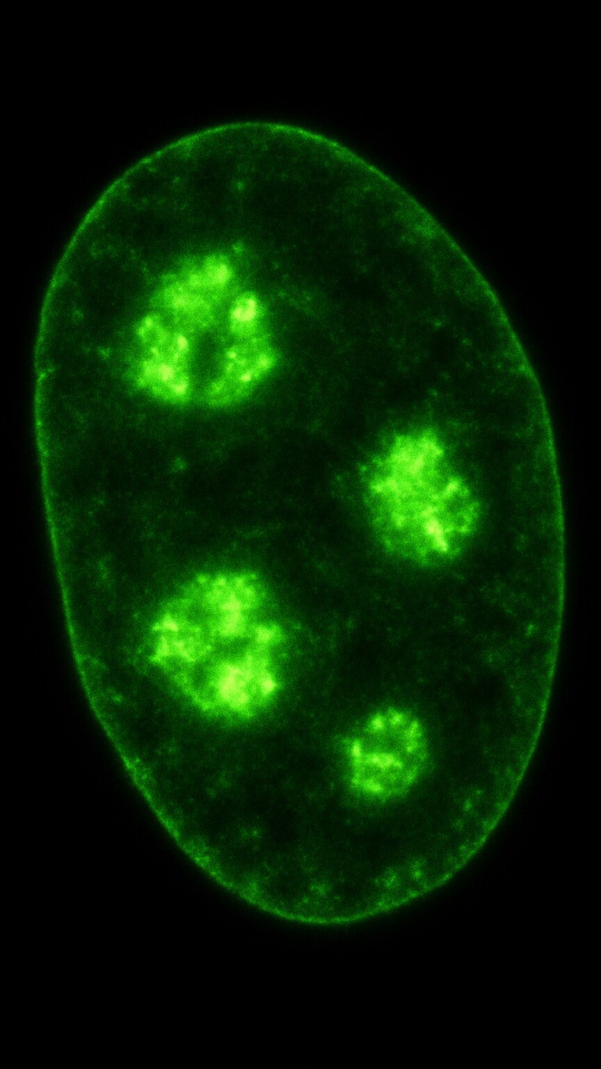
Figure 4. The nucleolar pattern shows a small number of large, clumped glowing regions corresponding to the nucleolus, the cell's dedicated structure for building ribosomes, and is strongly linked to scleroderma.
The nucleolar pattern shows only a small number of larger, distinct, well-defined glowing clumps within the nucleus, corresponding quite specifically to the nucleolus itself, a dense sub-structure inside the nucleus responsible for assembling ribosomes, the cellular machinery that builds proteins. This pattern looks visibly different from the fine, evenly distributed dots of a true speckled pattern, since the nucleolar regions are fewer in number and noticeably larger and more clumped in appearance.
Antibodies producing this pattern — including anti-RNA polymerase III, anti-fibrillarin, and anti-Th/To — are most strongly associated with systemic sclerosis, more commonly known as scleroderma, a condition involving progressive thickening and hardening of the skin and, in more severe cases, internal organs. Anti-RNA polymerase III specifically carries added clinical weight within this category, since it's associated with a higher risk of a serious scleroderma-related kidney complication, making its identification genuinely useful for guiding closer monitoring in patients who test positive for it.
Each of the three antibodies associated with this pattern actually carries its own somewhat distinct clinical profile within the broader scleroderma category, illustrating that even a single named staining pattern can conceal genuinely important differences once the specific responsible antibody is identified. Anti-fibrillarin, sometimes called anti-U3-RNP, is more frequently associated with a form of scleroderma that involves significant internal organ involvement, including the lungs and heart, and appears somewhat more often in certain patient populations than others. Anti-Th/To is generally associated with a comparatively milder disease course, more often limited primarily to skin involvement without the same degree of internal organ risk seen with the other two antibodies in this category.
This meaningful variation within a single staining pattern category is exactly why the nucleolar pattern, once identified, typically prompts a full, specific antibody workup rather than being treated as a single undifferentiated finding — the difference between anti-RNA polymerase III, anti-fibrillarin, and anti-Th/To carries real, practical weight for how closely a patient's kidneys, lungs, and heart need to be monitored going forward, even though all three antibodies would produce essentially the same visual pattern under the microscope.
Centromere Pattern: A Distinctive Marker Visible in Dividing Cells
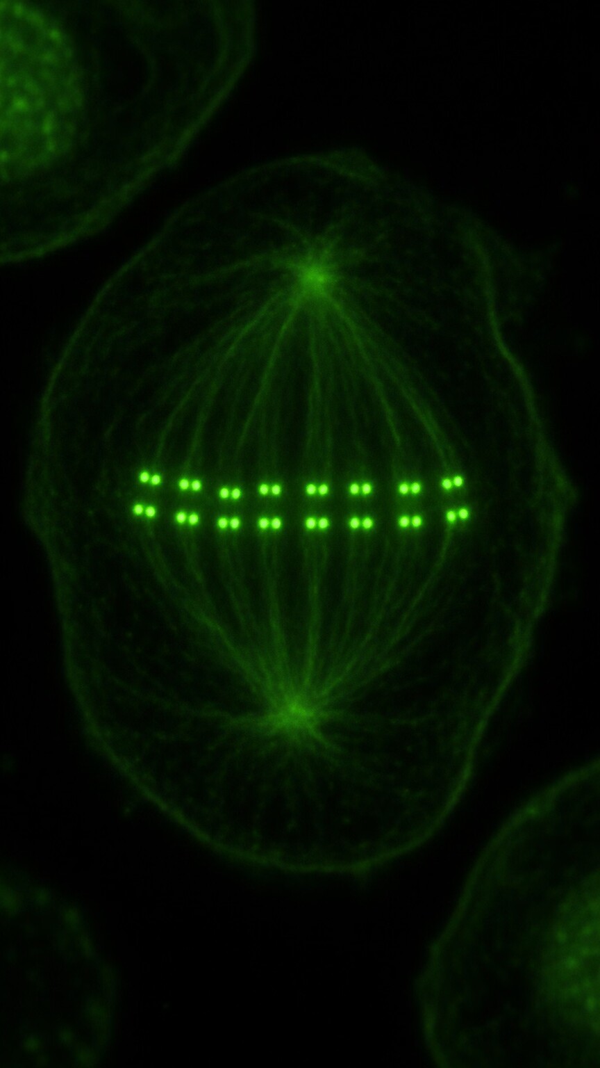
Figure 5. The centromere pattern is most clearly visible in dividing cells, appearing as a distinctive row of small paired dots aligned along the chromosomes at the exact location of each centromere.
The centromere pattern is one of the more visually distinctive and easiest patterns for a technologist to confidently identify, precisely because it's most clearly visible specifically in cells caught in the act of dividing. During cell division, chromosomes line up in the middle of the cell, and antibodies producing this pattern bind to the centromere — the pinched, central region of each chromosome where the two paired chromosome copies stay attached to each other during division — producing a highly organized row of small, discrete, evenly spaced paired dots that tracks the chromosomes' alignment.
This dependence on catching a cell mid-division is precisely why the choice of actively growing HEp-2 cells, discussed earlier in this article, matters so directly for this specific pattern. A resting, non-dividing cell simply doesn't display its chromosomes in this organized, aligned arrangement at all, meaning this pattern would be essentially invisible, or at least far less obvious, on a slide using a less actively dividing cell population. A skilled technologist specifically scans a slide for the handful of dividing cells present among the many resting ones, since these are the only cells where this particular pattern reveals itself clearly.
This pattern is strongly and specifically associated with a limited form of scleroderma historically known by the acronym CREST syndrome (calcinosis, Raynaud's phenomenon, esophageal dysfunction, sclerodactyly, and telangiectasia), now more often referred to simply as limited cutaneous systemic sclerosis. Unlike the broader, more diffuse form of scleroderma linked to the nucleolar pattern described above, this limited form tends to progress more slowly and affects the skin in a more restricted distribution, primarily the hands, face, and forearms, making the centromere pattern a genuinely useful early clue distinguishing between these two meaningfully different disease courses.
The specific antibody responsible for this pattern targets a group of proteins called CENP (centromere protein) that make up the physical structure of the centromere itself, with CENP-B being the most commonly tested and most clinically significant target within this group. Because this pattern is so visually distinctive and its underlying antibody target so well characterized, it's actually one of the more reliably identified patterns across different laboratories, with less of the inter-observer variability that can affect interpretation of some of the more subtle speckled sub-patterns discussed earlier.
Beyond its purely diagnostic value, the centromere pattern also carries genuine, real prognostic significance once an actual diagnosis of limited cutaneous systemic sclerosis has already been formally made. Patients with a confirmed centromere pattern and CENP-B antibody tend to have a somewhat more predictable long-term disease course than patients whose limited scleroderma diagnosis was reached without this specific antibody marker present, giving clinicians a useful additional data point when discussing expected disease trajectory and monitoring needs with a newly diagnosed patient. This particular pattern is also occasionally identified incidentally, well before any clinical symptoms of scleroderma become apparent, in patients being tested for an entirely unrelated reason — a finding that, when it happens, often prompts closer monitoring for the eventual emergence of Raynaud's phenomenon or other early scleroderma symptoms, even in the absence of a current diagnosis.
Nuclear Dot and Other Less Common Patterns
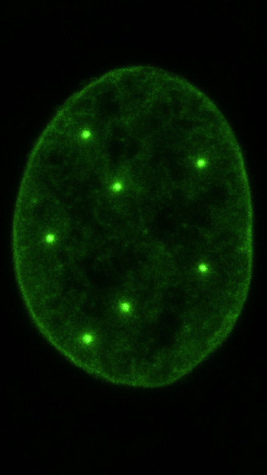
Figure 6. The nuclear dot pattern shows only a small handful of well-separated, discrete dots, distinct from the far more numerous fine dots of a true speckled pattern, and is linked to primary biliary cholangitis.
Beyond the four major patterns covered above, several rarer, more specific patterns exist, each tied to a narrower set of antibodies and conditions. The nuclear dot pattern shows a small number of discrete, well-separated dots scattered throughout the nucleus — visually distinct from the more numerous, finer dots of a true speckled pattern — produced by antibodies including anti-Sp100 and anti-gp210, both associated with primary biliary cholangitis, an autoimmune liver condition involving progressive damage to the small bile ducts within the liver. The number of dots seen in this pattern is genuinely diagnostic in itself — typically somewhere between two and twenty per nucleus, a range specific enough that a trained technologist can usually distinguish this pattern from a true speckled pattern, which typically shows dozens to hundreds of much finer dots, almost at a glance. Anti-gp210 specifically targets a protein embedded directly within the nuclear pore complex, the structure that regulates what molecules can pass into and out of the nucleus, while anti-Sp100 targets a different protein located in small structures within the nucleus called PML nuclear bodies, illustrating that even within this one narrowly defined pattern category, the underlying molecular targets involved are genuinely distinct from one another.
Cytoplasmic patterns, genuinely distinct from all of the nuclear patterns covered throughout most of this article, occur specifically when antibodies target structures located entirely outside the nucleus itself, within the surrounding cytoplasm of the cell. These patterns are associated with a range of different conditions depending on the exact structure targeted, including certain inflammatory muscle diseases and, notably, anti-mitochondrial antibodies, which produce a distinctive granular cytoplasmic pattern and are themselves one of the most specific and sensitive markers used to help diagnose primary biliary cholangitis.
The nuclear envelope pattern, another somewhat less common but still fully recognizable category, shows a genuinely distinctive glowing outline that traces closely along the outer rim of the nucleus itself, rather than filling its entire interior, produced by antibodies targeting proteins within the nuclear membrane. This particular pattern is also associated with primary biliary cholangitis, alongside the nuclear dot pattern already described, meaning a patient's overall clinical picture — including liver function tests and specific symptoms like persistent itching and fatigue — often carries as much diagnostic weight as the exact pattern reported when this particular liver condition is suspected.
A pattern called the PCNA (proliferating cell nuclear antigen) pattern deserves brief mention here specifically because it's one of the rarer patterns most classically, though not exclusively, linked to lupus, producing a distinctive appearance that varies depending on exactly which phase of the cell division cycle a given cell happens to be captured in at the moment of testing — a level of pattern complexity that makes this particular finding one of the more genuinely challenging for even experienced technologists to reliably and consistently identify.
Taken together, these rarer patterns illustrate a genuinely important broader principle running through this entire topic: the range of possible ANA staining patterns extends well beyond the four major categories most commonly discussed, and each additional, more specialized pattern represents its own specific molecular target with its own distinct clinical associations. A comprehensive, well-trained laboratory doesn't simply sort every positive result into one of a handful of broad buckets — it aims to identify the most precise pattern the sample actually displays, since that added precision genuinely does translate into more efficient, more targeted follow-up testing for the patient on the other end of that result.
Why the Pattern Alone Never Confirms a Diagnosis
A staining pattern is a genuinely useful clue, but it functions as a starting point for further, more specific testing rather than a standalone diagnosis, for a simple structural reason: each pattern reflects a broad category of possible antibodies rather than one single, uniquely identifiable antibody. This is a distinction worth sitting with for a moment, since it explains why two people can receive an identical pattern report and end up on entirely different diagnostic paths depending on which specific antibody the follow-up testing actually identifies.
It also helps to think of the pattern as analogous to a general description given at the scene of an incident, rather than a confirmed identification. A description like "medium height, dark clothing" narrows down who might be responsible far more than having no description at all, but it still requires further investigation — fingerprints, witness statements, additional evidence — before any specific individual can actually be confirmed. An ANA staining pattern functions in much the same investigative role: genuinely useful for narrowing the field, but intentionally not designed to serve as the final word on its own.
A speckled pattern, as covered earlier, could reflect anti-Sm, anti-RNP, anti-SSA, anti-SSB, or several other possibilities, each pointing toward a meaningfully different condition — confirming which one is actually present requires a separate, targeted test measuring that specific antibody by name, commonly grouped together and ordered as an extractable nuclear antigen, or ENA, panel.This is precisely, exactly why a positive ANA result reported alongside a specific pattern typically triggers what's commonly called a reflex panel — a coordinated set of follow-up tests specifically chosen based on which pattern was observed, designed to identify the exact antibody responsible rather than leaving the finding as a general, unresolved clue. The pattern's real clinical value lies in efficiently narrowing down which follow-up tests are actually worth running, rather than in providing a complete answer entirely on its own.
Many laboratories today have made this reflex-testing process largely automatic on the technical side, meaning a positive ANA screen with a specific pattern can trigger the relevant follow-up antibody panel without requiring the ordering physician to separately request each individual test by name. This kind of built-in workflow reduces the chance that a genuinely useful follow-up test gets accidentally missed simply because a busy clinician didn't think to order it manually, and it reflects a broader trend across modern laboratory medicine toward embedding this kind of clinical decision support directly into the testing process itself, rather than leaving every downstream step entirely up to individual physician memory.
It's also worth understanding that the reverse situation happens too: a meaningful number of patients with a genuinely confirmed autoimmune condition, including lupus, don't show the "classic" pattern most textbooks associate with their specific disease, and some patients with a textbook-classic pattern turn out, after full workup, not to have any autoimmune condition requiring treatment at all. This isn't a failure of the pattern system — it's simply a reflection of how biological reality rarely sorts itself as neatly into categories as a simplified reference chart might suggest, and it's precisely why an experienced clinician always weighs the pattern alongside the patient's actual symptoms, physical exam findings, and other laboratory results, rather than treating any single piece of the puzzle as decisive entirely on its own.
The overall titer, or concentration, at which a given pattern appears also meaningfully affects how much weight that pattern actually carries. The same homogeneous or speckled pattern reported at a very low titer, as covered in more depth in a related article on this site, carries considerably less diagnostic significance than that identical pattern reported at a high titer, since a low concentration of antibody can sometimes reflect a milder, non-specific immune response rather than a genuinely active autoimmune process. This is one of several reasons the full laboratory report — pattern and titer together, not either one in isolation — is what actually gets interpreted by a treating clinician.
A Worked Example: Following One Pattern to a Specific Diagnosis
Consider a fairly typical, realistic case: a 34-year-old with several months of joint pain, unexplained fatigue, and a distinctive facial rash, whose ANA test comes back genuinely positive at a meaningful titer with a homogeneous pattern specifically reported on the lab result. Given this particular pattern's genuinely strong, classic association with lupus specifically, her physician appropriately orders a targeted anti-dsDNA test as a direct, logical follow-up step, which comes back positive at a clinically significant level, along with low complement levels on a separate blood test — a combination that, together with her symptoms, supports a confident lupus diagnosis and specifically helps guide the choice of treatment, since anti-dsDNA levels are also useful for tracking disease activity over time in confirmed lupus.
These two cases together illustrate an important broader point worth carrying forward: the same overall diagnostic logic — pattern first, then targeted confirmatory testing, then clinical correlation with symptoms — applies consistently regardless of which specific pattern happens to be reported, even though the exact antibodies and conditions involved differ completely from one case to the next.
Now consider a different patient: a 52-year-old with progressively tightening, thickening skin on the hands and forearms, along with color changes in the fingers triggered by cold exposure. Her ANA test comes back positive with a centromere pattern specifically. Given this pattern's strong, specific association with limited cutaneous systemic sclerosis, her rheumatologist proceeds directly toward evaluating for that specific condition, including assessing for the other classic features of CREST syndrome, rather than pursuing a broader, less targeted workup — illustrating exactly how a specific pattern can meaningfully shorten and sharpen the diagnostic path when it points clearly toward one well-recognized condition.
A third, genuinely instructive scenario rounds out this comparison: a 45-year-old with mild, nonspecific fatigue undergoes an ANA test purely as part of a routine wellness panel, unrelated to any specific autoimmune concern. The result comes back positive with a fine speckled pattern at a modest titer. Because this particular pattern is so broadly associated with several different possible antibodies, and because her symptoms are vague and nonspecific rather than pointing toward any one particular condition, her physician reasonably orders a full ENA panel to check for the range of antibodies this pattern could represent, while also considering the possibility that this finding reflects nothing more than a mild, clinically insignificant immune quirk rather than any developing disease. Her ENA panel ultimately comes back entirely negative, and given her lack of any specific supporting symptoms, the finding is reasonably attributed to the kind of incidental, non-specific positivity that a meaningful percentage of otherwise healthy people show on this particular test — illustrating that not every positive pattern, even a genuinely correct laboratory finding, ultimately points toward an active disease process requiring treatment.
Frequently Asked Questions
Can I have more than one ANA staining pattern at once?
Yes. It's genuinely common for a sample to show a mixed pattern, reflecting the presence of more than one distinct antibody at the same time, which is one of several reasons follow-up testing is used to clarify exactly which antibodies are actually present.
Does a specific staining pattern confirm which autoimmune disease I have?
No. A pattern narrows down which specific antibody tests are worth ordering next, but confirming an actual diagnosis requires those targeted follow-up tests along with your symptoms and overall clinical picture.
Why is the speckled pattern considered less specific than the others?
Because several genuinely different antibodies, each associated with a different condition, can all produce a visually similar speckled appearance, meaning this particular pattern alone can't distinguish between them without additional testing.
Is the centromere pattern always a sign of scleroderma?
It's strongly and specifically associated with limited cutaneous systemic sclerosis, but like any single ANA finding, it's interpreted alongside symptoms and confirmatory testing rather than treated as a diagnosis on its own.
Why does the test use cells that are actively dividing?
Certain structures, including the centromere, are only clearly visible during cell division, which is why HEp-2 cells, a rapidly dividing cell line, are specifically used to make patterns like the centromere pattern identifiable at all.
Conclusion
An ANA staining pattern isn't decorative detail on a lab report — it's a direct, physical reflection of exactly where your antibodies are attaching inside the cell, and that specific location genuinely narrows down which underlying antibodies, and which autoimmune conditions, are actually worth investigating further. Homogeneous points toward lupus, speckled covers a broad and diverse group of conditions including lupus, mixed connective tissue disease, and Sjögren's, nucleolar and centromere both point toward different forms of scleroderma, and nuclear dot patterns point toward primary biliary cholangitis. Understanding what each pattern actually represents turns an unfamiliar laboratory term into a specific, meaningful clue guiding exactly what happens next.
The next time a lab report lists a specific pattern name alongside a titer, it's worth remembering that this single word represents an entire physical story playing out under a microscope — a specific antibody, produced somewhere in your immune system, finding and binding to a specific structure inside a cell, made visible through decades of refined laboratory technique. That story is genuinely worth understanding in its own right, even before the follow-up testing that eventually determines exactly what it means for your own health going forward.
Still Not Sure What Your Results Mean?
Upload your labs and get a complete, visual, plain-language interpretation of every biomarker — delivered to your inbox in under 15 minutes.
Get My ReportThis article is for educational purposes only and does not constitute medical advice. Always consult your healthcare provider regarding your specific lab results.