
What Does a High ESR Level Mean?
A high erythrocyte sedimentation rate, or ESR, means that inflammation is present somewhere in your body right now — but on its own, the test can't tell you where, how serious it is, or what's causing it. ESR is one of the oldest lab tests still in routine use, and it works on a surprisingly simple physical principle: it measures how quickly red blood cells settle to the bottom of a vertical tube over the course of one hour. When inflammation is present, certain proteins in the blood change how red blood cells interact with each other, causing them to clump together and fall faster than they normally would. A higher number simply means faster-falling cells, and faster-falling cells simply mean more of those inflammation-related proteins circulating in your blood. This article walks through exactly what's happening inside that tube, what different ranges of ESR results typically mean, the wide range of conditions — from the mundane to the genuinely serious — that can drive it up, and why doctors almost always interpret it alongside other information rather than treating it as a standalone answer on its own.
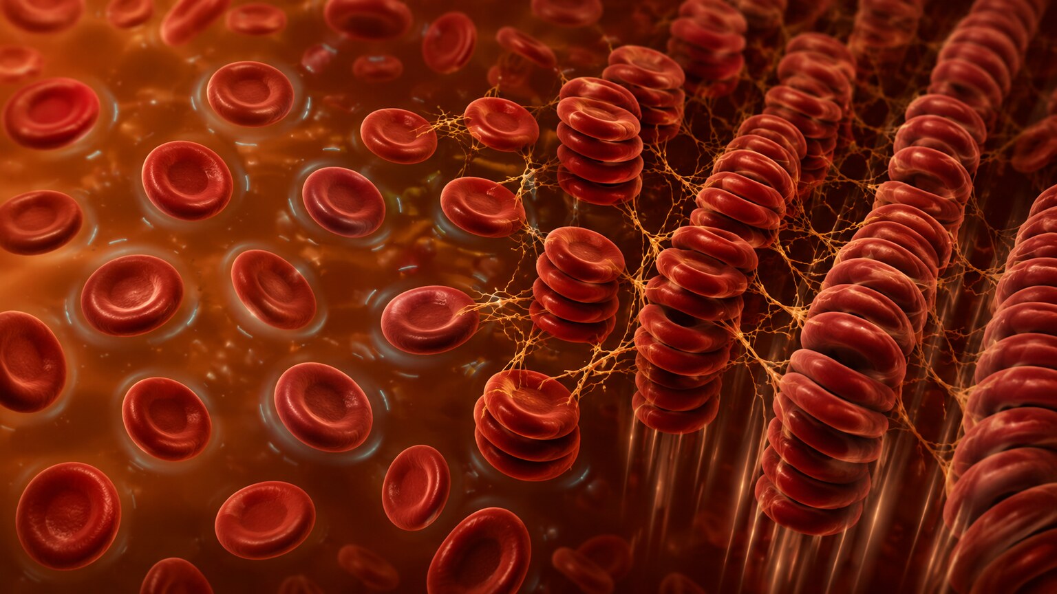
Figure 1. During inflammation, fibrinogen and other proteins coat red blood cells and cause them to stack together like coins, forming clumps called rouleaux that fall through plasma faster than individual cells.
What ESR Actually Measures — The Sedimentation Race Explained
The test itself, still commonly performed using a method called the Westergren technique first developed in the 1920s, is almost deceptively simple. A blood sample is drawn into a thin, vertical glass or plastic tube marked with a measurement scale and left standing perfectly upright, undisturbed, for exactly one hour. Left alone, red blood cells are naturally denser than the surrounding plasma and slowly sink under gravity, leaving a column of clear plasma above them. The result is reported as the number of millimeters that column of clear plasma measures after that hour — a higher number means the red blood cells sank faster and farther, leaving more clear space above them.
What makes this such a durable, still-relevant test almost a hundred years after its invention is precisely how little sophisticated technology it actually requires — a calibrated tube, an anticoagulant to keep the blood from clotting during the hour it sits, a level surface free of vibration, and a clock. That simplicity was a genuine practical advantage in the era before automated lab analyzers existed, and even today it keeps ESR testing inexpensive and widely accessible in settings that might not have access to more advanced inflammatory marker assays. The tradeoff, as later sections of this article get into, is that this same simplicity is also exactly why the test is so nonspecific: it's measuring an indirect physical consequence of inflammation rather than any single inflammatory molecule directly, which is part of the price paid for a test this straightforward and this cheap to run.
Under normal conditions, red blood cells carry a slightly negative surface charge that causes them to repel each other slightly, similar to how magnets with matching poles push apart, which keeps them relatively spread out and slows their descent through the plasma. Inflammation changes this picture by increasing the blood concentration of certain proteins — most importantly fibrinogen, along with immunoglobulins and other acute-phase proteins the liver ramps up production of during an inflammatory response. These proteins coat the surface of red blood cells and effectively neutralize some of that natural repulsion, allowing cells to stick together in stacked clusters called rouleaux, named for their resemblance to a stack of coins. Because rouleaux formations are larger and denser than individual cells, they sink through plasma considerably faster, which is the entire physical basis for why inflammation produces a higher, faster ESR result.
This mechanism also explains why ESR responds relatively slowly compared to some other inflammatory markers. Fibrinogen and the other proteins responsible for promoting rouleaux formation aren't released instantly the moment inflammation begins — they're manufactured by the liver in response to signaling molecules called cytokines that are dispatched once an inflammatory process is underway, a production process that takes time to ramp up and, just as importantly, time to wind back down once the trigger has resolved. This lag is why ESR is sometimes described as reflecting the cumulative, sustained state of inflammation over the preceding days to weeks rather than capturing a single, immediate snapshot — a distinction that becomes clinically relevant later in this article when comparing ESR to faster-moving markers like CRP.
It's also worth understanding what the test explicitly does not measure, since the name itself can be slightly misleading. ESR doesn't directly measure fibrinogen, immune cells, or any inflammatory molecule at all — it measures a purely physical, mechanical outcome (how far cells fall in an hour) that happens to be strongly influenced by those molecules. This indirect relationship is part of why a handful of unrelated factors, covered later in this article, can shift ESR up or down without any real change in inflammation, and it's also why some labs and specialists increasingly favor more modern, automated sedimentation-rate analyzers that mechanically standardize the test conditions to reduce the kind of small technical variability the century-old manual Westergren method can introduce between different labs and technicians.
Understanding the Numbers — Mild, Moderate, and Extremely High ESR
Normal ESR ranges vary somewhat by lab, sex, and age, but as a general guide, adult men typically fall below roughly 15 mm/hr and adult women below roughly 20 mm/hr, with normal ranges creeping modestly higher in older adults simply as a function of aging. Mildly elevated results, generally in the 20 to 40 mm/hr range, are common and often reflect low-grade, nonspecific inflammation — sometimes from something as ordinary as a recent minor infection, and sometimes from factors that aren't inflammatory at all, like pregnancy or anemia, both of which independently push ESR upward through mechanisms unrelated to active disease.
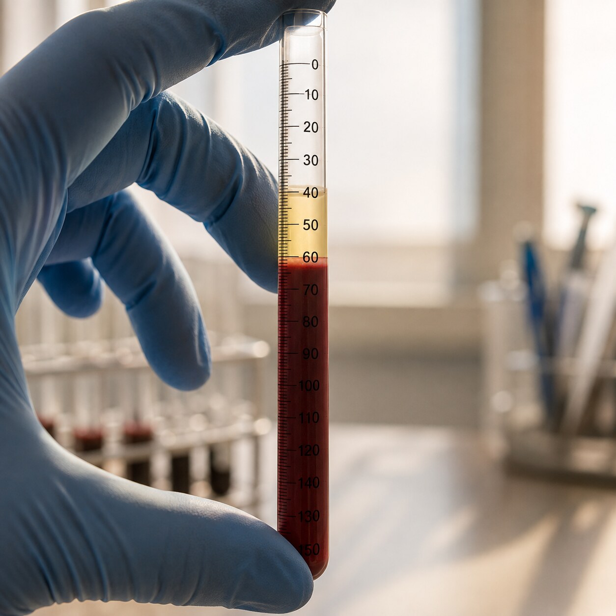
Figure 2. The ESR result is read directly as the height, in millimeters, of clear plasma that has separated above the settled red blood cells after exactly one hour.
Moderate elevations, roughly in the 40 to 100 mm/hr range, more often point toward an actual inflammatory process needing attention — active infections, flares of autoimmune conditions like rheumatoid arthritis or lupus, or inflammatory bowel disease, among others. Results climbing above 100 mm/hr are considered extremely high and, while they can occasionally still come from a straightforward infection, this range is disproportionately associated with a shorter, more specific list of serious conditions: giant cell arteritis, certain cancers (particularly multiple myeloma, which can push ESR to some of the highest levels ever recorded on this test), and severe infections like endocarditis or deep abscesses. This general pattern — the higher the number, the shorter and more serious the realistic list of causes becomes — is one of the more useful ways clinicians mentally triage an ESR result while waiting for further workup to clarify the picture.
Sex and age differences in normal ranges deserve a bit more explanation, since they're a frequent source of confusion when comparing results between two people. Women tend to run slightly higher baseline ESR values than men at every age, a pattern generally attributed to differences in average red blood cell count and certain hormonal effects on plasma proteins. Age has an even larger effect: a commonly used rough estimate for the upper limit of normal is a person's age divided by two for men, and age plus ten divided by two for women, which means a healthy 70-year-old man might have a normal ESR sitting close to 35 mm/hr — a number that would be flagged as clearly abnormal in a 25-year-old. Because of this drift, clinicians evaluating an older adult's ESR result routinely weigh it against these age-adjusted expectations rather than a single fixed cutoff, to avoid over-interpreting a mild elevation that may simply reflect the ordinary effects of aging on the vascular and immune system.
The Most Common Causes of a Mildly to Moderately High ESR
In everyday clinical practice, most elevated ESR results fall into the mild-to-moderate range and trace back to a fairly predictable set of causes. Infections of almost any kind — respiratory, urinary, skin, dental — can transiently raise ESR while the body's inflammatory response is active, typically returning to normal within weeks of the infection resolving. Autoimmune and inflammatory conditions are another major category: rheumatoid arthritis, lupus, inflammatory bowel disease, and various forms of vasculitis all commonly produce a persistently elevated ESR that tends to track, at least loosely, with how active the underlying disease is at any given time.
Rheumatoid arthritis deserves particular mention given how often ESR testing enters the picture specifically because of joint symptoms. In active rheumatoid arthritis, ongoing inflammation in the joint lining continuously releases the same signaling molecules responsible for driving up fibrinogen production elsewhere in the body, meaning ESR often rises and falls in step with how many joints are actively inflamed and how severe that inflammation is at any given time. This is part of why ESR, alongside CRP and a clinical joint count, is frequently incorporated into formal composite disease-activity scores used by rheumatologists to quantify, in a semi-standardized way, exactly how active a person's rheumatoid arthritis is at a given visit — turning a somewhat abstract, nonspecific lab value into one concrete input within a more structured, reproducible clinical assessment tool.

Figure 3. Morning joint stiffness and swelling lasting more than thirty minutes is a common early symptom prompting ESR testing when an inflammatory joint condition is suspected.
Beyond active disease, several ordinary physiological states independently raise ESR without any disease process at all. Pregnancy is one of the most notable, since normal pregnancy-related changes in plasma protein levels can push ESR meaningfully higher even in a completely healthy pregnancy, which is part of why ESR is rarely used as a standalone screening test during pregnancy. Anemia has a similar independent effect: because ESR partly depends on the ratio of red blood cells to plasma, having fewer red blood cells changes how they settle regardless of any inflammation, meaning a low red blood cell count can inflate an ESR result on its own. Obesity has also been associated with modestly elevated ESR, likely related to low-grade inflammation generated by fat tissue itself, and age is a factor too — ESR tends to drift upward gradually over the decades even in people without any specific illness, which is why age-adjusted reference ranges are sometimes used, particularly in older adults, to avoid over-interpreting a mild elevation that may simply reflect normal aging.
A handful of technical and situational factors can also nudge an ESR result without reflecting the patient's true underlying state at all. Blood samples that sit too long before being processed, are exposed to significant temperature swings, or aren't kept perfectly vertical and undisturbed during the one-hour settling period can all introduce measurement artifacts that skew the result in either direction. Certain medications, including oral contraceptives and some other hormone-containing drugs, have been associated with modest ESR changes independent of any inflammatory effect. Even something as simple as a tilted tube rack, tilting the tube by even a few degrees off vertical, can measurably speed up settling and produce a falsely elevated reading — which is part of why an unexpected result, especially one that doesn't fit the rest of the clinical picture, is often simply repeated before being acted upon, rather than immediately triggering an extensive workup.
Already have an ESR number from a recent test, but not sure if it's actually good or bad for you? Just plug it in and see instantly.
🧮 Try the Free CalculatorWhen ESR Is Extremely High — Giant Cell Arteritis and Other Serious Causes
An ESR result climbing above 100 mm/hr deserves particular attention, since this range is strongly associated with a shorter list of specific, often serious conditions. Giant cell arteritis, an inflammatory condition affecting medium and large blood vessels, most classically the arteries supplying the scalp and temples, is one of the most well-known causes of an extremely high ESR, frequently producing results well above 100 in affected individuals, often alongside a closely related condition called polymyalgia rheumatica, which causes significant pain and stiffness in the shoulders and hips.
The multiple myeloma connection is worth understanding mechanistically, since it illustrates just how directly ESR can respond to certain conditions even though the test itself is so indirect. In multiple myeloma, abnormal plasma cells in the bone marrow produce enormous quantities of a single, identical antibody protein, called a monoclonal protein, far in excess of what a normal, healthy immune response would generate. Because these antibody molecules are exceptionally effective at neutralizing red blood cells' natural surface charge and promoting rouleaux formation — often more effective, molecule for molecule, than the fibrinogen responsible for most ordinary inflammatory ESR elevations — even a moderate excess of this monoclonal protein can drive ESR to extraordinarily high levels, sometimes exceeding 130 or even 150 mm/hr. This is part of why an unexplained, extremely high ESR in an older adult, particularly one accompanied by unexplained bone pain, anemia, or kidney function changes, prompts specific testing for this kind of abnormal protein rather than being assumed to reflect ordinary inflammation.

Figure 4. A new, severe headache localized to the temple, especially in someone over 50, is a classic warning sign of giant cell arteritis — a condition strongly associated with an extremely high ESR.
Recognizing giant cell arteritis promptly matters enormously, since untreated inflammation in these vessels can, in a subset of cases, lead to sudden and permanent vision loss if blood flow to the eye is affected — which is exactly why a new headache localized to the temple in someone over 50, especially alongside scalp tenderness, jaw pain while chewing, or visual disturbances, combined with a markedly elevated ESR, is treated as a genuine medical urgency rather than something to schedule around. Beyond vasculitis, extremely high ESR values are also disproportionately associated with certain cancers, most notably multiple myeloma, a blood cancer in which abnormal antibody proteins produced in large quantities dramatically promote rouleaux formation, sometimes producing some of the highest ESR values seen in clinical medicine. Severe or deep-seated infections — bacterial endocarditis affecting the heart valves, osteomyelitis (bone infection), or deep abscesses that haven't yet produced obvious localized symptoms — round out the list of conditions clinicians specifically consider when an ESR comes back this high.
This clustering of serious causes at the extreme end of the ESR scale is sometimes referred to informally among clinicians as the "rule of 100," a loose but genuinely useful heuristic suggesting that an ESR above 100 mm/hr is significantly more likely to reflect one of this shorter list of serious conditions than the broader, more benign grab-bag of causes responsible for milder elevations. It isn't an infallible rule — a small percentage of people with results this high turn out to have a relatively benign or self-limited cause, and conversely, some serious conditions on this list can present with a lower ESR in certain individuals — but it captures a real statistical pattern well enough that an ESR in the triple digits reliably prompts a more urgent, more thorough workup than a moderate elevation would, including a careful search for symptoms of vasculitis, a broader infection workup, and consideration of blood or imaging studies aimed at ruling out an underlying malignancy.
Diagnosing giant cell arteritis specifically, once it's suspected based on symptoms and a strikingly high ESR, generally involves a temporal artery biopsy — a small tissue sample taken from the artery near the temple, examined under a microscope for the characteristic inflammatory changes that give the condition its name. Because starting corticosteroid treatment early is what actually protects against vision loss, treatment is typically begun as soon as the diagnosis is strongly suspected clinically, without waiting for biopsy confirmation first, since the biopsy can still reliably detect the diagnostic changes for a meaningful window of time even after treatment has already started. This willingness to treat first and confirm second is a fairly unusual approach in medicine generally, and it reflects just how seriously the potential consequence — permanent vision loss — is weighed against the comparatively low risk of a short course of corticosteroids in someone where the clinical suspicion, symptoms, and an extremely high ESR all point convincingly in the same direction.
Why ESR and CRP Are Often Ordered Together
ESR is frequently ordered alongside another inflammatory marker, C-reactive protein (CRP), and the two are genuinely complementary rather than redundant, because they behave differently over time. CRP is produced directly by the liver in response to inflammatory signals and can rise within just 6 to 8 hours of an inflammatory trigger, then fall relatively quickly, within days, once the trigger resolves. ESR, by contrast, depends on the buildup of fibrinogen and other proteins that accumulate more gradually and also clear more slowly, meaning it typically takes longer to rise after inflammation begins and, correspondingly, longer to fall back to normal once inflammation has actually resolved.
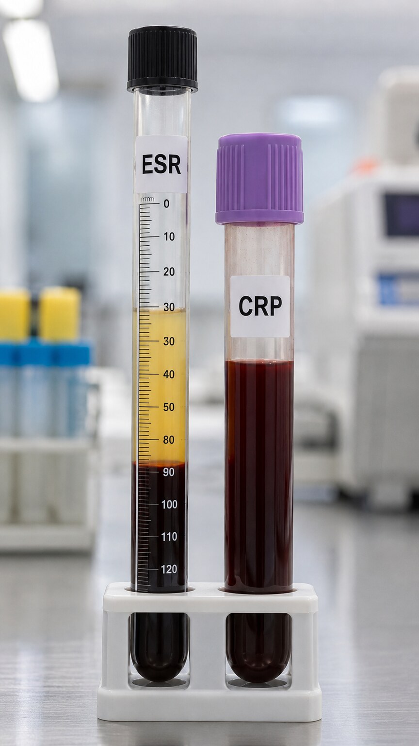
Figure 5. ESR and CRP are drawn together because they rise and fall on different timelines, giving clinicians a fuller picture of both how recent and how sustained an inflammatory process is.
This difference in timing is clinically useful: a high CRP with a still-normal ESR can suggest inflammation that's very recent or just beginning, while a high ESR with a normalizing CRP might suggest inflammation that peaked days ago and is now on its way down, even though the slower-moving ESR hasn't caught up yet. Some conditions also tend to produce a more pronounced rise in one marker than the other for reasons that aren't always fully understood — lupus, for example, sometimes produces a notably high ESR with a CRP that stays closer to normal, a pattern experienced clinicians sometimes use as one small additional clue when the two results don't move in lockstep. Ordering both tests together, rather than relying on just one, gives a more complete picture of an inflammatory process's timeline than either test could provide alone.
The two tests also differ in how directly they reflect the biology of inflammation, which affects how each is best used in practice. CRP is manufactured directly in response to a specific signaling molecule (interleukin-6) and its blood level tends to track fairly tightly and predictably with how much of that inflammatory signal is present at any given moment, making it a comparatively precise, fast-responding gauge. ESR, being an indirect physical measurement shaped by several different proteins along with non-inflammatory factors like anemia and plasma composition, is a noisier, slower signal, but one that captures a broader, more sustained sense of a person's overall inflammatory state. Neither characteristic makes one test simply "better" than the other; it's closer to using both a car's speedometer and its odometer together — one tells you what's happening right now, the other tells you about the sustained journey — which is exactly why clinicians managing chronic inflammatory conditions often want both numbers rather than picking a single favorite.
Cost and availability sometimes factor into which test gets used in practice as well, particularly outside major hospital systems. ESR requires only basic, inexpensive equipment and can be performed in almost any lab setting with minimal specialized machinery, while CRP testing, especially high-sensitivity CRP assays used for certain cardiovascular risk applications, generally requires more specialized automated analyzers. In resource-limited settings, or in situations where cost is a genuine consideration, ESR sometimes remains the more practical first-line inflammatory marker for exactly this reason, even in an era where CRP is often considered the more precise and faster-responding test on purely biological grounds.
What a High ESR Doesn't Tell You — Why It's Never Diagnostic Alone
Perhaps the single most important thing to understand about ESR is what it can't do: it cannot identify a specific disease, locate where in the body inflammation is happening, or distinguish between a minor, self-resolving issue and a serious underlying condition. This is precisely why ESR is described as a nonspecific marker — the test detects that something is triggering an inflammatory response somewhere, but the list of things capable of doing that is genuinely enormous, ranging from a minor cold to cancer to a rheumatologic condition to nothing more concerning than normal pregnancy. Two people with an identical ESR of 55 mm/hr could have completely different, unrelated conditions, or one could have no significant disease at all.
Because of this fundamental limitation, ESR is almost never ordered or interpreted in isolation. It's typically used alongside a detailed history and physical exam, other more specific blood tests targeted at particular suspected conditions, and often imaging, with the actual diagnosis emerging from that combined picture rather than from the ESR number by itself. A single elevated ESR in someone with no symptoms and an otherwise normal exam is frequently a false alarm in the sense that it doesn't reflect any condition requiring treatment, while the same elevated number in someone with a specific, matching symptom pattern can be an important supporting piece of evidence for a particular diagnosis a clinician is already suspecting based on the clinical picture.
This is also why an isolated, incidentally discovered high ESR — found, say, on bloodwork ordered for an entirely unrelated reason, in someone who feels completely well — is generally handled with a measured, stepwise approach rather than an immediate cascade of testing. A common first step is simply repeating the test after a few weeks; a result that has fallen back toward normal on its own strongly suggests the original elevation reflected something transient and self-resolving, like a minor viral illness the person may not have even noticed. A result that remains persistently elevated on repeat testing carries more weight and typically prompts a more structured evaluation, guided heavily by whatever symptoms, however subtle, might be present, rather than by the ESR number driving the workup on its own.
A basic, structured initial workup for a persistently elevated ESR of unclear cause typically includes a complete blood count (to check for anemia or other blood abnormalities), kidney and liver function tests, urinalysis, and a chest X-ray, alongside a thorough review of systems asking about fevers, weight loss, night sweats, joint pain, and any other subtle symptoms the person might not have thought to mention unprompted. This kind of broad but standardized initial screen is designed to efficiently catch the more common explanations — an unrecognized infection, early inflammatory arthritis, kidney disease — before moving on to more specialized, more invasive, or more expensive testing aimed at rarer causes, reflecting a general medical principle of working from common and easily detected conditions outward toward rarer ones, rather than jumping immediately to the most serious possibility on the differential.
How ESR Is Used to Track Disease Activity Over Time
Beyond its role in initial diagnosis, ESR has a genuinely valuable ongoing use once a specific inflammatory condition has already been identified: tracking how active that condition is over time and how well it's responding to treatment. In conditions like rheumatoid arthritis, lupus, and especially giant cell arteritis, clinicians often use a falling ESR, measured through repeat blood draws over weeks to months, as one piece of evidence that treatment is working and inflammation is genuinely coming under control, while a rising ESR during treatment can prompt a closer look at whether the current approach needs adjustment.

Figure 6. For conditions like rheumatoid arthritis and giant cell arteritis, tracking ESR across repeat visits helps clinicians judge whether treatment is genuinely controlling inflammation over time.
This monitoring role matters especially for giant cell arteritis, where treatment with corticosteroids is typically tapered gradually over many months, and ESR trends, alongside symptoms, help guide how quickly that taper can safely proceed. It's worth noting, though, that ESR's slower response time — the same feature that makes it useful for tracking sustained trends — also means it isn't ideal for catching very rapid, short-term changes, which is one more reason it's typically tracked alongside CRP and clinical symptoms rather than used as the sole measure of how a condition is progressing at any given moment.
An important practical nuance in this monitoring role is that ESR trends are almost always interpreted relative to a person's own individual baseline and disease pattern rather than compared against the general population reference range. Someone with well-controlled rheumatoid arthritis might have a personal baseline ESR of 30 mm/hr even when feeling entirely well, meaning a rise to 45 might represent a meaningful flare worth investigating, even though 45 would sit comfortably within a broader population's "moderately elevated" category rather than looking dramatically abnormal on its own. This is part of why clinicians managing chronic inflammatory conditions build a longitudinal record of a patient's ESR over repeated visits, since that personal trend line, tracked over months and years, often carries more diagnostic and monitoring value than any single reading compared against a generic population range, turning what looks like an unremarkable number in isolation into a genuinely meaningful early warning sign when read in the context of that individual's own established pattern.
Frequently Asked Questions
Can a high ESR happen without any underlying disease?
Yes. Pregnancy, anemia, obesity, and normal aging can all independently raise ESR without reflecting active disease. This is part of why a mildly elevated ESR in someone with no symptoms and a normal exam often turns out to be a false alarm rather than a sign of a specific illness.
How high does ESR need to be before it's considered concerning?
There's no single fixed cutoff that applies equally to everyone, since normal ranges naturally vary by age and by sex, but results climbing above 100 mm/hr are generally considered extremely high and are disproportionately associated with a shorter list of more specific conditions, including giant cell arteritis, certain cancers, and serious infections, all of which warrant prompt further evaluation.
Why would a doctor order both ESR and CRP instead of just one?
Because the two tests genuinely rise and fall on different timelines. CRP responds within just a few hours and clears back down quickly, while ESR rises and falls far more gradually over a span of days to weeks. Ordering both together gives a noticeably more complete picture of whether inflammation is very recent, still ongoing, or already resolving, than either single test could reliably provide entirely on its own.
If my ESR is high, does that mean I have an autoimmune disease?
Not necessarily, no. A high ESR reflects inflammation somewhere in the body but genuinely can't identify its underlying cause on its own. Autoimmune disease is only one possible explanation among many others, including ordinary infection, certain cancers, pregnancy, and anemia. A high ESR is typically interpreted carefully alongside symptoms and other, more specific tests rather than used to diagnose autoimmune disease all by itself.
How quickly does ESR return to normal once the underlying cause is treated?
It varies quite a bit from person to person, but generally more slowly than CRP does. Because ESR depends on proteins that both accumulate and clear gradually over time, it can take several full weeks for ESR to fully normalize even after successful treatment of the underlying condition, which is why clinicians don't expect an immediate drop and instead track the overall trend across multiple follow-up visits.
Should I be worried about a mildly elevated ESR found on routine bloodwork with no symptoms?
Usually not urgently. A mild, isolated elevation with no accompanying symptoms and a normal physical exam is often repeated after a few weeks rather than immediately investigated further, since it frequently reflects a transient, self-resolving cause that clears up entirely on its own. Persistent elevation on repeat testing, or the appearance of new symptoms in the meantime, is generally what prompts a more thorough evaluation from that point forward.
Conclusion
A high ESR is best understood as a genuine but nonspecific signal: it reliably tells you that something is prompting an inflammatory response somewhere in your body, but it can't say what that something is, where it's happening, or how serious it might be on its own. The size of the number offers a useful rough guide — mild elevations are common and often benign or self-limited, while results climbing above 100 mm/hr narrow the realistic list of causes toward a shorter set of conditions, including giant cell arteritis, certain cancers, and serious infections, that generally warrant prompt further evaluation. Whether it's being used for initial diagnosis or to track how well an already-identified condition is responding to treatment, ESR does its most useful work not in isolation, but as one piece of a larger clinical picture built from symptoms, exam findings, and other more specific tests working together.
What makes ESR worth understanding, despite its imprecision, is exactly that combination of simplicity and longevity — a test invented nearly a century ago, based on nothing more sophisticated than gravity and a graduated tube, still earning its place on modern lab requisition forms alongside far more technologically advanced assays. That staying power exists precisely because the physical principle it relies on, inflammation altering how red blood cells interact and settle, remains genuinely informative even now, as long as the number is read for what it actually is: a starting point for investigation, not a finish line. Paired with the right symptoms, the right exam findings, and the right follow-up tests, a high ESR often turns out to be one of the more efficient, low-cost ways a clinician can confirm that a hunch about inflammation is genuinely worth pursuing further, rather than simply a stray, meaningless number sitting on an otherwise ordinary lab report.
Still Not Sure What Your Results Mean?
Upload your labs and get a complete, visual, plain-language interpretation of every biomarker — delivered to your inbox in under 15 minutes.
Get My ReportThis article is for educational purposes only and does not constitute medical advice. Always consult your healthcare provider regarding your specific lab results.