
What Does a High Alkaline Phosphatase Level Indicate?
Alkaline phosphatase, usually shortened to ALP on a lab report, is a genuinely unusual test result in one specific way: unlike most of the enzymes on a standard liver panel, a high ALP doesn't automatically point to the liver at all. This one enzyme is manufactured in meaningful quantities by two entirely different organ systems — the liver and bile ducts on one side, bone tissue on the other — plus smaller amounts from the placenta during pregnancy and the intestines. That means an elevated ALP is really only half an answer on its own; the real diagnostic work is figuring out which of those sources is actually responsible, since a liver problem and a bone problem call for completely different next steps, completely different specialists, and completely different levels of urgency. This article walks through exactly how ALP works in the body, the specific detective work doctors use to trace an elevated result back to its actual source, and the full range of conditions — from a routine pregnancy to a rare bone disease — that can be behind the number.
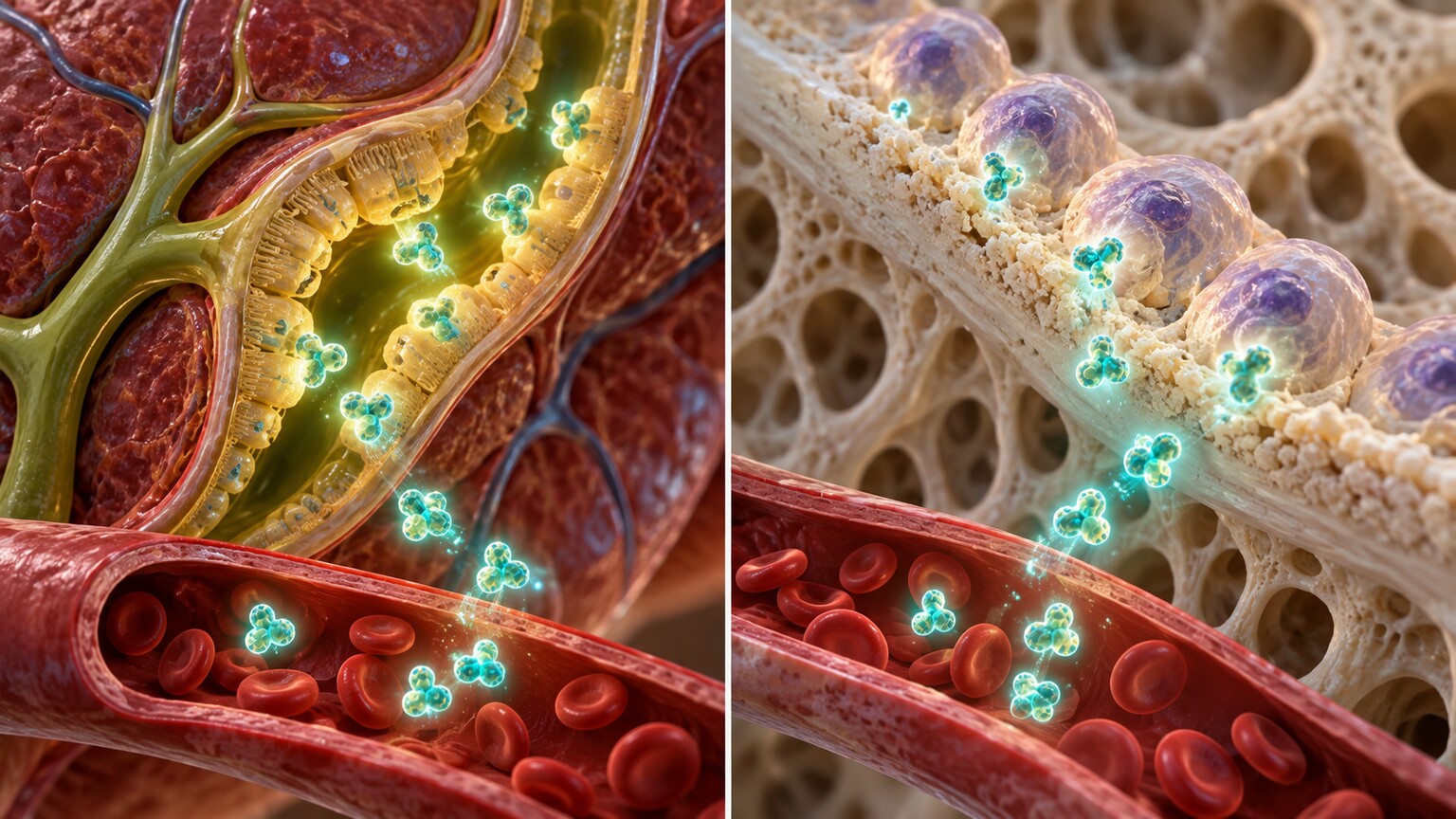
Figure 1. Alkaline phosphatase is produced by two structurally unrelated tissues, the liver's bile duct system and bone, meaning the same elevated lab value can point to either one.
What Alkaline Phosphatase Actually Does in the Body
Alkaline phosphatase belongs to a family of enzymes whose job is to remove phosphate groups from other molecules, a chemical reaction that turns out to be essential in two seemingly unrelated biological processes. In the liver, ALP sits along the membranes lining the small bile ducts — the tiny tubes that carry bile, a digestive fluid, out of the liver and into the intestine — and it plays a role in transporting substances across those membranes. In bone, ALP is produced by osteoblasts, the specialized cells responsible for building new bone tissue, where it helps create the right chemical environment for calcium and phosphate to be deposited into the bone matrix, a process called mineralization.
These two roles have nothing to do with each other functionally, but they happen to be carried out by the same enzyme family, which is precisely the biological quirk that makes interpreting a single ALP number so much trickier than interpreting most other lab values. A test result showing "high alkaline phosphatase" is, in effect, two different questions compressed into one number: is the liver's bile drainage system under stress, or is bone tissue being actively built or broken down at an unusual rate? Sorting out which question is actually being answered is the entire point of the diagnostic process this article walks through.
It genuinely helps to understand that "alkaline phosphatase" isn't technically one single molecule at all, but a family name covering several closely related enzyme variants, called isoenzymes, that share the same basic chemical function but are each produced by a different tissue and encoded by a subtly different gene. The liver/bone/kidney isoenzyme, despite its name, accounts for the overwhelming majority of both the liver-associated and bone-associated ALP measured on a standard blood test, while separate genes produce the distinct placental and intestinal isoenzymes. A standard ALP blood test reports the combined total of all these isoenzymes together as one number, which is exactly why a single elevated result can't, by itself, say which specific tissue is responsible — that's a question the rest of this article's testing strategies exist specifically to answer.
The Two Dominant Sources, and Why the Distinction Matters So Much

Figure 2. Because GGT is produced almost exclusively by the liver and bile ducts, not bone, checking it alongside an elevated ALP is the fastest, most common way to determine which organ system is actually responsible.
The single fastest, most widely used way to sort a high ALP into "liver" or "bone" doesn't require any exotic testing at all — it relies on a companion enzyme called gamma-glutamyl transferase, or GGT, which is produced almost exclusively by the liver and bile ducts and isn't meaningfully made by bone tissue at all. When ALP and GGT are elevated together, that pattern points strongly toward a liver or bile duct origin. When ALP is elevated but GGT comes back completely normal, that combination points away from the liver and toward bone as the far more likely source — a distinction that can spare someone an unnecessary liver workup entirely, redirecting attention toward bone-specific causes instead.
This GGT-pairing approach works well enough that it's the default first step in the vast majority of cases, but it isn't infallible, and a small percentage of results land in a genuinely ambiguous middle ground where GGT is only mildly elevated or the clinical picture doesn't fit cleanly either way. For those specific situations, labs have a more direct, more technically involved option available: ALP isoenzyme testing, which physically separates the different molecular forms of alkaline phosphatase from each other using a laboratory technique called electrophoresis, based on subtle structural differences between the liver form and the bone form of the enzyme. This test is more expensive and less commonly ordered than the simple GGT-pairing approach, reserved specifically for the cases where the faster method doesn't provide a clear enough answer on its own — the full mechanics of that isoenzyme technique are covered in more detail later in this article.
Beyond GGT, doctors also lean heavily on the broader clinical context surrounding an elevated ALP, since the same lab value read against a different backstory points in genuinely different directions. A high ALP in someone with known gallstones, recent abdominal pain, and jaundice tells a very different story than the identical number in someone with no symptoms at all but a family history of Paget's disease, or in a fourteen-year-old in the middle of a growth spurt. This is exactly why a lab report alone, stripped of any accompanying history, is a genuinely incomplete tool for interpreting ALP specifically — more so than for most other individual values on a routine blood panel, precisely because of the dual-source ambiguity this entire article is built around.
Curious what your own liver panel or lab report is actually telling you? Upload your results and get a complete, plain-language breakdown in under 15 minutes.
Analyze My ResultsWhen the Liver Is Responsible: Bile Duct Obstruction
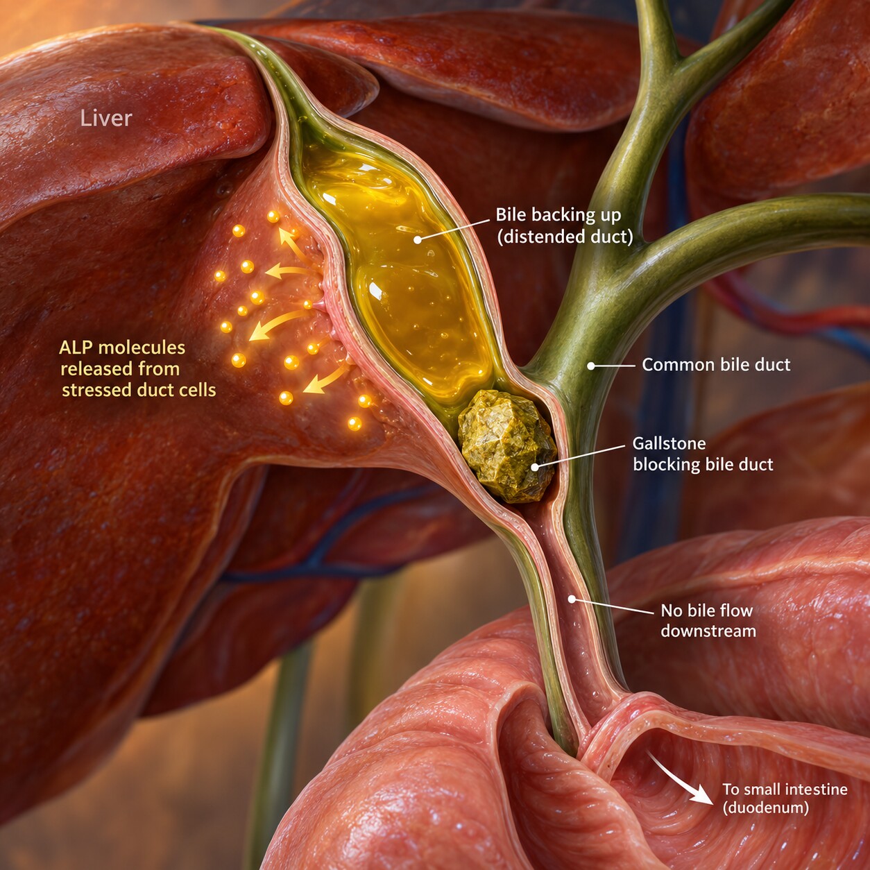
Figure 3. A gallstone or tumor physically obstructing the bile duct forces bile to back up, mechanically stressing the duct lining and releasing large amounts of ALP into the bloodstream.
When the liver is the true source of a high ALP, the underlying mechanism is usually mechanical rather than the kind of direct cell injury that raises ALT and AST. A gallstone lodged in the common bile duct, a tumor pressing on the duct from outside, or scarring that narrows the duct's internal diameter all physically obstruct the normal outward flow of bile, and that backup puts direct pressure on the cells lining the bile duct walls, triggering them to produce and release considerably more ALP than usual — a pattern doctors call cholestatic, referring to impaired bile flow, as opposed to the hepatocellular pattern seen when liver cells themselves are being directly damaged and ALT and AST dominate the picture instead.
This distinction between cholestatic and hepatocellular patterns is one of the more useful organizing concepts in liver lab interpretation, because it often points toward an entirely different diagnostic pathway before any imaging or biopsy is even ordered. A markedly elevated ALP with only mildly elevated ALT and AST suggests a mechanical obstruction problem, prompting an ultrasound or other imaging study specifically to look for a gallstone, a mass, or a structural narrowing somewhere along the bile duct system. A markedly elevated ALT and AST with only mildly elevated ALP suggests direct liver cell injury instead, prompting a different set of tests entirely — for viral hepatitis, alcohol-related injury, or fatty liver disease, the causes covered in more depth in related articles on this site about ALT and AST specifically.
Bilirubin, the yellow-orange pigment that gives bile its color and, when it builds up in the blood, causes the yellowing of skin and eyes known as jaundice, tends to rise alongside ALP in a genuine bile duct obstruction, since the same physical blockage preventing bile from draining normally also traps bilirubin that would otherwise be excreted through that same pathway. Seeing ALP and bilirubin climb together, particularly with a patient reporting the classic combination of yellowing skin, dark urine, and pale, clay-colored stool, paints a fairly specific clinical picture pointing toward an obstruction somewhere along the bile duct system, often prompting more urgent imaging than an isolated ALP elevation without jaundice would call for, since a complete bile duct blockage can progress toward serious complications, including infection of the bile ducts themselves, if it isn't identified and relieved promptly.
Primary Biliary Cholangitis and Primary Sclerosing Cholangitis
Two specific autoimmune conditions deserve particular attention because ALP, more than any other single lab value, is the classic hallmark that first raises suspicion for them. Primary biliary cholangitis is a chronic autoimmune condition in which the immune system gradually attacks and destroys the small bile ducts within the liver itself, occurring predominantly in middle-aged women, and it typically presents with a markedly elevated ALP, often well before any other symptom appears, sometimes discovered entirely incidentally on routine bloodwork years before fatigue or itching (two of its more classic symptoms) ever develop.
Primary sclerosing cholangitis is a related but distinct condition, causing progressive inflammation and scarring of the bile ducts, both inside and outside the liver, and it's strongly associated with inflammatory bowel disease, particularly ulcerative colitis — a connection significant enough that many patients newly diagnosed with ulcerative colitis get their liver enzymes checked specifically to screen for this overlap. Like primary biliary cholangitis, a persistently elevated ALP is often the first clue that prompts further evaluation, typically a specific antibody blood test for primary biliary cholangitis or specialized bile duct imaging for primary sclerosing cholangitis, since neither condition can be definitively diagnosed from the ALP elevation alone. Both conditions are considerably less common than the everyday causes of a mildly elevated liver panel, but they're specifically why a persistently high, isolated ALP with a normal ALT and AST — rather than being dismissed as a lab quirk — often prompts this more targeted, dedicated bile-duct-focused workup.
The specific antibody test used to help confirm primary biliary cholangitis deserves a brief, concrete mention, since it's one of the more distinctive diagnostic markers in all of hepatology: a blood test for anti-mitochondrial antibodies comes back positive in the substantial majority of people who actually have the condition, making it one of the more reliable single antibody tests used anywhere in liver disease diagnosis. Primary sclerosing cholangitis, by contrast, doesn't have an equivalently reliable blood antibody marker, which is exactly why direct imaging of the bile duct system — typically a specialized MRI technique that maps out the duct anatomy in detail — plays the larger diagnostic role for that particular condition instead, looking directly for the characteristic pattern of alternating narrowed and widened segments along the bile ducts that gives the disease its name.
When Bone Is Responsible: Paget's Disease
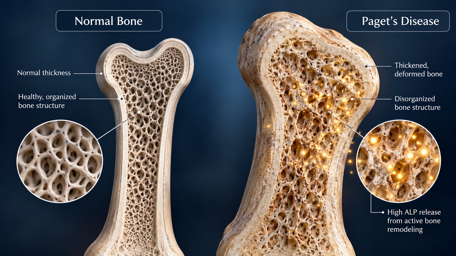
Figure 4. Paget's disease disrupts the normal, orderly cycle of bone breakdown and rebuilding, causing bone to be remodeled chaotically and often producing some of the highest ALP levels seen in clinical practice.
When bone turns out to be the source, Paget's disease of bone is one of the most classic and instructive examples, precisely because it can produce some of the most dramatically elevated ALP levels seen anywhere in medicine — sometimes many times higher than even a significant liver obstruction would typically cause. Paget's disease disrupts the normal, tightly coordinated cycle in which old bone is broken down by cells called osteoclasts and new bone is built back up by osteoblasts; in Paget's disease, this cycle becomes chaotic and accelerated in specific, localized areas of the skeleton, producing bone that's larger, less organized, and structurally weaker than normal, and the intense osteoblast activity involved in that disorganized rebuilding releases correspondingly enormous amounts of ALP into the bloodstream.
Paget's disease most commonly affects older adults, frequently develops with no symptoms at all in its early stages, and is often discovered incidentally when a markedly high ALP on routine bloodwork prompts further investigation, or when an X-ray taken for an unrelated reason happens to show the characteristic bone changes. When symptoms do occur, they typically include localized bone pain, and in more advanced cases, visible bone deformity or an increased fracture risk in the affected area, since the disorganized new bone, despite being produced rapidly, isn't nearly as structurally sound as normal bone. A bone-specific imaging study, along with an X-ray of the affected area, confirms the diagnosis once an unusually high ALP with a normal GGT points a doctor toward bone as the likely source in the first place.
Treatment for Paget's disease centers on a class of medications called bisphosphonates, the same broad drug family used to treat osteoporosis, which work by slowing down the overactive osteoclast activity driving the chaotic remodeling process in the first place. Because ALP directly reflects how actively the affected bone is being rebuilt, it also happens to be one of the most useful values for tracking whether treatment is actually working — a falling ALP over the weeks and months following treatment is a genuinely reassuring sign that the disease's activity is settling down, giving both the patient and the treating physician a concrete, measurable number to follow rather than relying on symptoms alone, which can be subtle or entirely absent even in bone that's still being actively remodeled.
Bone Metastases and Other Bone-Related Causes

Figure 5. During the third trimester of pregnancy, the placenta itself produces its own distinct ALP isoenzyme, meaning a moderately elevated result during this specific window is usually a normal, expected finding rather than a sign of illness.
Cancer that has spread to bone — most commonly originating from prostate, breast, or lung cancer — is another significant bone-related cause of elevated ALP, since the bone's response to invading tumor cells, whether that response is bone destruction or, in some cases, an unusual amount of new reactive bone formation around the tumor, drives up osteoblast activity and, with it, ALP production. In someone with a known cancer diagnosis, a rising ALP is one of several markers doctors watch as part of routine monitoring for possible bone spread, and in someone without a known cancer history, an unexplained high ALP combined with bone pain or other concerning symptoms can occasionally be the finding that prompts the imaging that ultimately leads to a new cancer diagnosis.
It's worth being specific about which cancers tend to produce this particular pattern, since bone metastases behave somewhat differently depending on the original tumor type. Prostate cancer that spreads to bone characteristically produces what's called an osteoblastic pattern, where the bone reacts by building new, if structurally disorganized, tissue around the tumor deposits, which is exactly the kind of process that drives ALP up substantially. Breast cancer bone metastases more often produce the opposite pattern, called osteolytic, where bone is destroyed rather than built, though a mixed pattern with elements of both is also common. This distinction matters clinically beyond just explaining the ALP pattern — it directly shapes fracture risk and the specific medications used to manage bone metastases, since the class of drugs used to strengthen bone against destructive lytic lesions works through a different mechanism than what's needed to manage the disorganized new bone formation seen in an osteoblastic pattern.
Away from disease entirely, it's worth understanding the genuinely normal, physiological reasons ALP rises from a bone source, since these situations shouldn't be mistaken for anything worrisome. Pregnancy is the clearest example: during the third trimester specifically, the placenta itself produces its own distinct isoenzyme of ALP, and it's entirely normal and expected for total ALP to rise moderately during this window as a result, with levels returning to a person's pre-pregnancy baseline within a few weeks after delivery. A healing fracture is another entirely benign bone-source cause, since the same bone-building process responsible for normal growth ramps up locally at a fracture site during healing, sometimes measurably raising overall ALP for several weeks to months while the bone knits back together.
Osteomalacia, a condition in adults caused by severe, prolonged vitamin D deficiency that leaves bone under-mineralized and structurally soft (the adult counterpart to rickets in children), represents another genuine bone-source cause worth knowing about, since it sits in an interesting middle ground between the benign and the more concerning causes covered elsewhere in this article. Without adequate vitamin D, the body can't properly absorb and deposit calcium and phosphate into newly formed bone matrix, and osteoblasts respond to this mineralization failure by working overtime, driving ALP upward even though the underlying problem is a nutritional deficiency rather than a tumor or an inherited bone disease. This particular cause is genuinely encouraging from a treatment standpoint, since correcting the vitamin D deficiency through supplementation typically allows bone mineralization to normalize over subsequent months, with ALP gradually falling back toward baseline as a direct, measurable sign that the underlying deficiency is being properly corrected.
Why Children and Teenagers Have Much Higher Normal Ranges
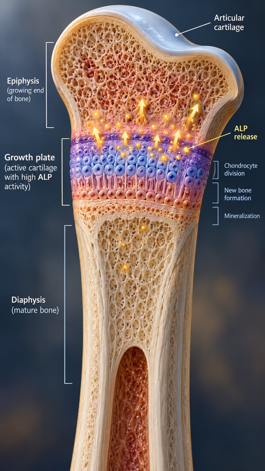
Figure 6. Active growth plates at the ends of a child's or teenager's long bones drive intense, entirely normal bone-building activity, which is why pediatric ALP reference ranges run several times higher than adult ranges.
One of the most important context clues for interpreting an ALP result correctly is age, and it's a detail that catches a lot of parents off guard when they see their child's lab report. Children and adolescents, particularly during growth spurts, have ALP levels that run routinely two to three times higher than a healthy adult's normal range, simply because their growth plates — the active zones of bone-building tissue at the ends of long bones responsible for height growth — are producing large amounts of ALP as a completely normal, expected part of a body that's still actively growing.
This means a child's ALP result has to be interpreted against a pediatric, age-specific reference range, never against the adult range that most general lab report templates default to displaying — a mismatch that can otherwise make an entirely healthy, normally growing child's bloodwork look alarmingly abnormal to a parent reading it without that specific context. ALP levels typically peak during the fastest phase of adolescent growth (generally somewhat earlier in girls than in boys, tracking each child's individual growth timeline) and then gradually decline back down to adult levels once bone growth plates fully close in early adulthood, a transition called growth plate fusion that marks the end of height growth for good.
This same age-related pattern also plays out, in a smaller and less dramatic way, at the other end of the age spectrum. Older adults, particularly those with age-related bone density loss, can sometimes show a modest baseline increase in ALP compared to a younger, middle-aged adult, reflecting ordinary bone turnover rather than any specific disease process. This is one more reason a single ALP value is generally most useful when interpreted against a person's own established baseline over time, when one is available, rather than compared only to a single population-wide reference range that necessarily has to accommodate this kind of normal variation across the entire human lifespan, from active childhood growth through the gradual bone changes that come with older age.
What Testing Comes Next
Once GGT (and, when needed, isoenzyme testing) has pointed toward a likely source, the workup that follows is targeted specifically at that source rather than continuing as a broad, undirected search. A liver or bile-duct source typically leads to an abdominal ultrasound, looking directly for gallstones, duct narrowing, or a mass; if the ALP pattern and other clinical clues suggest an autoimmune bile duct condition specifically, a dedicated antibody blood test or specialized bile duct imaging follows instead. A bone source typically leads to a plain X-ray or a bone-specific imaging study of the affected area if bone pain or a prior finding points to a specific location, along with basic calcium, phosphate, and vitamin D testing, since deficiencies in these can also modestly affect bone ALP production.
Throughout this process, a doctor is also weighing how markedly elevated the result is and whether it's isolated or accompanied by other abnormal findings. A mildly elevated ALP with an otherwise normal panel and no symptoms is often simply rechecked in a matter of weeks, since transient, benign elevations are common and frequently resolve without ever needing the more extensive workup described above. A markedly elevated result, especially one accompanied by other abnormal liver values, unexplained bone pain, or jaundice, moves more quickly toward the targeted imaging and specialist referral this section describes.
Medications and certain everyday supplements are also worth ruling out early in this process, since they're a common and easily overlooked contributor to a mildly elevated ALP. Certain anti-seizure medications, some antibiotics, and a range of other prescription drugs have documented associations with mild ALP elevation, generally through effects on the liver's bile-handling machinery rather than direct cell injury, and reviewing a person's complete medication and supplement list is a standard, low-cost early step that can sometimes explain an elevation without needing to proceed to imaging or more specialized blood work at all. This is exactly why a thorough medication review, alongside the GGT-pairing approach described earlier, is typically among the very first things addressed once an elevated ALP is confirmed on a repeat test, well before more involved investigations are considered.
When the Simple Test Isn't Enough: Isoenzyme Electrophoresis

Figure 7. Isoenzyme electrophoresis physically separates the liver and bone forms of alkaline phosphatase into distinct bands based on subtle structural differences, resolving cases the GGT-pairing method leaves ambiguous.
For the minority of cases where GGT doesn't provide a clean answer — say, a mildly elevated GGT alongside a markedly high ALP, where it's genuinely unclear whether the liver alone explains the full picture — isoenzyme electrophoresis offers a more direct, physical answer. The technique works by placing a small blood sample onto a specialized gel or membrane and applying an electric current, which pulls the different molecular forms of ALP through the material at different speeds based on subtle differences in their size and electrical charge. Because the liver form and the bone form of ALP, despite being chemically similar enough to share the same basic enzymatic function, have distinct enough structural differences, they separate into distinct, visible bands positioned at different points along the gel, and the relative intensity of each band shows roughly what proportion of the total ALP is coming from each source.
This test isn't run as a routine first step for most people, both because it's more expensive and technically involved than a simple GGT comparison, and because the GGT-pairing method already resolves the vast majority of cases without needing this additional layer of testing. It's reserved specifically for genuinely ambiguous situations — a person with a history that could plausibly explain either a liver or bone source, or a result pattern that doesn't cleanly fit the usual GGT-based logic — where knowing the exact proportional breakdown between liver and bone contribution meaningfully changes what happens next, rather than being ordered reflexively alongside every elevated ALP result.
An older, low-tech alternative to full electrophoresis, called heat stability testing, is occasionally still used in settings where electrophoresis isn't readily available, and it works on a genuinely clever biochemical principle: the bone form of ALP is considerably less stable under heat than the liver form, meaning that briefly heating a blood sample to a specific controlled temperature destroys a much larger proportion of the bone isoenzyme than the liver isoenzyme. Comparing the ALP activity measured before and after this heat exposure gives a rough estimate of how much of the total came from each source, without requiring the specialized electrophoresis equipment isoenzyme testing depends on. It's a less precise method than full electrophoresis, which is why it's been largely superseded in most modern labs, but it remains a useful illustration of just how differently the liver and bone forms of this one enzyme actually behave at the molecular level, despite performing the identical basic chemical function in the body.
Common Misconceptions Worth Addressing
A common misconception is assuming any elevated ALP automatically points to a liver problem, simply because it's grouped with ALT, AST, and GGT on a standard "liver panel." As this article has covered at length, bone is just as legitimate and, in some populations like children and pregnant women, an even more likely source than the liver — the panel groups these enzymes together for testing convenience, not because they all originate from the same organ. The name "liver panel" itself is, in this specific sense, a slightly misleading holdover from how these tests are typically ordered together in practice, rather than an accurate description of where each individual enzyme on that panel actually comes from.
A second misconception is assuming a very high ALP always signals something serious. While conditions like Paget's disease and bone metastases can produce dramatic elevations, so can entirely normal, benign situations — a healthy pregnancy, a growing teenager, or a fracture quietly healing. The magnitude of the number alone, without the context of GGT, age, symptoms, and the rest of the clinical picture, simply isn't enough information to draw a conclusion either way.
A third, related misconception worth naming directly is assuming that once GGT has pointed toward the liver as the likely source, the specific cause must automatically be something serious like cancer or an autoimmune bile duct disease. In reality, the overwhelming majority of liver-source ALP elevations trace back to considerably more common and more readily managed causes — a gallstone, a medication effect, or ordinary fatty liver disease — with the rarer autoimmune and malignant causes covered in this article representing a genuine, statistical minority of cases overall rather than the typical or expected outcome. The purpose of covering those less common causes in this kind of depth isn't to suggest they're the most likely explanation for any given result, but to make sure a persistently unexplained elevation, once the far more common causes have been reasonably excluded, doesn't get dismissed indefinitely without ever considering them.
Frequently Asked Questions
How do doctors know if high ALP is from the liver or from bone?
The fastest method checks GGT alongside ALP, since GGT comes almost exclusively from the liver and bile ducts, not bone. If both are elevated together, the liver is the likely source; if only ALP is high, bone is more likely. Isoenzyme testing is used when this doesn't give a clear answer.
Is high ALP during pregnancy something to worry about?
Usually not. During the third trimester, the placenta produces its own ALP isoenzyme, which is a normal, expected cause of moderately elevated levels that typically returns to baseline within a few weeks after delivery.
Why is my teenager's ALP so much higher than the adult reference range?
Active growth plates at the ends of long bones produce large amounts of ALP as a normal part of height growth. Pediatric ALP levels can run two to three times higher than adult levels and should be compared against age-specific reference ranges, not adult ones.
Can a high ALP mean cancer has spread to bone?
It can be one sign, particularly in someone with a known cancer history, since bone metastases increase osteoblast activity and ALP production. It's one of several markers used in monitoring, not a standalone diagnostic test on its own.
What is Paget's disease, and why does it raise ALP so dramatically?
Paget's disease disrupts the normal cycle of bone breakdown and rebuilding, causing bone to be remodeled chaotically in specific areas. The intense, disorganized rebuilding activity releases large amounts of ALP, often producing some of the highest levels seen in clinical practice.
Conclusion
Alkaline phosphatase is one of the more genuinely two-sided numbers in laboratory medicine, produced in meaningful amounts by two structurally and functionally unrelated tissues that happen to share the same enzyme. That dual origin is exactly what makes a high ALP, on its own, an incomplete answer rather than a diagnosis — the real clinical work lies in figuring out whether the liver's bile drainage system or bone tissue is actually behind the number, using a companion test like GGT, occasionally supported by isoenzyme testing, and always interpreted against the full context of a person's age, symptoms, and the rest of their lab panel.
Once that source is correctly identified, an elevated ALP stops being an ambiguous, slightly unsettling line on a report and becomes exactly what it's meant to be: a specific, useful clue pointing toward one of two very different, very traceable parts of the body.
If you're looking at your own report and see an elevated ALP without any further context, the single most useful next step is checking whether GGT was measured on the same panel, and if it wasn't, asking whether it should be added before drawing any conclusions from the ALP number alone. That one additional data point, paired with basic context about your age, any recent pregnancy, growth, fracture, or new medication, resolves the great majority of cases without needing anything more elaborate than a conversation with whoever ordered the test in the first place — turning what can feel like a confusing, open-ended result into a specific, answerable question with a clear next step attached to it.
Still Not Sure What Your Results Mean?
Upload your labs and get a complete, visual, plain-language interpretation of every biomarker — delivered to your inbox in under 15 minutes.
Get My ReportThis article is for educational purposes only and does not constitute medical advice. Always consult your healthcare provider regarding your specific lab results.