
Why Might Insulin Levels Be Tested Alongside C-Peptide?
If your doctor ordered a C-peptide test to be run alongside a standard insulin test, there's a genuinely elegant piece of underlying biology behind that particular decision: your pancreas manufactures insulin and C-peptide together, in exactly equal amounts, as two pieces cut from the very same parent molecule. But once released into your bloodstream, these two identical-in-quantity molecules behave completely differently — insulin gets cleared out of circulation quickly, largely by the liver, while C-peptide lingers much longer and gets cleared by the kidneys instead. This mismatch, which might sound like a technical footnote, is actually the whole reason C-peptide testing exists as a clinical tool: it lets a doctor see how much insulin your pancreas is truly producing, even in situations where measuring insulin directly would give a misleading or incomplete answer. This article explains where insulin and C-peptide come from, why their different clearance rates matter so much, how the tests are actually performed, and the specific clinical situations — from distinguishing types of diabetes to investigating unexplained low blood sugar to monitoring beta cell function over time — where testing them together provides an answer that neither test could give reliably on its own.
What Insulin and C-Peptide Actually Are, and Where They Come From
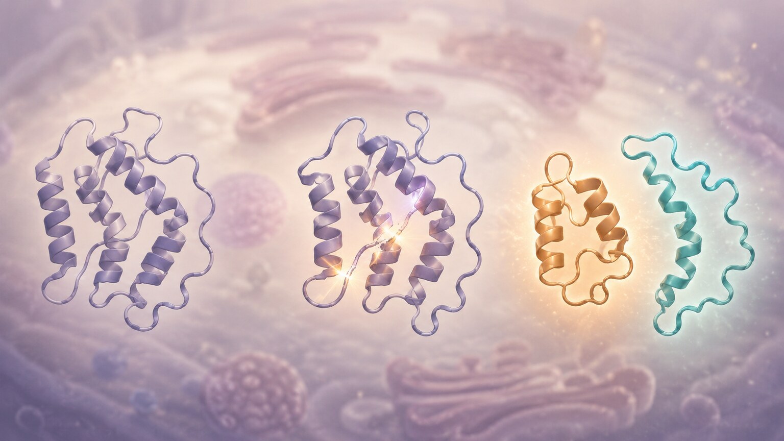
Figure 1. Insulin and C-peptide begin as a single molecule, proinsulin, which splits into two separate pieces released into the bloodstream in exactly equal amounts.
Inside specialized cells in your pancreas called beta cells, insulin isn't manufactured directly as the finished, active hormone. It's first produced as a single, longer precursor molecule called proinsulin — think of it as one continuous strand folded into a specific shape. Before proinsulin is released from the beta cell, an internal cutting process called cleavage splits it into two separate pieces: the insulin molecule itself, which goes on to do the well-known job of helping cells absorb glucose from the blood, and a second, biologically much less metabolically active fragment called C-peptide (short for "connecting peptide," since it originally physically connected the two separate ends of the insulin portion within the larger proinsulin structure before cleavage occurred). Because this splitting happens on a strict one-to-one basis — every single proinsulin molecule that gets cleaved produces exactly one insulin molecule and exactly one C-peptide molecule — the two are released into your bloodstream in precisely equal quantities, every single time, with no exceptions under normal physiology. This precise, predictable relationship is genuinely rare among hormone systems in the body, and it's exactly this predictability that laboratory medicine has learned to exploit for diagnostic purposes, turning a basic fact of protein biochemistry into a practical clinical tool. Few other hormone pairs in the body offer this same kind of built-in, one-to-one internal reference point, which is part of why the insulin-C-peptide relationship has become such a well-established and specifically named diagnostic pairing rather than simply one test among many unrelated options a clinician might separately choose to order.
This equimolar relationship (equal molar amounts) is the entire foundation of why C-peptide testing is clinically useful at all. If insulin and C-peptide always entered the bloodstream in identical amounts and behaved identically once there, measuring either one would tell you the same story, and there would be no reason to ever test both. But that's not what happens after release — and understanding exactly how their paths diverge from that point forward is what turns this seemingly simple biological detail into a genuinely powerful diagnostic tool, one that lets clinicians answer questions a single insulin measurement, taken alone, simply cannot answer with confidence.
How Insulin and C-Peptide Testing Is Actually Performed
Understanding the mechanics of how these tests are actually done helps explain why the results need to be interpreted carefully rather than taken at face value in isolation. Both insulin and C-peptide are typically measured from the same blood draw, usually after a period of fasting (commonly 8 to 12 hours), since eating triggers a substantial, temporary rise in both values that would otherwise obscure the baseline pattern a clinician is trying to assess. In certain specific clinical situations — most notably when investigating suspected hypoglycemia — a standard fasting draw isn't enough, and a supervised extended fast (sometimes lasting up to 72 hours, conducted in a monitored clinical setting) may be used instead, with blood drawn for insulin, C-peptide, and glucose together at the moment symptoms of low blood sugar actually appear, capturing the exact physiological snapshot that matters most for diagnosis.
Laboratory measurement of both insulin and C-peptide relies on immunoassay techniques, which use antibodies designed to specifically bind to each respective molecule and generate a signal proportional to its concentration in the sample. Because insulin and C-peptide are structurally distinct molecules, despite originating from the same parent proinsulin, these assays can measure each one independently and accurately without meaningfully cross-reacting with the other — a technical detail that matters because it's precisely this independent, accurate measurement of two separate molecules that makes the comparison between them meaningful in the first place. Reference ranges for both tests can vary somewhat between laboratories depending on the specific assay technology used, which is another reason the relationship and pattern between the two values often carries more diagnostic weight than either absolute number compared against a generic, non-lab-specific range.
Why C-Peptide Is a More Reliable Marker of the Body's Own Insulin Production
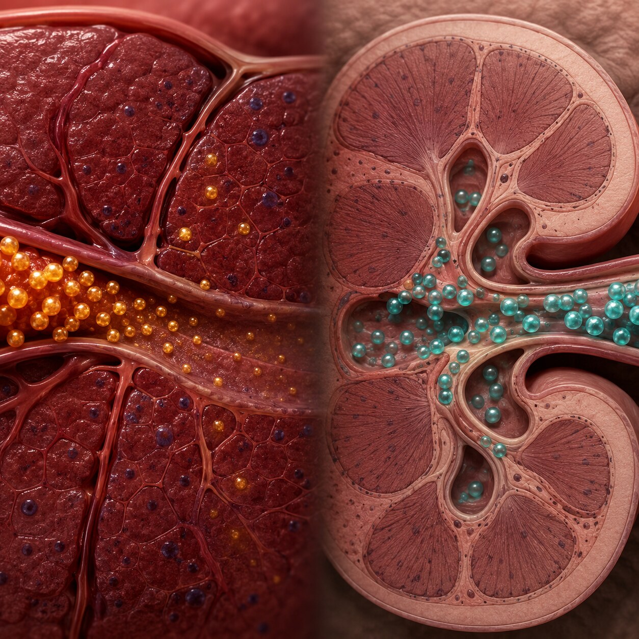
Figure 2. Insulin is cleared from blood quickly, largely by the liver, while C-peptide lingers far longer, cleared more slowly by the kidneys.
Once actually released into circulation, insulin has a remarkably short, quite fleeting lifespan in the bloodstream — its half-life, meaning the time it takes for half of any given amount to be cleared away, is only about 4 to 6 minutes, largely because your liver aggressively removes a substantial portion of insulin from blood on essentially its first pass through, in a process called first-pass hepatic extraction. C-peptide, by clear contrast, has a considerably longer half-life of roughly 20 to 30 minutes — several times longer than insulin's own — because it's cleared much more slowly and gradually, primarily by the kidneys rather than the liver, and simply doesn't undergo that same rapid, aggressive first-pass removal process. The practical result of this difference is that a blood insulin measurement reflects a narrow, recent snapshot heavily shaped by exactly how much the liver happened to be clearing at that precise moment, while a C-peptide measurement instead reflects a steadier, more representative, more genuinely trustworthy average of how much insulin the pancreas has actually been producing over a somewhat longer preceding time window.
This particular distinction becomes especially, practically important in anyone with underlying liver disease or otherwise altered liver blood flow, since a sluggish, impaired, or otherwise compromised liver clears insulin considerably less efficiently than a healthy one would, which can make blood insulin levels look artificially elevated even if the pancreas's actual production rate hasn't changed at all — a distortion that C-peptide, cleared through a completely different organ system altogether, isn't subject to in that same way, making it a more dependable choice specifically in this clinical context. This is also why C-peptide is generally considered the more stable, more reproducible, and in many clinical contexts more trustworthy reflection of true beta cell output, even though insulin remains the molecule doing the actual biological work of managing blood sugar. In short: insulin tells you what's actively circulating and available to act on cells right now, while C-peptide tells you, more reliably, how much your pancreas has genuinely been making — and in several important clinical scenarios, that second question turns out to be the one that actually needs answering.
Distinguishing Type 1 From Type 2 Diabetes
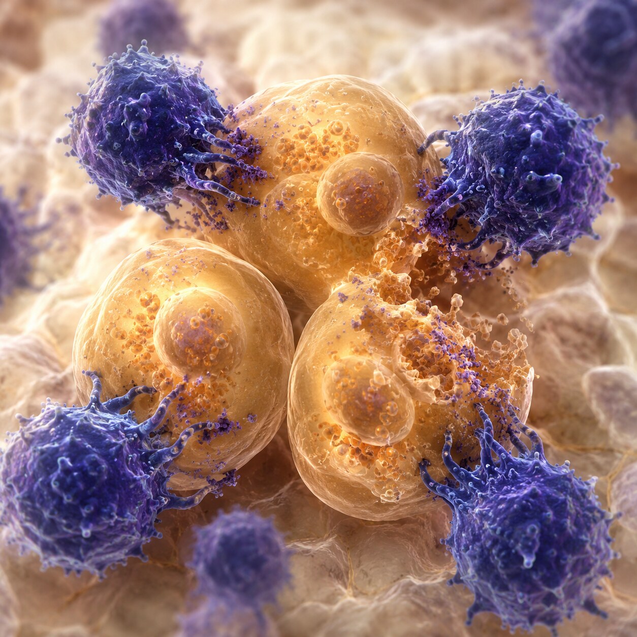
Figure 3. In type 1 diabetes, the immune system progressively destroys insulin-producing beta cells, which C-peptide testing can reveal through a low or absent result.
One of the most clinically valuable uses of combined insulin and C-peptide testing is helping distinguish type 1 from type 2 diabetes, a distinction that isn't always obvious from symptoms or blood sugar readings alone, particularly in adults, since type 1 diabetes can occasionally develop later in life (sometimes called latent autoimmune diabetes in adults, or LADA) rather than exclusively in childhood as is commonly assumed. In type 1 diabetes, the immune system mistakenly identifies the pancreas's beta cells as a threat and progressively destroys them, meaning the pancreas gradually loses its physical capacity to produce insulin at all — as this destruction advances, C-peptide levels fall, often becoming very low or essentially undetectable, since there are simply fewer and fewer functioning beta cells left to produce either insulin or its C-peptide byproduct.
In type 2 diabetes, the underlying problem is fundamentally different: the pancreas typically still produces insulin, often in normal or even elevated amounts, but the body's cells have become less responsive to that insulin's signal, a condition called insulin resistance. Because beta cell function is generally preserved, at least in earlier stages of type 2 diabetes, C-peptide levels in this scenario tend to be normal or elevated rather than low — reflecting a pancreas that's still actively manufacturing plenty of insulin, even though that insulin isn't managing blood sugar as effectively as it should. This contrast — low C-peptide pointing toward type 1's loss of production capacity, versus normal-to-high C-peptide pointing toward type 2's resistance to otherwise-adequate production — is often the single clearest piece of evidence a clinician can use to correctly classify an ambiguous diabetes diagnosis, which matters enormously for treatment, since type 1 diabetes requires insulin replacement therapy essentially from diagnosis onward, while type 2 diabetes often responds initially to other treatment approaches — oral medications, lifestyle changes, and sometimes newer non-insulin injectable therapies — that wouldn't be appropriate, or sufficient, for someone whose pancreas has genuinely lost its own production capacity and depends entirely on external insulin to survive.
Curious what your own insulin, C-peptide, or glucose results are really telling you? Upload your results and get a complete, plain-language breakdown in under 15 minutes.
Analyze My ResultsDetecting Insulin Resistance and Early Metabolic Dysfunction
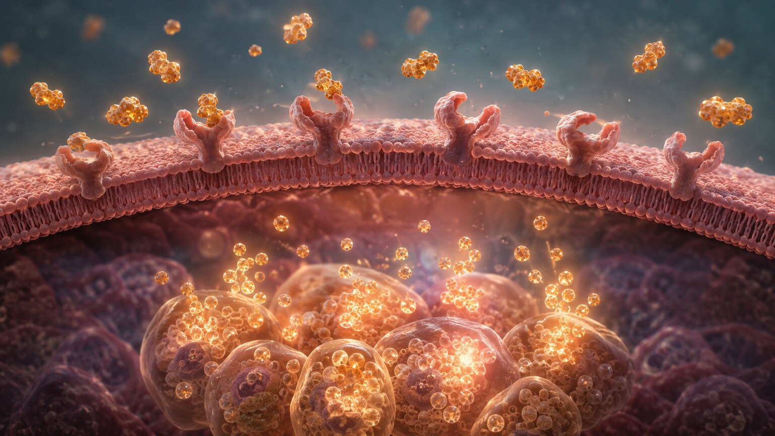
Figure 4. When cells resist insulin's signal, the pancreas compensates by producing more of both insulin and C-peptide, often years before blood sugar itself becomes abnormal.
Beyond distinguishing established diabetes types, combined insulin and C-peptide testing plays an important role in detecting insulin resistance well before it progresses to full diabetes — a stage where identifying the problem early carries real, meaningful value for prevention. When cells throughout the body become less responsive to insulin's signal, the pancreas initially compensates by producing more insulin to force the same glucose-lowering effect, a state called compensatory hyperinsulinemia. During this compensated phase, blood glucose and even hemoglobin A1c can remain entirely normal, since the extra insulin is successfully doing its job — but elevated insulin and elevated C-peptide, rising together in tandem, reveal that the pancreas is already working significantly harder than it should need to, often years before glucose control begins to break down and the person actually crosses into a "prediabetes" or diabetes diagnosis based on glucose values alone.
This pattern — both insulin and C-peptide elevated together, moving in the same direction — is what confirms that an elevated insulin reading genuinely reflects increased pancreatic output rather than some other, less common cause of an unusual insulin result, since both markers rising together rules out several alternative explanations that might otherwise need to be considered. This early detection window is clinically valuable because insulin resistance, caught at this compensated stage, often responds well to lifestyle interventions — changes in diet, physical activity, and weight management — that can meaningfully reduce the burden on the pancreas before beta cells become progressively exhausted from years of chronic overproduction, a process increasingly understood as one of the pathways that eventually leads toward type 2 diabetes in people who don't intervene during this earlier, more reversible stage. Some researchers and clinicians specifically use elevated fasting insulin, confirmed by a correspondingly elevated C-peptide, as an early warning sign worth acting on well before glucose or A1c values themselves cross into any officially abnormal range, treating this compensated phase as a genuine opportunity for prevention rather than waiting for a formal prediabetes or diabetes diagnosis to prompt action.
Investigating Unexplained Low Blood Sugar
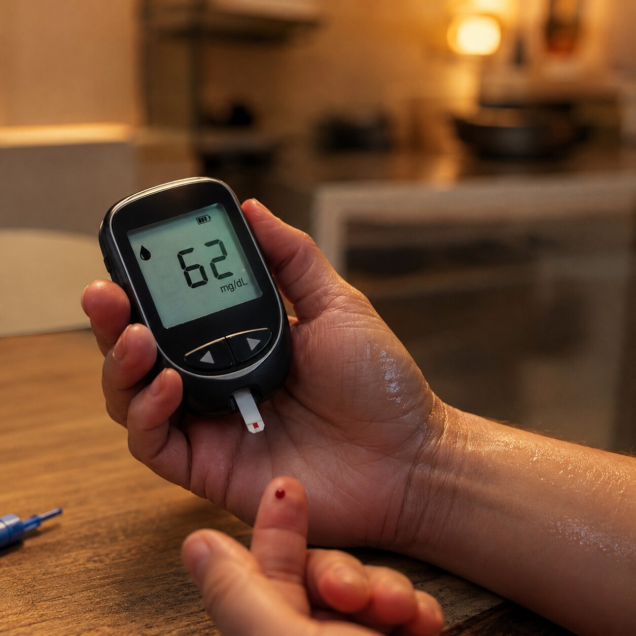
Figure 5. When someone experiences unexplained low blood sugar, the combined insulin and C-peptide pattern helps identify whether the cause is internal overproduction or an external source of insulin.
Perhaps the single most elegant clinical application of combined insulin and C-peptide testing is investigating unexplained hypoglycemia (low blood sugar) in someone who isn't taking insulin medication for diagnosed diabetes. A rare but important cause of spontaneous hypoglycemia is an insulinoma, a typically benign tumor of the pancreas's insulin-producing cells that secretes insulin inappropriately, without regard to the body's actual glucose needs. In this scenario, since the excess insulin is genuinely being manufactured by the person's own pancreas through the normal proinsulin-splitting process, both insulin and C-peptide will be elevated together, appropriately reflecting real, if inappropriately excessive, endogenous (self-produced) insulin production. Insulinomas are frequently small enough to escape detection on routine imaging until specifically searched for, which is exactly why establishing the correct biochemical pattern first — elevated insulin alongside elevated C-peptide — is so important before committing to the more involved, more expensive imaging workup needed to actually locate the tumor.
This is where the test pair becomes genuinely diagnostic in a way few other lab combinations can match: if someone is instead secretly or accidentally taking insulin medication from an external, injected source (a scenario clinicians must sometimes consider, whether from mislabeled medication, factitious disorder, or unintentional exposure), that injected insulin will show up as elevated on a blood insulin test, but because it bypassed the pancreas's normal proinsulin-splitting process entirely, it arrives in the bloodstream with no accompanying C-peptide. In this specific situation, insulin appears elevated while C-peptide is simultaneously low or suppressed — a mismatched, internally inconsistent pattern that would be essentially impossible to detect using either test alone, but that becomes immediately, unambiguously clear the very moment both results are viewed carefully side by side, together. This single diagnostic pattern is a textbook example of why testing both markers together can answer a question that neither one could answer independently, no matter how carefully each individual result was interpreted on its own. It's worth noting that this particular application of the test isn't merely theoretical — it's a well-established, standard part of the diagnostic workup for hypoglycemia specifically because this exact scenario, while relatively uncommon, carries significant clinical consequences if missed or misattributed, making the added clarity this test pair provides genuinely valuable rather than an academic curiosity.
Special Consideration: Kidney Disease and C-Peptide Interpretation
Because C-peptide is cleared from the bloodstream primarily by the kidneys, a fact established earlier in this article, it follows logically that impaired kidney function changes how C-peptide results need to be interpreted — and this is a genuinely important caveat clinicians must actively account for, rather than an obscure technical footnote. In someone with reduced kidney function, whether from chronic kidney disease, acute kidney injury, or any other cause, C-peptide clearance slows down considerably, causing it to accumulate in the bloodstream at a higher concentration than the pancreas's actual production rate would otherwise produce. This means a C-peptide level in someone with significant kidney impairment can appear falsely elevated, reflecting reduced clearance rather than increased production, which is the exact opposite kind of distortion from what happens with insulin in liver disease, described earlier in this article.
This is precisely why a complete clinical picture — including kidney function tests like creatinine and estimated glomerular filtration rate (eGFR) — is essential context whenever C-peptide results are being interpreted in someone with any degree of known or suspected kidney impairment, since failing to account for this can lead to a mistaken conclusion that beta cell function is better preserved than it truly is. In practice, this means a clinician evaluating a diabetic patient with coexisting kidney disease has to hold two pieces of information in mind simultaneously: what the C-peptide number itself says, and how much of that number might be explained by reduced clearance rather than genuine pancreatic output — a nuance that underscores, once again, why these lab values are tools for building a complete clinical picture rather than numbers meant to be read in isolation from the rest of a person's health status. Clinicians managing diabetic patients who also have kidney disease often lean more heavily on trends over time, and on other markers less affected by kidney clearance, rather than treating a single C-peptide value as a definitive, standalone answer in this particular population.
Monitoring Beta Cell Function in People Already Taking Insulin
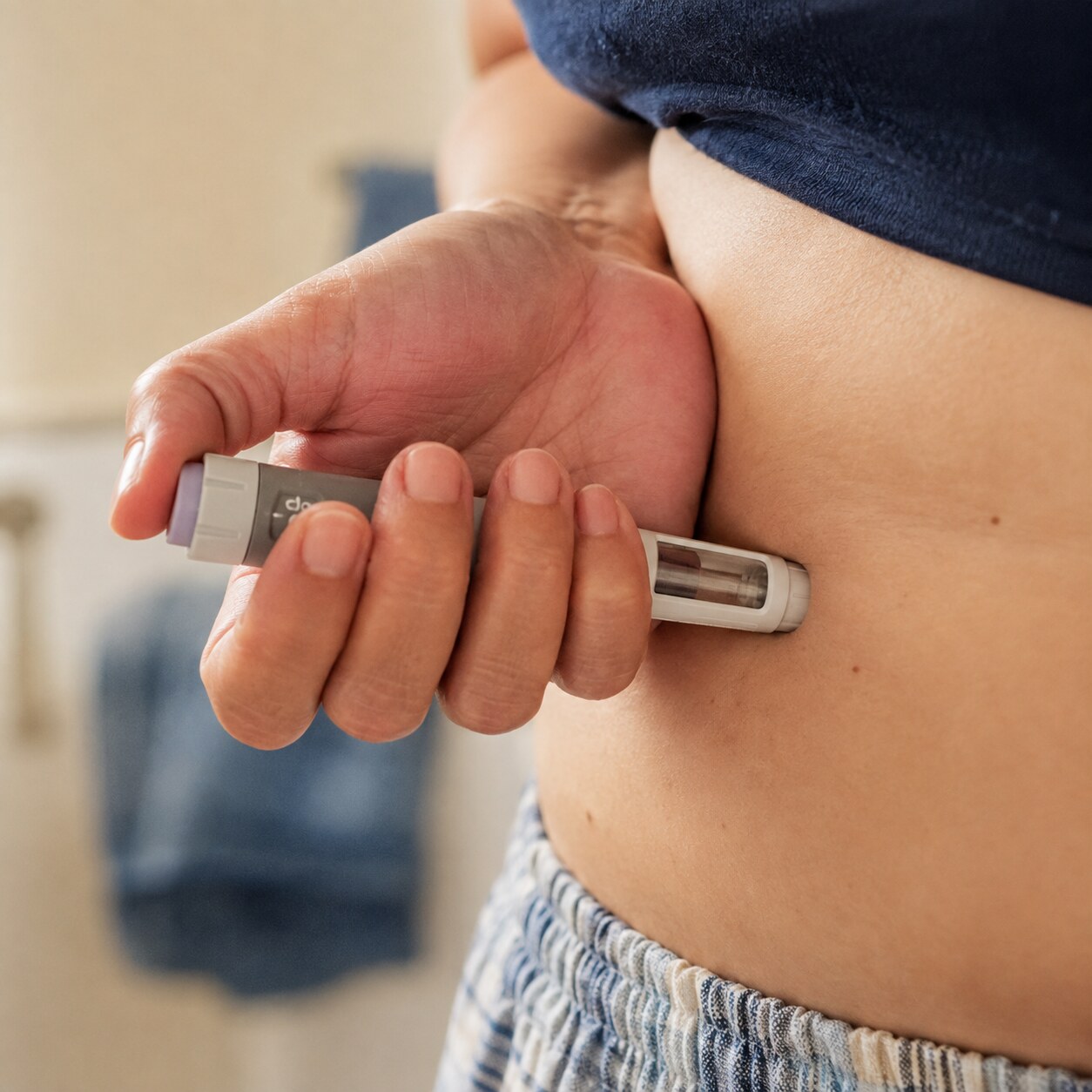
Figure 6. In people already taking insulin medication, C-peptide becomes the only reliable way to measure how much insulin the pancreas is still producing on its own.
For anyone already being actively treated with injected insulin — whether they have type 1 diabetes or more advanced, insulin-requiring type 2 diabetes — a standard blood insulin test becomes essentially useless as a genuine measure of the pancreas's own remaining function, since it can no longer distinguish between insulin the body made itself and insulin that was injected from an external source; both look identical on a standard insulin assay. This is precisely the scenario where C-peptide testing becomes not just useful but genuinely irreplaceable: because injected insulin medication contains no C-peptide at all (it's manufactured synthetically or extracted separately, without the connecting peptide fragment attached), a C-peptide measurement in someone on insulin therapy reflects only their own remaining endogenous production, completely unaffected by however much medication they've injected that day or that week.
This makes C-peptide the standard, go-to tool for answering a genuinely important clinical question that comes up repeatedly in ongoing diabetes management: is this particular person's pancreas still producing any meaningful, clinically relevant amount of its own insulin? A newly diagnosed type 1 diabetic with a still-measurable C-peptide may be in what's sometimes called the "honeymoon phase," where some residual beta cell function persists for a period after diagnosis before eventually declining further — information that can meaningfully influence treatment decisions and expectations. In longer-standing type 2 diabetes, declining C-peptide over years of monitoring can signal progressive beta cell exhaustion, helping guide the decision about when oral medications alone are no longer sufficient — a signal that the pancreas's own insulin production has fallen enough that starting or increasing insulin therapy has become clinically necessary, rather than making that call based on glucose control alone. This kind of objective, biological evidence of declining pancreatic reserve can also help patients and clinicians alike understand why a treatment plan needs to change, offering a clearer, more concrete rationale than simply pointing to a rising A1c number, which reflects the downstream consequence of declining beta cell function rather than the underlying mechanism itself.
Tracking Insulin and C-Peptide Over Time
Because beta cell function tends to change gradually — declining slowly over years in progressive type 1 diabetes, or shifting in response to lifestyle changes and treatment in insulin resistance — a single insulin and C-peptide measurement is often less informative than tracking both values across multiple visits over time. For someone in the early stages of suspected type 1 diabetes or LADA, serial C-peptide measurements can help chart the pace of beta cell decline, informing decisions about when insulin therapy should begin and how aggressively to plan for it, since a rapidly falling C-peptide suggests a faster-progressing autoimmune process than one that's declining only gradually over several years.
For someone working to address insulin resistance through lifestyle changes, a declining insulin and C-peptide level at a stable, healthy glucose reading over successive visits is a genuinely encouraging sign — it suggests the pancreas no longer needs to work as hard to maintain normal blood sugar, meaning the underlying resistance is actually improving rather than simply being compensated for more aggressively. This kind of longitudinal tracking transforms what might otherwise be two isolated numbers into a meaningful trend line, offering insight into the trajectory of someone's metabolic health that a single snapshot, no matter how carefully interpreted, simply cannot provide on its own. This is part of why some clinicians recommend rechecking both values at set intervals — every six to twelve months in many cases — specifically for patients whose treatment plan or risk profile might reasonably shift based on the direction that trend is heading.
Related Tests Often Ordered Alongside This Pair
Insulin and C-peptide rarely stand entirely alone in a diagnostic workup — they're usually part of a broader panel designed to build a complete picture rather than relying on any two values by themselves. Fasting glucose is essentially always measured at the same time, since the relationship between glucose and insulin (how much insulin is needed to manage a given glucose level) is itself diagnostically meaningful, forming the basis of calculations like the HOMA-IR score, a formula that estimates insulin resistance from paired fasting glucose and insulin values. Hemoglobin A1c, reflecting average blood sugar over roughly the preceding three months, adds a longer-term view that a single fasting snapshot can't provide on its own, helping distinguish a temporary metabolic disturbance from a more established, longstanding pattern.
When type 1 diabetes or LADA is a genuine possibility, autoantibody testing is frequently ordered alongside insulin and C-peptide, since these antibodies (directed against specific proteins in beta cells, such as GAD65, IA-2, or insulin itself) provide direct evidence of the autoimmune process driving beta cell destruction, complementing the functional information C-peptide provides about how much beta cell capacity actually remains. When an insulinoma is suspected based on the combined insulin and C-peptide pattern, additional testing typically includes proinsulin levels (since insulinomas sometimes release a disproportionate amount of unprocessed proinsulin alongside mature insulin) and a check for sulfonylurea medications in the blood or urine, ruling out another possible cause of inappropriately high insulin and C-peptide together — medication-induced hypoglycemia from a class of diabetes drugs that stimulate the pancreas to release more insulin, which can produce a lab pattern that looks similar to a true insulinoma without imaging and further workup to distinguish the two.
What Happens After These Results Come Back
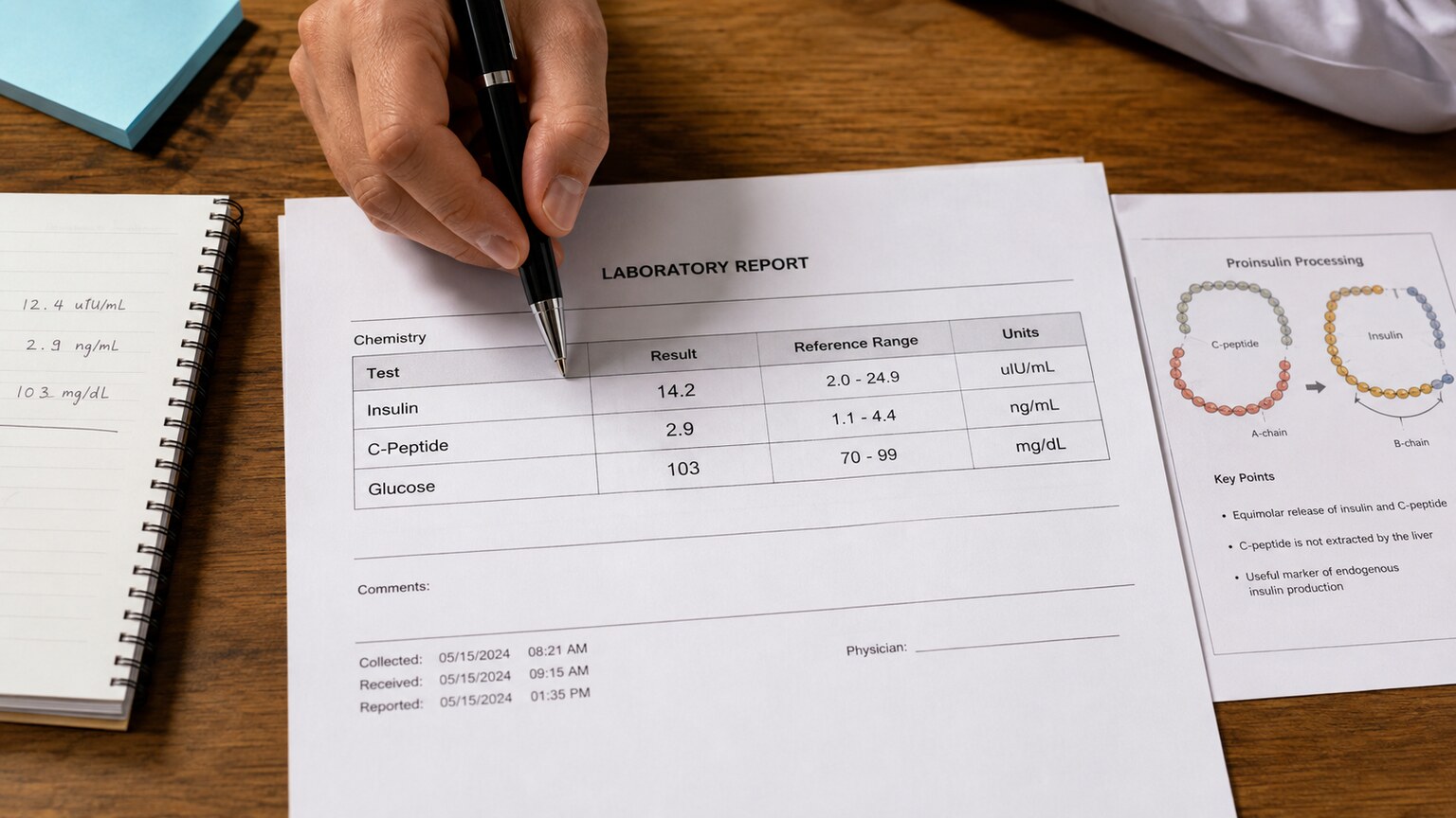
Figure 7. Interpreting paired insulin and C-peptide results always involves reading the relationship between the two values together, alongside glucose, rather than either number in isolation.
Once both results come back, the practical next step depends entirely on which of the patterns described throughout this article the values actually form together. Low C-peptide, found together with symptoms and other lab findings consistent with diabetes, generally points toward either type 1 diabetes or a late-stage, severely advanced case of type 2 diabetes involving substantial, cumulative beta cell loss, prompting a meaningful shift toward insulin-based treatment planning going forward. Elevated insulin and C-peptide together, found in someone without diabetes but with other recognized metabolic risk factors, often prompts a proactive conversation about insulin resistance and lifestyle-focused prevention strategies well before more serious glucose abnormalities have the chance to develop. A mismatched pattern — elevated insulin paired with a suppressed, inappropriately low C-peptide — in someone with unexplained hypoglycemia raises the genuine possibility of external insulin exposure and typically prompts a more detailed, sometimes understandably sensitive, clinical conversation and further evaluation to clarify exactly what's happening. And elevated levels of both, together with symptoms of hypoglycemia, may prompt imaging studies — such as a CT scan, MRI, or specialized endoscopic ultrasound — specifically aimed at locating a suspected insulinoma so it can be surgically removed, which is typically curative.
In every one of these scenarios, what actually drives the clinical decision isn't either number in isolation, but the specific relationship between them, interpreted alongside the person's glucose level, symptoms, medication history, and broader clinical picture, since the same absolute number can point toward entirely different conclusions depending on everything else surrounding it. This is really the throughline of the entire combined insulin and C-peptide test: it exists specifically because certain critically important clinical questions — is this diabetes type 1 or type 2, is this hypoglycemia coming from inside the body or from an outside source, is this pancreas still functioning — simply cannot be answered with confidence by either marker alone, no matter how precisely each one is measured.
What Symptoms Prompt This Combined Testing in the First Place
Combined insulin and C-peptide testing isn't part of routine annual bloodwork for most people — it's typically ordered when a specific clinical question has already emerged from other findings or symptoms, and it's worth understanding what those triggering situations usually look like. Someone newly diagnosed with diabetes whose presentation doesn't clearly fit the typical pattern for either type — perhaps an adult with a lean body type and rapidly rising blood sugar, more typical of type 1, or a child with obesity and gradually worsening glucose control, more typical of type 2 — is a common candidate, since the combined test helps resolve genuine diagnostic ambiguity rather than assuming a diagnosis based on age or body type alone, both of which are far from perfectly reliable indicators on their own.
Recurrent, unexplained episodes of low blood sugar, particularly ones that occur without any clear trigger like skipped meals or excessive exercise, are another common reason this combined testing gets ordered, since distinguishing an insulinoma from other causes of hypoglycemia, including the possibility of external insulin exposure, depends heavily on exactly the kind of insulin-versus-C-peptide comparison described throughout this article. People with strong risk factors for insulin resistance — significant weight gain concentrated around the abdomen, a family history of type 2 diabetes, or related conditions like polycystic ovary syndrome (PCOS) — sometimes have this testing ordered proactively, as part of an effort to catch developing metabolic dysfunction before it progresses to overt diabetes. And people already established on insulin therapy, particularly those with an uncertain original diagnosis or those whose treatment plan is being reevaluated, often have C-peptide checked periodically simply to reassess how much natural insulin production remains.
Frequently Asked Questions
Do I need to fast before an insulin and C-peptide test?
Usually, yes. Most insulin and C-peptide testing is done in a fasting state, typically after 8 to 12 hours without food, since both values are affected by recent eating. Some specific evaluations, particularly for hypoglycemia, may instead involve a supervised extended fast or a test timed around symptoms, so it's important to follow the specific instructions given for your particular test.
Can C-peptide be low for reasons other than type 1 diabetes?
Yes. Very low insulin secretion from any cause of significant beta cell loss or dysfunction, including advanced type 2 diabetes, certain pancreatic diseases, or pancreatic surgery, can also produce a low C-peptide result. This is why C-peptide alone isn't used to diagnose type 1 diabetes definitively — it's interpreted alongside clinical history, antibody testing, and the overall presentation.
If I'm on insulin therapy, will taking my insulin dose affect my C-peptide result?
No, and this is exactly the point of the test. Because injected insulin medication doesn't contain C-peptide, your C-peptide result reflects only your own pancreas's natural production, regardless of how much insulin medication you've taken. This is precisely why C-peptide, rather than insulin, is used to assess remaining natural function in people already on insulin therapy.
Is a normal C-peptide result enough to rule out diabetes entirely?
Not on its own. A normal or even elevated C-peptide can still occur alongside diabetes if insulin resistance is present, since the pancreas may be producing plenty of insulin that the body simply isn't responding to effectively. C-peptide is best interpreted together with glucose, A1c, and clinical context, not as a standalone screening test for diabetes itself.
Does kidney disease affect how C-peptide results should be interpreted?
Yes. Because C-peptide is cleared from the blood primarily by the kidneys, reduced kidney function can cause it to accumulate and appear falsely elevated, even if the pancreas's actual insulin production hasn't changed. Anyone with known kidney impairment should have that factored into how their C-peptide result is read, alongside kidney function tests like creatinine and eGFR.
Can insulin and C-peptide levels change based on what I ate the night before?
Yes, to some degree, which is why fasting is generally required before these tests. A large, carbohydrate-heavy meal the night before typically doesn't meaningfully distort a properly fasted morning result, but not following the fasting instructions given by your provider, or eating close to the actual blood draw, can produce misleadingly elevated values that don't reflect your true baseline.
Conclusion
Ultimately, this test pairing is a good reminder that lab values rarely exist in a vacuum.
Insulin and C-peptide start out as two halves of the exact same molecule, released into the bloodstream in perfectly equal amounts — but the way they're cleared afterward, on completely different timelines through completely different organs, is what turns this simple, almost elegant biological pairing into one of the more genuinely clever and clinically useful diagnostic tools available in modern metabolic medicine today. Testing them together answers questions that neither one can reliably answer alone: whether a pancreas has genuinely lost its production capacity or is simply being resisted by insulin-insensitive cells, whether elevated insulin reflects true internal overproduction or an external, injected source, and how much natural beta cell function actually remains in someone already relying on insulin medication for daily glucose control. Understanding this relationship — rather than looking at either number in isolation — is what turns two individually limited lab values into a combined result genuinely capable of answering some of the more important, and sometimes more nuanced, questions in diabetes and metabolic health, offering clarity that neither test could reliably provide standing entirely on its own.
Still Not Sure What Your Results Mean?
Upload your labs and get a complete, visual, plain-language interpretation of every biomarker — delivered to your inbox in under 15 minutes.
Get My ReportThis article is for educational purposes only and does not constitute medical advice. Always consult your healthcare provider regarding your specific lab results.