
What Causes a Culture to Show Mixed Bacterial Growth?
A culture report showing exactly one clearly identified bacterium alongside a clean, simple sensitivity panel generally feels fairly straightforward to interpret — you clearly have an infection, here's precisely what's causing it, and here's exactly what treats it effectively. A report instead showing "mixed bacterial growth," listing two, three, or even more genuinely different organisms all at once, feels considerably murkier by comparison, and it's genuinely a real fork in the road with two very different explanations sitting on either side of it. Sometimes mixed growth means the sample picked up bacteria that were never actually part of an infection at all, just innocent bystanders from skin or normal body flora that got swept up during collection. Other times, it means several different organisms really are working together to cause a genuine, and sometimes more serious, infection. This article explains exactly how to tell these two situations apart, why the distinction matters so much for treatment, and what typically happens next once a lab reports growth this way.

Figure 1. Mixed growth means a culture plate shows more than one visually and biochemically distinct type of bacterial colony, each identified separately by shape, color, and texture.
What "Mixed Growth" Actually Means on a Lab Report
When a clinical sample is first plated in the lab, the individual bacteria that happen to land on the growth medium multiply outward into visible, countable clusters called colonies, and a trained technician can often tell several different species apart just by carefully examining how those colonies actually look — their color, overall shape, size, surface texture, and precisely how they interact with specific chemical indicators deliberately built into the growth medium itself. "Mixed growth" simply means the technician identified more than one distinct colony type growing on the same plate, indicating more than one kind of bacteria was present in the original sample.
This single finding alone genuinely doesn't tell you very much of anything meaningful yet about what those multiple organisms actually mean clinically — that interpretation depends entirely on where the sample came from, how it was collected, and what the overall pattern of growth looks like, which is exactly what the rest of this article walks through.
It genuinely helps to picture a culture plate the way a gardener might picture a small plot of soil deliberately seeded with several entirely different kinds of seeds all at once. Some seeds sprout quickly into small, uniform clusters; others take longer and spread differently; and a trained eye can often tell the resulting plants apart from a distance just by their shape and growth pattern, long before any deeper testing confirms exactly what each one is. Bacterial colonies work in a broadly similar way — each species tends to have a fairly characteristic appearance on a given growth medium, and an experienced laboratory technician can often make an educated first guess about how many distinct organisms are present just by looking at the plate, before running any of the more specific biochemical or genetic tests that provide a definitive identification.
It's genuinely worth being clear from the very outset here that "mixed growth" is really only a purely descriptive laboratory term, never a diagnosis in and of itself. It simply states an observed fact about the plate — more than one organism grew — without saying anything yet about whether that fact matters clinically. Everything that follows throughout the rest of this article is really about how doctors and microbiologists take that one single neutral observation and figure out precisely which of two very different underlying stories it's actually telling in any given case.
The Most Common Explanation: Contamination During Collection
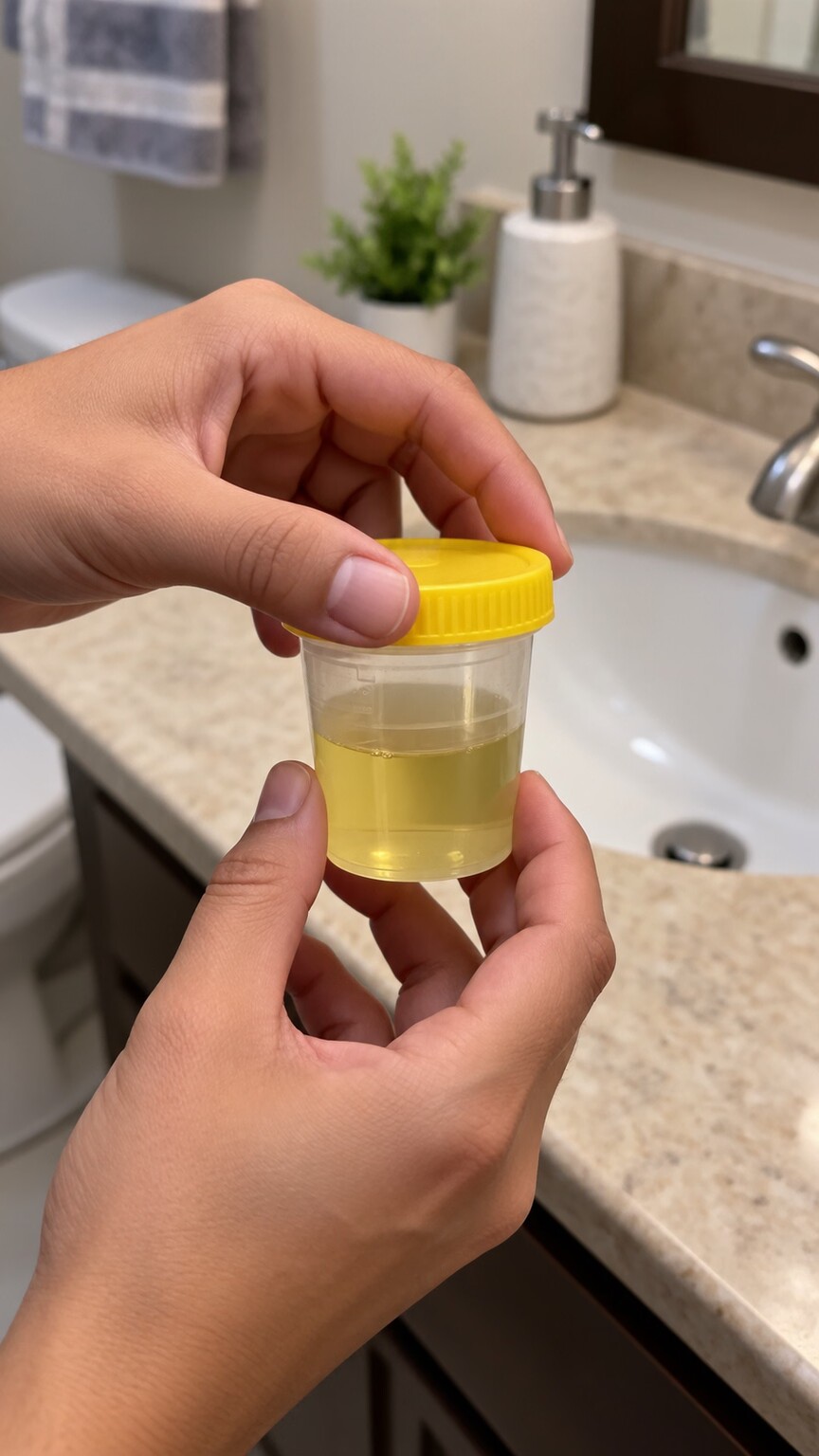
Figure 2. Skin, vaginal, or perineal bacteria that are normally present on the body can easily contaminate a urine sample during collection, producing mixed growth that reflects the collection process rather than a genuine urinary infection.
By quite a wide margin, the single most common reason any culture shows mixed growth in the first place, especially for urine samples specifically, is genuine contamination picked up somewhere during the actual collection process itself, rather than reflecting any real underlying infection whatsoever. Human skin generally, and in women specifically the vaginal and perineal area surrounding the urethral opening, is naturally and permanently covered in a genuinely diverse resident community of largely harmless, everyday bacteria. A urine sample that brushes against skin during collection, rather than being caught mid-stream after the area has been properly cleaned, can easily pick up several of these normal residents at once, producing exactly the multi-organism pattern seen in mixed growth, without any of those organisms actually being present inside the bladder or causing any infection at all.
Labs are generally quite familiar with this pattern and often report it explicitly as "mixed growth, likely representing skin/perineal flora" or similar wording, particularly when the colony counts for each organism are low and none of them individually reach the threshold considered significant for a genuine urinary infection. This is precisely why a doctor receiving this kind of report often requests a repeat sample using more careful technique rather than treating any of the organisms listed, since treating contamination with antibiotics provides no benefit and contributes unnecessarily to antibiotic resistance.
The specific organisms typically implicated in this kind of urinary contamination tend to be recognizable, everyday residents of human skin and the genital area rather than aggressive pathogens: various species of coagulase-negative staphylococci, lactobacilli (a normal and generally beneficial component of vaginal flora), diphtheroids, and occasionally low counts of common gut bacteria that migrated a short distance from the perianal area during collection. Seeing a familiar cast of these organisms, especially in low numbers and especially in combination, is itself a clue pointing toward contamination — a genuine urinary pathogen behaving badly tends to show up alone and at high numbers, not as one of several low-level entries in a crowded mixed report.
Collection technique isn't the only contributor to this kind of contamination, though it's by far the most common one. The container itself matters — a non-sterile cup, or one that's been left open to the air for an extended period before or after collection, can pick up environmental bacteria that have nothing to do with the person providing the sample at all. Delayed processing after collection compounds this problem further: bacteria of any kind, including contaminants that arrived in only small numbers, continue multiplying at room temperature the longer a sample sits before reaching the lab, which is exactly why urine samples are supposed to be refrigerated promptly or processed quickly, and why a delayed or improperly stored sample is more likely to show misleadingly higher colony counts across multiple organisms than a sample handled correctly from the start.
Curious what your culture report or the rest of your labs are really telling you? Upload your results and get a complete, plain-language breakdown in under 15 minutes.
Analyze My ResultsHow Labs Use Colony Counts to Separate Contamination From Infection
Rather than relying on subjective judgment alone, laboratories use a specific, well-established numeric approach specifically designed to help reliably sort genuine contamination apart from real infection whenever a given culture shows multiple distinct organisms present together. Each distinct colony type is counted separately, and the count for any single organism is compared against an established threshold — for a urine culture, that threshold is typically 100,000 colony-forming units per milliliter for a clean-catch sample, though a lower threshold applies when a sample is collected more invasively, such as through a catheter.
When a mixed urine culture shows two or three organisms, each present below this significant threshold, the overall pattern reads as consistent with contamination — a scattering of normal flora, none of which reached numbers suggesting they were actually multiplying inside the urinary tract itself. When one organism in a mixed culture clearly dominates, present at a colony count well above the significant threshold while the others remain low, that dominant organism is far more likely to represent the genuine infecting organism, with the low-level others still reflecting incidental contamination alongside it.
This colony-count approach exists because a genuine urinary tract infection behaves in a specific, recognizable way: a single organism that has actually established itself in the bladder or urinary tract multiplies rapidly and essentially takes over, quickly outcompeting whatever small amount of contaminating flora might also be present in the same sample. This is fundamentally different from what happens with pure contamination, where several different organisms are present in comparably modest, roughly similar numbers, none of them having had the specific opportunity or environment to multiply aggressively the way a true urinary pathogen would inside the bladder itself.
The number of distinct organisms identified also carries its own weight in this interpretation, independent of the specific colony counts involved. A urine culture showing exactly two organisms, each present in meaningful numbers, is interpreted quite differently from one showing four or more organisms scattered across lower counts. Most laboratory protocols specifically flag a culture growing three or more distinct organism types, regardless of their individual counts, as very unlikely to represent a genuine single urinary infection and far more likely to reflect a poorly collected specimen — a rule of thumb sometimes informally referred to among laboratory staff as ruling out anything looking like a "three or more organism" contaminated plate before considering it representative of a true infection.
It's worth understanding why this numeric threshold approach, rather than simple visual judgment, became the accepted standard in the first place. Decades of research comparing culture results against clinical outcomes established that colony counts above roughly 100,000 per milliliter in a clean-catch sample correlated strongly with an actual, symptomatic urinary infection, while counts below that threshold correlated poorly with genuine infection and much more often reflected contamination or, in some cases, a real but low-grade colonization that didn't require treatment. This research-backed threshold gives clinicians a reasonably objective number to lean on, rather than having to rely purely on subjective impression when deciding whether a given mixed result deserves treatment.
When Mixed Growth Reflects a Genuine Polymicrobial Infection

Figure 3. Chronic wounds and deep tissue infections, particularly in people with diabetes, frequently involve several bacterial species genuinely coexisting and contributing together to a single infection.
Not every instance of mixed growth reflects contamination — in certain body sites and certain kinds of infections, multiple organisms genuinely do coexist and contribute together to a single infectious process, a situation called a polymicrobial infection. Chronic wounds, particularly diabetic foot ulcers, are a classic example: the combination of poor blood flow, reduced sensation, and repeated minor trauma common in diabetes creates an environment where several different bacterial species, often including a mix of skin bacteria and organisms from the surrounding environment, can establish themselves together in the same wound bed simultaneously.
What makes chronic wounds particularly prone to this pattern is the loss of several of the body's usual defenses against bacterial buildup all at once. Reduced blood flow limits how effectively immune cells and antibiotics can reach the wound site in the first place, nerve damage common in diabetes means minor trauma to the area often goes unnoticed and unaddressed for far longer than it otherwise would, and the moist, protein-rich environment of an open wound bed provides an ideal growing surface for whichever bacteria happen to establish themselves there first. Once one species successfully colonizes this kind of compromised tissue, it often becomes easier, rather than harder, for additional species to join afterward, since the first colonizers begin altering the local environment in ways that can favor further bacterial buildup rather than resisting it.
Intra-abdominal infections following a ruptured appendix or bowel injury are another well-recognized example, since the gut naturally harbors an enormous, diverse population of bacteria, and any breach of the bowel wall spills a genuinely mixed community of organisms into a normally sterile space at once. Aspiration pneumonia, which occurs when material from the mouth or stomach is inhaled into the lungs, similarly tends to involve multiple organisms together, reflecting the mixed bacterial population naturally present in the mouth and upper airway.
The human gut alone is estimated to host many hundreds of distinct bacterial species living in a normally balanced, contained ecosystem, and this sheer diversity is exactly why any breach of the intestinal wall — whether from a ruptured appendix, a perforated ulcer, or a penetrating abdominal injury — almost never releases just one organism into the surrounding abdominal cavity. Surgeons and infectious disease specialists managing these infections routinely expect and plan for a polymicrobial picture from the outset, often starting broad-spectrum antibiotic coverage targeting several likely categories of gut-associated bacteria before culture results even come back, precisely because waiting for a single, cleanly identified organism would mean leaving several likely contributors untreated in the meantime.
Dental and deep neck space infections represent another body region where genuinely polymicrobial infections are the expected norm rather than the exception. The mouth harbors one of the most bacterially dense environments anywhere on the human body, and infections originating from an abscessed tooth or diseased gum tissue that spread into deeper facial or neck tissue planes typically carry along a representative mix of the mouth's normal bacterial community, again requiring antibiotic coverage broad enough to address that full mixed population rather than a single targeted organism.
Necrotizing soft tissue infections, though genuinely considerably rarer overall than any of the examples already given above, still clearly illustrate just how severe a truly genuine polymicrobial infection can ultimately become once several sufficiently aggressive organisms combine forces together within the very same tissue space. These infections, sometimes involving a specific combination of one or more anaerobic bacteria alongside aerobic organisms, can spread through tissue planes with alarming speed precisely because of the combined destructive capability of the different organisms working together, which is part of why these particular infections are treated as surgical and medical emergencies requiring both aggressive antibiotic coverage and, often, urgent surgical intervention rather than antibiotics alone.
Synergistic Infections: When Different Bacteria Actually Help Each Other

Figure 4. In some polymicrobial infections, aerobic bacteria consume available oxygen in a way that creates favorable conditions for anaerobic bacteria, allowing the two groups to genuinely reinforce each other's growth.
Some of the single most clinically important polymicrobial infections encountered in practice involve a very specific, genuinely mutually reinforcing relationship between different distinct types of bacteria, rather than merely simple, coincidental coexistence happening to occur in the same space. Aerobic bacteria, which need oxygen to thrive, can consume the available oxygen within an infected tissue space faster than the body can resupply it, creating a low-oxygen environment that anaerobic bacteria, which actually prefer or require the absence of oxygen, find ideal for their own growth. This relationship works in both directions in many cases, with certain byproducts of anaerobic metabolism in turn supporting aerobic bacterial growth, creating a genuine synergy where the combination causes more aggressive tissue damage than either group of organisms would produce on its own.
This synergistic mechanism is part of why certain severe soft tissue infections are specifically associated with mixed aerobic-anaerobic growth on culture, and why these particular infections are treated with combination antibiotic regimens deliberately chosen to cover both categories of organism at once, rather than a single antibiotic aimed at whichever organism seems most prominent. Missing one half of a synergistic pair on the treatment plan can allow the infection to continue progressing even while the other organism is being effectively treated.
Anaerobic bacteria specifically present their own distinct laboratory challenge that's worth understanding, since it partly explains why anaerobic involvement in a mixed infection sometimes gets missed or underrepresented on an initial culture report. Because these organisms are, by definition, harmed or killed by exposure to oxygen, a sample that isn't collected, transported, and processed using specialized anaerobic technique can lose its anaerobic bacterial population before the sample ever reaches the growth plate, even though those anaerobic organisms were genuinely present and contributing to the infection at the time of collection. This is exactly why deep wound and abscess cultures suspected of anaerobic involvement are often collected with special anaerobic transport containers and processed differently in the lab than a standard aerobic culture, specifically to avoid this kind of false-negative loss of an organism that was truly present.
Because of this collection sensitivity, a clinician suspecting a synergistic aerobic-anaerobic infection based on the clinical picture — a foul odor, gas visible on imaging, or a wound appearance consistent with this pattern — will sometimes begin empiric antibiotic coverage for likely anaerobic involvement even before culture results confirm it, precisely because of how easily anaerobic organisms can be under-detected on a standard culture despite genuinely being present and contributing to the infection.
How Labs Decide Whether to Fully Work Up Every Organism
Because fully identifying every organism in a mixed culture and running individual sensitivity testing on each one is time-consuming and expensive, labs use judgment, guided by established protocols, to decide how much workup a given mixed culture actually deserves. A urine culture showing three low-count organisms consistent with likely contamination is typically reported with a brief note rather than pursued further, since fully identifying incidental skin flora provides no clinical value. A wound or deep tissue culture from a normally sterile body site showing two or three organisms at meaningful colony counts, by contrast, usually does get each organism individually identified and tested for antibiotic sensitivity, since any of them could plausibly be a genuine contributor to a real infection requiring targeted treatment.
This decision also factors in how urgently a treatment decision actually needs to be made. In a stable outpatient with a mild, non-urgent presentation, a lab may reasonably choose a more conservative, lower-cost workup and simply recommend a repeat sample if the picture is ambiguous, since there's no immediate pressure to commit to a specific treatment plan. In a hospitalized patient who is clearly sick and already receiving empiric antibiotics based on the overall clinical picture, the same ambiguous mixed result may prompt a more complete workup of every organism present, since the treating team genuinely needs to know as much as possible to confirm or adjust the antibiotic regimen already underway rather than waiting for a cleaner, less ambiguous specimen that may take additional days to obtain.
The specific body site sampled plays a large role in this decision. Samples from sites that are never sterile in a healthy person — skin, throat, and, to a significant degree, urine collected non-invasively — are interpreted with the expectation that some normal flora will inevitably be present, and mixed growth from these sites is judged more skeptically before being labeled a genuine infection. Samples from normally sterile sites — blood, spinal fluid, deep surgical wounds, and fluid drawn directly from a joint — carry a very different starting assumption, since any bacterial growth at all from these locations, mixed or not, is inherently more likely to represent a real, clinically significant finding rather than incidental contamination.
Throat and skin cultures deserve their own specific mention here, since they sit in an interesting middle ground between the two extremes described above. A throat swab, for instance, is expected to grow a wide array of normal oral and pharyngeal flora essentially every time it's collected, which is exactly why a throat culture ordered to check for a specific pathogen, like the bacteria responsible for strep throat, is typically evaluated by specifically looking for that one particular organism against the expected mixed background, rather than reporting or reacting to every organism present on the plate. Skin and superficial wound cultures follow similar logic: some degree of mixed normal skin flora is expected and clinically uninteresting, while a specific organism present in unusually high numbers, or a recognized pathogen not normally part of healthy skin flora, is what actually draws clinical attention against that expected mixed backdrop.
Respiratory cultures from deeper in the lungs, such as sputum samples, occupy their own particular gray zone worth understanding. Because a sputum sample necessarily passes through the mouth and upper airway on its way out of the body, it almost inevitably picks up some of that region's normal bacterial population along the way, even when the sample is genuinely representative of an infection occurring deeper in the lungs themselves. This is one of several reasons sputum culture results are interpreted with real caution and alongside the broader clinical picture — chest imaging, symptoms, and how much the sample actually looked like genuine lung secretion under the microscope rather than mostly saliva — rather than reacting to every organism identified on the plate as though it were necessarily the cause of a lung infection.
What Happens If You're Asked to Provide a Repeat Sample

Figure 5. Careful clean-catch technique — thorough handwashing, cleaning the area, and collecting mid-stream urine — meaningfully reduces the chance of a repeat sample showing the same contamination-driven mixed growth.
If a first sample comes back with mixed growth suggestive of contamination, a repeat collection using more careful technique is the standard, straightforward next step, rather than any kind of alarming escalation. For urine specifically, this generally means thoroughly washing hands, cleaning the genital area with the provided wipe, and collecting only the middle portion of the urine stream rather than the very beginning, which is the portion most likely to have picked up surface bacteria on its way out. Following these steps meaningfully reduces the odds of a second sample showing the same contamination pattern.
A few additional, less commonly mentioned details can further improve the reliability of a repeat sample. Collecting the sample first thing in the morning, when urine has had several hours to concentrate in the bladder overnight, generally provides a more concentrated, easier-to-interpret specimen than a sample collected after drinking large amounts of fluid shortly beforehand. Getting the sample to the lab or refrigerator promptly after collection, ideally within an hour, prevents the kind of room-temperature bacterial multiplication discussed earlier from artificially inflating colony counts and muddying an otherwise clean collection. Small, practical details like these, easy to overlook amid the more obvious cleaning instructions, genuinely do make a measurable difference in how interpretable the resulting culture turns out to be.
If a repeat sample using careful technique still shows mixed growth, that's a genuinely more informative result, since proper technique makes incidental contamination considerably less likely to explain the finding a second time. At that point, a doctor is more likely to consider whether the mixed growth reflects a true, if unusual, polymicrobial infection, or whether some anatomical or technical factor is making clean collection unusually difficult for that particular person, prompting a different collection method such as catheterization to get a cleaner sample.
Certain groups of people are genuinely more prone to persistent contamination-pattern results despite good technique, and it's worth understanding why rather than assuming something has gone wrong with the collection process itself. Older adults, particularly those with limited mobility or difficulty positioning themselves for a clean mid-stream collection, women during pregnancy, when anatomical changes can make clean collection more physically awkward, and anyone with significant obesity, where positioning the collection cup correctly relative to the urethral opening can be genuinely more difficult, all face a higher baseline likelihood of repeat contamination-pattern results even with a good-faith effort at proper technique.
For people in these situations, or anyone who has had two or more consecutive samples come back with a similar contamination-suggestive pattern, catheterized collection is often the most practical next step rather than a third or fourth attempt at an increasingly frustrating clean-catch process. A single, briefly inserted catheter used purely to obtain a sample bypasses the external contamination sources entirely, generally producing a far more reliably interpretable result, and is a routine, low-risk procedure specifically designed for exactly this situation rather than something reserved only for more serious circumstances.
A Worked Example: The Same Report Read Two Different Ways
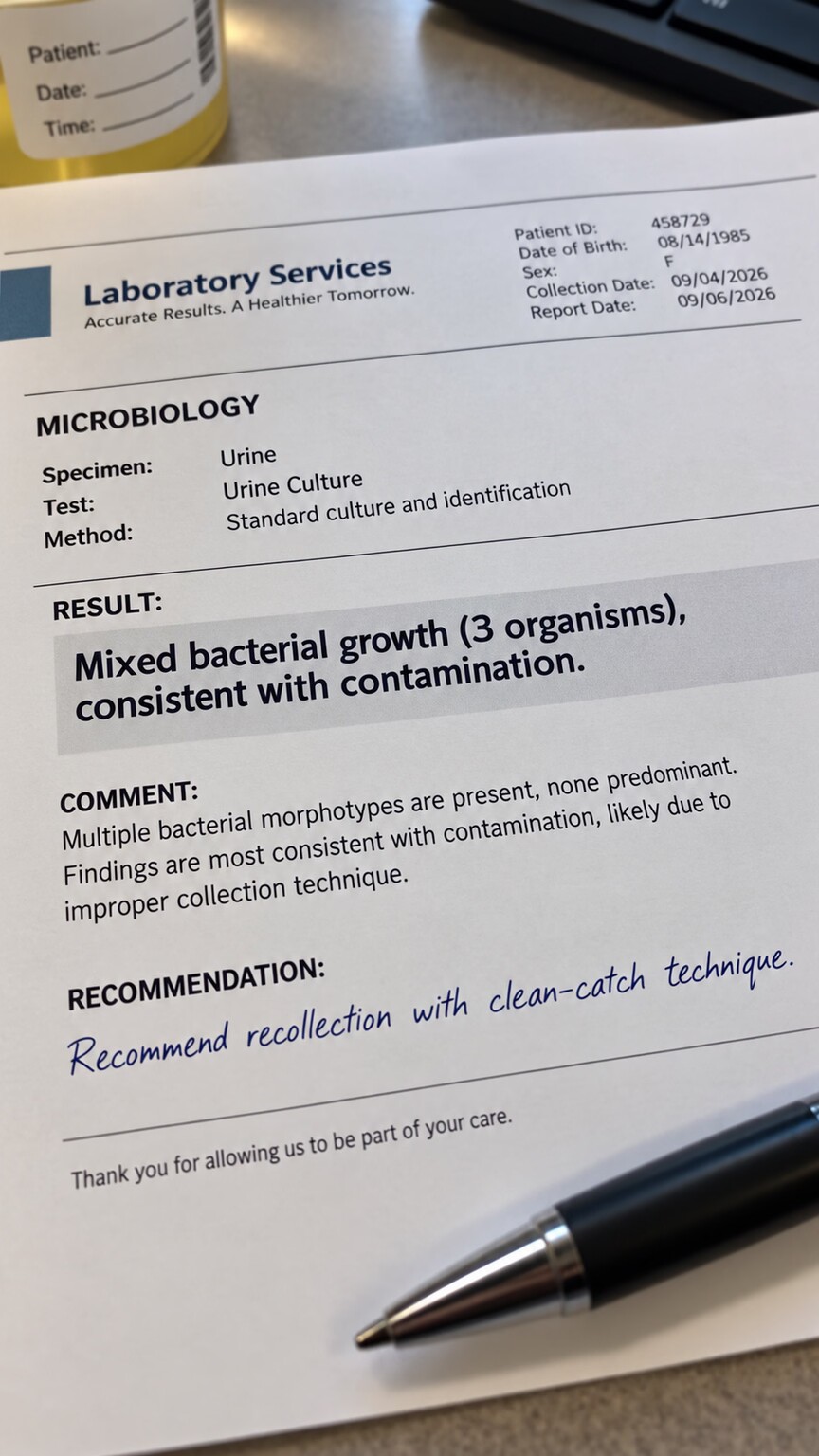
Figure 6. A mixed culture report showing multiple low-count organisms and a recommendation to recollect the sample is one of the most common and least concerning versions of this finding.
Consider a 32-year-old with mild urinary symptoms whose urine culture comes back showing three organisms, each at a colony count of around 10,000 per milliliter, well below the significant threshold. Her doctor reasonably interprets this as likely contamination from imperfect collection technique, provides clearer instructions for a clean-catch sample, and requests a repeat culture rather than starting antibiotics for any of the three organisms listed.
She follows the clearer, more detailed collection instructions carefully and precisely this time, and her repeat sample comes back entirely clean and unambiguous, growing a single organism at a genuinely significant colony count consistent with an actual urinary tract infection. The original mixed result is retrospectively confirmed as exactly what it always appeared to be — a simple collection artifact rather than any real, meaningful reflection of what was actually happening inside her urinary tract — and she's treated appropriately based on this second, considerably more reliable result instead.
Now consider a very different patient: someone hospitalized with a deep surgical wound infection following bowel surgery, whose wound culture shows two organisms — one aerobic, one anaerobic — both present at high colony counts consistent with active infection. Here, the mixed growth reflects a genuine, expected polymicrobial infection of exactly the kind that follows bowel surgery, and the treatment plan appropriately includes antibiotic coverage targeting both organisms together, since missing either one could allow the infection to persist despite treatment. Same laboratory language, "mixed growth," but two completely different clinical stories requiring two entirely different responses.
A third scenario helps round out this comparison: an elderly patient with a chronic, non-healing diabetic foot ulcer has a wound swab sent that grows four different organisms, each present at only modest colony counts. Unlike the first two scenarios, this one doesn't cleanly resolve into either "clearly contamination" or "clearly a genuine dominant infection" — chronic wounds are frequently colonized on their surface by a broad community of bacteria that may or may not be meaningfully driving active infection at any given moment. Here, the clinical picture surrounding the wound itself, rather than the culture result alone, does most of the real interpretive work: if the wound shows clear signs of worsening infection — increasing redness, warmth, drainage, or the patient developing fever — that same mixed result is treated as clinically significant and guides a broader antibiotic regimen. If the wound instead looks stable or slowly improving with routine wound care, that identical mixed culture result is far more likely interpreted as reflecting normal surface colonization rather than active infection requiring systemic antibiotics, illustrating once again that the same laboratory finding can mean genuinely different things depending on everything surrounding it.
Frequently Asked Questions
Does mixed bacterial growth always mean the sample was contaminated?
No. While contamination is the most common explanation, especially for urine samples, certain infections — particularly wounds, intra-abdominal infections, and aspiration pneumonia — genuinely and expectedly involve multiple bacterial species at once.
Why does my doctor want a repeat sample instead of treating the mixed growth?
When colony counts for each organism are low and the sample site isn't normally sterile, the pattern usually reflects contamination rather than infection. Treating contamination with antibiotics offers no benefit and contributes to antibiotic resistance.
Are polymicrobial infections harder to treat than single-organism infections?
Often, yes. They typically require antibiotic combinations covering every organism involved, and in synergistic infections where aerobic and anaerobic bacteria reinforce each other, missing coverage for either type can allow the infection to persist.
Why do labs treat mixed growth differently depending on where the sample came from?
Sites that are never sterile, like skin, throat, or non-invasively collected urine, are expected to sometimes show normal flora. Samples from normally sterile sites, like blood or deep wounds, are interpreted with much less tolerance for incidental bacteria.
What colony count is considered significant for a urine culture?
For a clean-catch sample, 100,000 colony-forming units per milliliter is the typical threshold used to distinguish a likely genuine infection from lower counts more consistent with contamination, though a lower threshold applies to catheter-collected samples.
Conclusion
A culture report showing mixed bacterial growth genuinely sits at a fork in the road, and figuring out which path applies depends on a handful of concrete clues: where the sample came from, how many organisms are present and at what colony counts, and whether the specific combination fits a recognized pattern of genuine polymicrobial infection. Most of the time, especially with urine samples, mixed growth reflects a collection issue easily solved with a repeat sample and better technique. When it does reflect a genuine infection involving multiple organisms, recognizing that pattern correctly is exactly what allows treatment to actually cover everything contributing to the problem.
If there's a single practical takeaway worth carrying forward from everything covered here, it's that the word "mixed" on a lab report should prompt a specific question rather than either automatic dismissal or automatic alarm: mixed compared to what expectation, from what body site, at what colony counts, and alongside what clinical picture? Answered honestly, that question almost always points clearly toward one of the two paths this article has laid out — and knowing which one applies is what actually determines whether the right next step is a repeat sample or a targeted course of treatment.
Still Not Sure What Your Results Mean?
Upload your labs and get a complete, visual, plain-language interpretation of every biomarker — delivered to your inbox in under 15 minutes.
Get My ReportThis article is for educational purposes only and does not constitute medical advice. Always consult your healthcare provider regarding your specific lab results.