
What Does Antibiotic Sensitivity Testing Actually Show?
Antibiotic sensitivity testing — also called a "culture and sensitivity" or "C&S" — is a lab process that takes the actual bacteria growing in your infection and exposes them, one by one, to a panel of real antibiotics to see which ones stop them cold and which ones do nothing at all. It's the difference between guessing and knowing. A doctor can prescribe an antibiotic based on what usually works for an infection like yours, but sensitivity testing tells them what will actually work for the specific bacteria living in your body, right now, today. If you've ever seen a lab report with a long list of drug names next to the letters S, I, or R, this article walks through exactly what those results mean, how the lab arrives at them, and why your doctor sometimes changes your prescription days into treatment based on what this test finds.
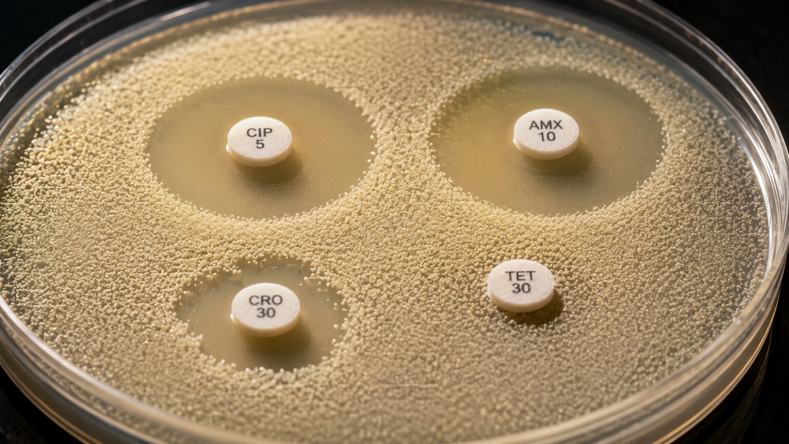
Figure 1. Zones of inhibition form where an antibiotic diffuses outward from a paper disk and stops bacterial growth; a clear ring means the drug worked, growth reaching the disk's edge means it didn't.
Curious what the rest of your lab results are telling you? Upload your results and get a complete, plain-language breakdown in under 15 minutes.
Analyze My ResultsWhy This Test Exists in the First Place
Not every antibiotic works on every bacterium, and even antibiotics that usually work on a certain type of infection can fail against a specific strain that happens to have picked up resistance. Think of bacteria the way you'd think of a lock, and antibiotics as keys. Most locks of a certain brand accept a certain key — that's why doctors can often start treatment with a "best guess" antibiotic before any lab results are back, based on which key usually fits which lock. But locks get re-cut over time. Bacteria evolve, swap genetic material, and pick up small mutations that change the shape of the proteins an antibiotic needs to grab onto. A key that opened a thousand identical locks can suddenly not turn in the one thousand-and-first, even though it looks exactly the same lock from the outside. Sensitivity testing exists because there is no way to look at an infection and simply know, with certainty, which keys still work on this particular lock — the only way to know is to try the keys and watch what happens.
This matters more than it might seem. Prescribing an antibiotic that doesn't actually work against the bacteria causing your infection doesn't just delay recovery — it gives the bacteria more time to multiply, spread, and potentially cause more serious illness, while also needlessly exposing your body to a drug's side effects for no benefit. On a larger scale, every time an ineffective antibiotic is used, it also nudges the overall bacterial population toward more resistance, since the bacteria that do survive that drug are the ones left to reproduce. Sensitivity testing is one of the main tools medicine has for keeping antibiotic use accurate and targeted, rather than a shot in the dark.
Where the Bacteria Actually Come From
Sensitivity testing never happens on its own — it's always the second half of a two-part process that starts with a culture. First, a sample is taken from wherever the suspected infection is: urine, blood, a wound swab, sputum coughed up from the lungs, a stool sample, or fluid drawn from a joint or the spine, depending on the symptoms. That sample gets placed onto a nutrient-rich gel called agar, inside a shallow round dish, and left in a warmed incubator, usually around human body temperature, for 18 to 24 hours. If bacteria are present in the sample, they multiply rapidly under those ideal conditions, forming visible clusters called colonies — each one technically a colony of millions of genetically identical bacterial cells that all grew from a single original cell.
Once colonies appear, a microbiologist examines their shape, color, and growth pattern, and often runs a handful of quick chemical or genetic identification tests to determine the exact species involved — is this E. coli, Staphylococcus aureus, Klebsiella pneumoniae, or one of hundreds of other possibilities? Only once the specific organism has been identified does the lab move on to sensitivity testing, because different bacterial species are tested against different antibiotic panels — there's no point testing a urinary bacterium against an antibiotic that's only ever used for skin infections, and some drugs aren't even relevant to certain organisms at all. This is also why sensitivity results almost never come back on day one: growing enough bacteria to test takes a full day by itself, and the sensitivity testing that follows typically takes another 16 to 24 hours on top of that.
Not All Infections Get Tested the Same Way
The exact antibiotics included on a sensitivity panel aren't a fixed universal list — they shift depending on both the bacterial species identified and where in the body the infection is located. A urine sample growing E. coli will typically be tested against drugs like nitrofurantoin or trimethoprim-sulfamethoxazole, which concentrate heavily in urine and work extremely well there even at doses that wouldn't reach useful levels elsewhere in the body. That same organism found instead in the bloodstream would prompt a different panel entirely, built around drugs proven to reach effective concentrations in blood and deep tissue, since a drug that works beautifully in urine can be nearly useless against a bloodstream infection if it simply doesn't circulate at high enough levels outside the urinary tract.
Location also changes which results actually matter clinically, even when the underlying lab process is identical. A skin and soft-tissue infection is judged primarily by whether a drug reaches the skin and superficial tissue in sufficient concentration, while an infection inside the fluid surrounding the brain and spinal cord — meningitis — depends on whether a drug can cross the blood-brain barrier at all, a filtering system that blocks many otherwise effective antibiotics from ever reaching that space in meaningful amounts. This is part of why microbiology labs don't hand back a single, one-size-fits-all "susceptible" or "resistant" verdict for a given organism; the same bacterium, with the exact same MIC value, can be interpreted differently by a physician depending on whether it was found in urine, blood, spinal fluid, or a wound, because "enough drug to work" means a different concentration in each of those places.
Method One: Disk Diffusion, the Classic "Zone of Clearing" Test
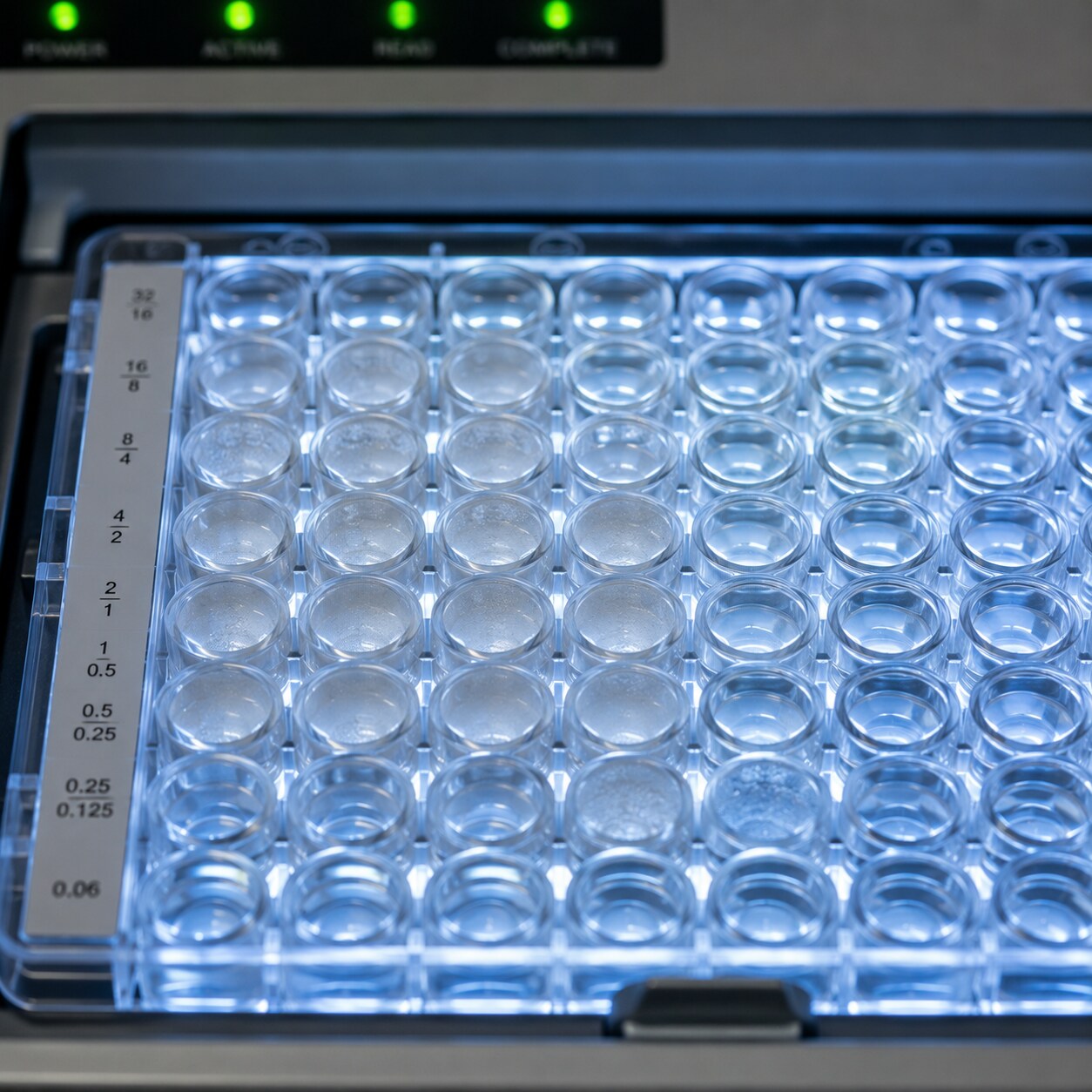
Figure 2. Automated broth microdilution systems test a single bacterial isolate against a grid of antibiotics at multiple concentrations simultaneously, reporting the minimum inhibitory concentration for each drug.
The oldest and most visually intuitive method is called disk diffusion, or the Kirby-Bauer method, named after the two scientists who standardized it in the 1960s. A technician takes a swab loaded with a precisely measured concentration of the identified bacteria and spreads it evenly across the entire surface of a fresh agar plate, so that if left alone, the bacteria would grow into one uniform, unbroken sheet covering the whole dish. Small paper disks, each pre-soaked with a specific antibiotic at a standardized dose, are then placed at even intervals across that plate — a single dish might carry a dozen or more different disks, each testing a different drug at once.
As the plate incubates, two things happen simultaneously: the bacteria begin multiplying across the agar surface, and each antibiotic disk begins releasing its drug outward into the gel in a shrinking gradient — highest concentration right next to the disk, progressively weaker the farther out it spreads. Wherever the antibiotic concentration is still strong enough to stop that particular strain of bacteria from growing, a clear, bacteria-free ring appears around the disk — this is the "zone of inhibition." Wherever the bacteria can tolerate the drug even at its strongest concentration near the disk, there's no clear ring at all — the bacterial lawn grows right up to the paper's edge as if the disk weren't even there. After incubation, the technician measures the diameter of each zone in millimeters with a ruler or an automated imaging camera. A bigger zone generally means the drug is more effective against that particular strain; a small or absent zone means the drug barely slowed the bacteria down, if at all.
Method Two: Minimum Inhibitory Concentration (MIC) Testing
Disk diffusion is fast and cheap, but it only tells you whether a drug worked or didn't at one fixed dose — it doesn't tell you exactly how much of the drug it actually took. For that, most modern labs also run, or exclusively run, a more precise method that determines the Minimum Inhibitory Concentration, or MIC: the lowest concentration of a given antibiotic that still manages to visibly stop the bacteria from growing. Picture a row of small wells or tubes, each containing the exact same bacterial sample but a different, progressively diluted concentration of the same antibiotic — full strength in the first well, half that in the next, a quarter of that in the one after, and so on, cut in half each time. After incubation, the lab checks each well for visible cloudiness, which signals bacterial growth. The MIC is the concentration in the very last clear well before the bacteria start growing again in the more dilute wells next to it — the lowest dose that still held the line.
Today this dilution process is almost always automated. Machines with names like VITEK, Phoenix, or MicroScan use small plastic cards or plates pre-loaded with dozens of tiny wells, each containing a different antibiotic at a different concentration, and an optical sensor reads bacterial growth in each well automatically over several hours, calculating the MIC for every drug on the panel at once. This is why a single sensitivity report can list 15 or 20 different antibiotics with results returned together, rather than the lab running each one separately by hand.
Why Standardization Keeps Results Trustworthy Across Different Labs
One detail that rarely gets mentioned but matters enormously is how tightly controlled every variable in this process has to be for the results to mean anything at all. The concentration of bacteria smeared onto the plate, the exact thickness and composition of the agar, the potency and diameter of each paper disk, the incubation temperature, and even the length of time before the zones are measured are all set to strict, published standards — largely by CLSI in the United States — specifically so that a result generated in a hospital lab in one state means the same thing as a result generated in a completely different lab a thousand miles away. If a lab used a slightly more concentrated bacterial suspension, or a slightly warmer incubator, zones of inhibition would measure differently even for the identical bacteria and identical drug, and a result that should read "Susceptible" could shift toward "Intermediate" or "Resistant" purely from a technical inconsistency that had nothing to do with the actual biology of the infection.
Because of this, accredited clinical microbiology labs run regular quality-control checks using bacterial reference strains with known, published, expected results — essentially a built-in answer key. If a control strain that should reliably produce a 22-millimeter zone around a particular antibiotic disk instead produces an 11-millimeter zone on a given day, that's a signal something in the process has drifted, and results from that batch get investigated before they're ever reported to a physician. This standardization is also exactly why the breakpoints discussed earlier can be trusted and applied nationally, rather than needing to be re-derived locally by every individual hospital lab — the whole system depends on every lab running the exact same test the exact same way, so that the same three-letter interpretation carries the same clinical meaning no matter where a patient happens to be treated.
How a Number Becomes a Verdict: Susceptible, Intermediate, and Resistant
An MIC value on its own — say, an MIC of 2 micrograms per milliliter for a particular drug — doesn't mean anything to most readers without context. What makes it useful is that researchers have already determined, through decades of clinical studies, roughly how much of each antibiotic the human body can actually deliver to an infection site when the drug is dosed normally and safely. Comparing the bacteria's MIC against that achievable drug level is what produces the three-letter interpretation that actually appears on your report:
Susceptible (S) means the concentration of antibiotic that a standard, safe dose can realistically achieve in the body is comfortably higher than what it took to stop this particular bacterial strain in the lab. In plain terms: at a normal dose, this drug should work.
Intermediate (I) — sometimes labeled "Susceptible-Dose Dependent" on newer reports — means the bacteria required a higher concentration to stop than the lowest end of what's typically achievable, but a drug might still work if it's dosed more aggressively, given more frequently, or used at a site in the body (like urine, where many antibiotics concentrate heavily) where higher local levels are easier to reach than they are in the bloodstream.
Resistant (R) means the bacteria kept growing even at concentrations well beyond what could be safely delivered to a patient. Giving this drug wouldn't meaningfully suppress the infection no matter how the dose is adjusted, and in some cases could do more harm than good by delaying effective treatment while side effects accumulate for nothing.
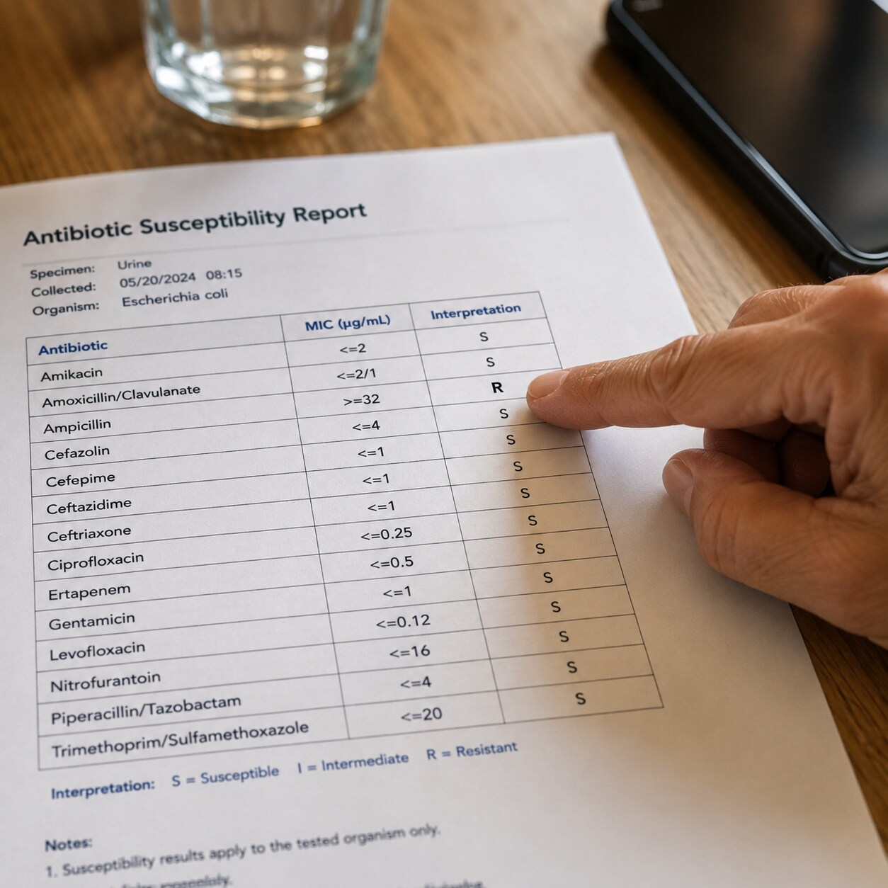
Figure 3. A susceptibility report lists each antibiotic tested alongside its interpretation — Susceptible, Intermediate, or Resistant — which the treating physician matches against the specific infection being treated.
These cutoff points — called breakpoints — aren't arbitrary, and they aren't permanent. They're set and periodically revised by expert panels, most notably the Clinical and Laboratory Standards Institute (CLSI) in the United States and the European Committee on Antimicrobial Susceptibility Testing (EUCAST) internationally, using enormous datasets that combine lab MIC values with real-world data on how patients with those MICs actually responded to treatment. When resistance patterns shift across a population, or when new research clarifies how a drug behaves in the body, these organizations can and do move the breakpoints — meaning the exact same MIC value for the exact same bacteria could theoretically be labeled "Susceptible" one year and "Intermediate" a few years later, purely because scientists learned more about what that number actually means for real patients, not because the bacteria itself changed.
Why Your Doctor Didn't Wait for This Result Before Starting Treatment
If you've ever started antibiotics the same day you saw a doctor, but the sensitivity report didn't come back for two or three days, you've already experienced the practical tension this test creates. Infections, especially serious ones, don't wait politely for a 48-hour lab process to finish — untreated bacterial infections can worsen significantly in that window, so doctors typically start what's called empiric therapy immediately: an antibiotic chosen based on the most statistically likely bacteria for that type of infection and the general local resistance patterns in that hospital or region, sometimes summarized in a reference document called an antibiogram that hospitals update every year from their own lab data.
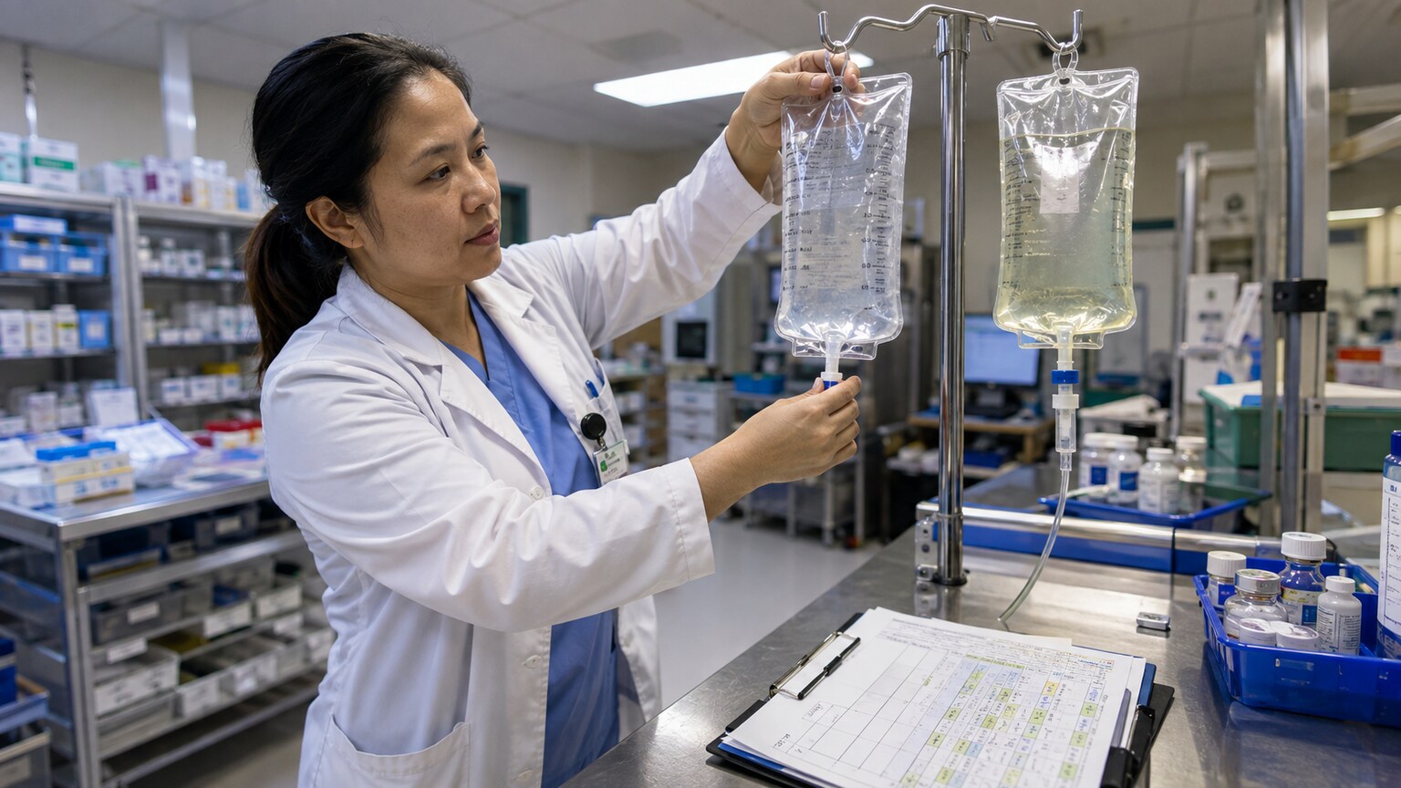
Figure 4. Once susceptibility results return, clinicians often switch a patient from a broad empiric antibiotic to a narrower, targeted drug shown to work against the specific organism identified.
Once the sensitivity results come back and reveal exactly which bacteria are present and which drugs actually work against them, doctors often "de-escalate" — switching from a broad-spectrum antibiotic that covers many possible bacteria at once (and disrupts a lot of the harmless bacteria in the body along the way) to a narrower drug specifically shown to work against the confirmed organism. This isn't a sign that anything went wrong with the initial treatment; it's the intended, deliberate design of the whole process. Broad-spectrum drugs buy time safely while the lab works, and the sensitivity result then lets the treatment get sharper, often with fewer side effects, a narrower disruption to the body's normal bacterial flora, and less pressure pushing other bacteria in the body toward resistance.
What It Means When the Report Shows Resistance to Several Drugs
Seeing multiple "R" results on a report can be alarming to read, but it's important to understand what the lab is actually testing for. Most sensitivity panels include a broad range of drug classes specifically so that if common first-line options fail, the lab has already identified backup options further down the list — the panel is built defensively, expecting some resistance, not as a worst-case scenario. A report showing three or four antibiotics marked "Resistant" alongside several others marked "Susceptible" isn't a crisis; it's the test doing exactly its job, steering treatment away from drugs that won't work and toward ones that will.
What genuinely does concern clinicians is when an organism shows resistance to nearly everything on a standard panel, sometimes called multidrug-resistant (MDR), extensively drug-resistant (XDR), or, in the rarest and most serious cases, pandrug-resistant. When this happens, labs often expand testing to include reserve antibiotics that aren't used routinely — partly because they're more toxic or expensive, and partly to preserve their effectiveness for exactly these situations by not overusing them on infections that respond fine to standard drugs. Infectious disease specialists frequently get involved directly in these cases, since choosing among a shrinking list of options requires balancing effectiveness against side effects and dosing complexity that goes beyond routine care.
Names You've Probably Heard: MRSA, ESBL, and VRE
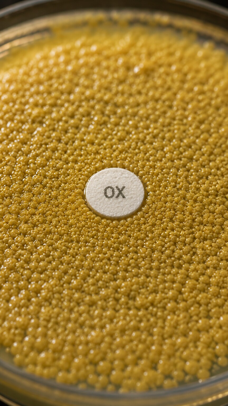
Figure 5. Staphylococcus aureus colonies showing no zone of clearance around an oxacillin disk, the classic laboratory signature of methicillin-resistant Staphylococcus aureus (MRSA).
Certain resistance patterns come up often enough in everyday medicine that they've earned their own shorthand names, and sensitivity testing is exactly how each one gets identified. MRSA, or methicillin-resistant Staphylococcus aureus, describes a strain of a very common skin and soft-tissue bacterium that has acquired a gene allowing it to resist an entire class of antibiotics related to penicillin, including drugs like oxacillin and methicillin itself. Because that resistance gene alters the very protein those drugs are designed to bind to, no amount of extra dosing helps — the lock has genuinely changed shape, not just gotten harder to pick. Labs specifically test for this using an oxacillin or cefoxitin disk, and a strain that shows no zone of inhibition around it is flagged as MRSA, which then guides the choice toward entirely different drug classes, such as vancomycin or newer alternatives.
ESBL, short for Extended-Spectrum Beta-Lactamase, refers to bacteria — often E. coli or Klebsiella species commonly involved in urinary tract infections — that produce an enzyme capable of chemically breaking apart a wide range of penicillin- and cephalosporin-family antibiotics before those drugs ever get the chance to act. Sensitivity testing catches this because the bacteria will show resistance across an entire pattern of related drugs rather than just one, which is itself a recognizable signature that prompts the lab to run confirmatory tests for the enzyme specifically. VRE, or vancomycin-resistant Enterococcus, works similarly, describing a strain that has become resistant to vancomycin — often used as a reliable backup drug for other resistant infections — which narrows the safe treatment options further still. In every one of these cases, the abbreviation isn't a separate diagnosis from the sensitivity test; it's simply a well-known nickname for a specific, recognizable resistance pattern the test reveals.
How This Test Fits Into the Bigger Picture of Antibiotic Resistance
Every individual sensitivity report is also, quietly, a small data point in a much larger public health picture. Hospitals compile thousands of these individual results over the course of a year into the antibiogram mentioned earlier — a running summary of what percentage of a given bacterial species tested susceptible to each major antibiotic at that specific facility. Public health agencies do something similar at a regional and national level, tracking how resistance patterns shift over time across entire populations. This is how the medical community first notices when a resistant strain is spreading rather than appearing in isolated, unconnected cases — a gradual year-over-year decline in the percentage of a bacterium testing susceptible to a once-reliable drug, visible only once enough individual sensitivity results are pooled together.
This is also why sensitivity testing and antibiotic stewardship — the deliberate effort to use antibiotics only when needed and to choose the narrowest effective option — are closely linked in modern hospital practice. Every time a broad-spectrum antibiotic is used when a narrower one would have worked just as well, it adds a small amount of selective pressure that favors resistant bacteria surviving and multiplying, not just in the person being treated but potentially in the wider bacterial population they come into contact with. Sensitivity results give doctors the specific evidence needed to de-escalate to a narrower drug with confidence, which is one of the most concrete tools medicine has for slowing the pace at which resistance spreads, one individual treatment decision at a time.
What Sensitivity Testing Does Not Tell You
It's worth being clear about the limits of this test, because it's easy to read more into a report than it actually says. A sensitivity result tells you how the bacteria behaved against a drug in a lab dish, under standardized, ideal conditions — it does not directly measure how your particular body will absorb, distribute, or clear that drug, and it doesn't account for other health conditions, allergies, drug interactions, kidney or liver function, or pregnancy, all of which a physician still has to weigh separately when actually choosing your prescription. A drug marked "Susceptible" in the lab is a strong, evidence-based signal that it should work — not an unconditional guarantee, since factors like how deeply an infection has penetrated into tissue, whether it involves a hard-to-reach location like bone or a medical implant, or whether the patient's immune system is compromised can all still affect real-world outcomes even when the lab result looks favorable.
One specific limitation worth understanding on its own is biofilms — thin, slimy layers that some bacteria build around themselves when they colonize a surface like a catheter, an artificial joint, a heart valve, or even a wound bed. Bacteria growing loosely in a lab dish behave very differently from that same species once it has organized itself into a biofilm, which can physically shield the bacteria from an antibiotic that would otherwise kill them easily in open culture. This is a major reason why infections tied to implanted medical devices are notoriously difficult to clear with antibiotics alone, even when a straightforward sensitivity report shows the organism as broadly susceptible — sometimes the device or affected tissue genuinely has to be removed or surgically addressed before antibiotics, however well-matched, can finish the job.
It's also worth remembering that this test only reflects the bacteria that were actually present in the sample and grew successfully in culture. If antibiotics were already started before the sample was collected, or if the sample wasn't handled or transported correctly, bacteria that were genuinely present in the infection can sometimes fail to grow, leading to a "no growth" result that doesn't necessarily mean there's no infection — it can also mean the test simply couldn't capture what's really there. This is one of several reasons doctors interpret culture and sensitivity results alongside your symptoms, exam findings, and other labs, rather than treating the report as the single, standalone source of truth.
How to Actually Read One of These Reports Yourself
If you request a copy of your own culture and sensitivity results, the layout is usually more approachable than it first looks once you know what each column represents. Near the top, you'll typically find the identified organism spelled out by name — something like "Escherichia coli" or "Staphylococcus aureus" — along with the specimen source, such as "urine" or "wound swab," and sometimes a rough colony count indicating how much bacteria grew. Below that sits the main table: one row per antibiotic tested, usually with two columns next to each drug name — an MIC value (a number, often followed by a comparison symbol like "<=" or ">=") and an interpretation column showing S, I, or R.
A practical way to scan it: start with the interpretation column rather than the raw MIC numbers, since those letters are already the finished, physician-usable conclusion — the MIC values exist mainly for clinicians who want the underlying precision, not something most readers need to personally calculate against a breakpoint table. Notice which drugs are marked "S," since those are the options your doctor is most likely already weighing, and don't be alarmed by a long list of "R" results elsewhere on the same report, since panels deliberately include many drugs precisely so that some will show resistance without leaving the list of workable options empty. If anything on the report is confusing or seems to conflict with what you've been prescribed, that's a reasonable, specific question to bring directly to your doctor or pharmacist — they're reading the exact same three letters you are, just with the added clinical context of your full medical picture layered on top.
Frequently Asked Questions
How long does it take to get antibiotic sensitivity results back?
Most conventional culture and sensitivity testing takes about 48 to 72 hours from when the sample is collected — roughly a day to grow enough bacteria to identify the species, then another day or so to run the sensitivity panel itself. Some hospitals now use faster molecular or automated methods that can shorten parts of this timeline, but the classic combined process still generally spans two to three days.
Why did my doctor start antibiotics before the sensitivity results came back?
This is standard practice, called empiric therapy. Waiting two to three days to start treatment could allow a genuine infection to worsen significantly, so doctors choose a broad-spectrum antibiotic likely to work based on the type of infection and local resistance patterns, then narrow or adjust that choice once the specific sensitivity results are available.
Does "Resistant" on a report mean the infection can't be treated?
No. A "Resistant" result next to one antibiotic simply means that particular drug won't work — it doesn't mean nothing will. Sensitivity panels are deliberately designed to test many drug options at once specifically so that if some show resistance, others on the same list are still available and effective.
Can sensitivity results change if I get tested again later?
Yes. Sensitivity reflects the specific bacterial sample tested at that moment, and results can differ on a repeat test — either because the infection is caused by a different strain the second time, because the bacteria acquired new resistance in between, or occasionally because the original infection has genuinely cleared and a new one has developed.
Is antibiotic sensitivity testing the same thing as a resistance gene test?
No, though the two are related. Traditional sensitivity testing is a functional test — it actually grows the bacteria and exposes them to real antibiotics to see what happens. Genetic resistance tests, often run by PCR, instead look for the presence of specific known resistance genes in the bacteria's DNA. Genetic tests can return results faster, but they only detect resistance mechanisms that are already known and specifically being searched for, while functional sensitivity testing captures the bacteria's actual real-world behavior against a drug, including resistance mechanisms that haven't been identified at the genetic level yet.
Can bacteria be resistant to an antibiotic they were never exposed to before?
Yes. This is called intrinsic resistance, and it's different from acquired resistance. Some bacterial species are naturally, permanently resistant to entire classes of antibiotics simply because of their basic cell structure — for example, certain drugs can't penetrate the outer membrane that some bacteria are built with, regardless of whether that particular strain has ever encountered the drug before. This is one reason sensitivity panels are tailored to the bacterial species identified rather than testing every organism against every antibiotic that exists, since including a drug the species can never respond to would only add clutter without adding any useful information to the report.
Conclusion
Antibiotic sensitivity testing takes the guesswork out of one of medicine's most consequential decisions: which drug will actually beat this specific infection. By growing the exact bacteria involved and testing them directly against a panel of real antibiotics, the lab can tell your doctor — with real evidence, not statistical probability — which drugs will work, which won't, and which sit somewhere in between. Understanding what those S, I, and R letters actually represent, and why your treatment might shift once they arrive, turns what can feel like a confusing lab report into a clear, logical story about how modern infection treatment is actually built to work.
It's also a good reminder of just how much careful, standardized science sits quietly behind a single prescription. Every disk placed on an agar plate, every automated microplate reading a color change in real time, and every breakpoint table revised by an international panel of experts exists for one practical reason: so that the antibiotic you're handed at the pharmacy has the best possible chance of actually working against the exact bacteria making you sick, on the first try, rather than the second or third. The next time a lab report lands in your inbox with a column of unfamiliar drug names next to the letters S, I, and R, you'll have a concrete picture of everything that happened behind the scenes to produce that single line of text — a swab, a warm incubator, a ring of cleared bacteria, and a careful comparison against decades of accumulated clinical evidence, all working together to point your treatment in the right direction.
Still Not Sure What Your Results Mean?
Upload your labs and get a complete, visual, plain-language interpretation of every biomarker — delivered to your inbox in under 15 minutes.
Get My ReportThis article is for educational purposes only and does not constitute medical advice. Always consult your healthcare provider regarding your specific lab results.