
What Does a KOH Prep Test Actually Show?
A KOH prep is one of the fastest tests in all of medicine — a doctor can have an answer in under ten minutes, right there in the exam room, without sending anything to an outside lab. Here's the trick behind it: potassium hydroxide, the "KOH" in the name, is a strong alkaline chemical that dissolves human skin cells, keratin, and hair proteins almost completely, but it barely touches the tough, chitin-reinforced cell walls of fungi. So when a technician drips it onto a scraping of skin, nail, or hair and looks under a microscope, everything human in the sample essentially melts away into a clear, faint background — and if a fungus is present, its branching threads or round budding cells are left standing out sharply, almost like the sample was designed to disappear around them. What a KOH prep actually shows, in the end, is a yes-or-no answer to a very specific question: is there fungal material physically present in this sample, visible right now, under this microscope. It does not tell you which fungus, how it got there, or how to treat it — and understanding exactly where that line sits is what actually matters once you're the one waiting on the result.
Curious what the rest of your lab results are really telling you? Upload them and get a complete, plain-language breakdown in under 15 minutes.
Analyze My ResultsThe Test, Step by Step
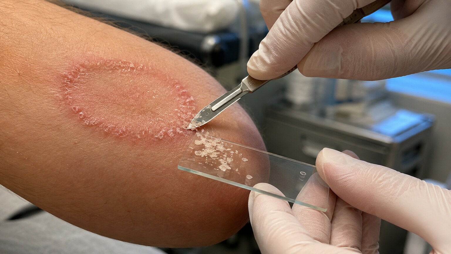
Figure 1. A KOH prep begins with a physical scraping of the sample — skin scale, nail clippings, or plucked hair — taken specifically from the active, leading edge of a lesion, where fungal growth tends to be most concentrated.
The process is refreshingly low-tech compared to most lab testing. A clinician uses the dull edge of a scalpel blade, a curette, or sometimes the edge of a glass slide itself to gently scrape the surface of the affected skin, nail, or scalp. The goal is to collect flakes of the outermost skin layer — the stratum corneum, a layer made almost entirely of dead, keratin-packed cells — because that's the layer fungi actually colonize when they cause conditions like athlete's foot, ringworm, or nail fungus. Where the sample is taken from matters enormously: scraping from the center of a healed-looking patch often comes back falsely negative, because the fungus has typically already moved outward to keep feeding on fresh keratin, leaving the center relatively fungus-free. That's why a clinician who knows what they're doing scrapes from the active, often slightly raised or scaly border of a lesion, not its middle.
Once the scale is collected, it's transferred onto a glass microscope slide and a drop or two of potassium hydroxide solution — typically 10% to 20% concentration — is placed directly on top, followed by a glass coverslip pressed gently down over it. At this point, the slide looks like nothing at all: a smear of skin flakes suspended in a small pool of clear liquid. The chemistry that makes the test work hasn't happened yet — it needs a few minutes, and often a little heat, to do its job.
The amount of sample matters more than people assume. Too little scale on the slide, and even a real infection may not have enough fungal material present to guarantee that any of it ends up directly under the coverslip where the microscope is actually pointed — a fungus can be genuinely present in the affected skin while still being physically absent from the specific few square millimeters being examined, purely by chance. This is one of the less obvious reasons two samples taken from the same rash, just a few minutes apart, can occasionally give different results: not because the fungus wasn't there, but because of how unevenly it can be distributed across even a small area of visibly affected skin.
Why Potassium Hydroxide Dissolves Everything Except the Fungus
To understand what a KOH prep is really showing you, it helps to understand why the chemical works the way it does. Potassium hydroxide is a strong base — the same broad chemical family as lye — and strong bases are excellent at breaking down keratin, the fibrous structural protein that makes up the bulk of skin cells, hair, and nails. Keratin is held together by a dense network of disulfide bonds, chemical crosslinks that give it its toughness. KOH attacks and breaks those bonds, causing the keratin structure to swell, soften, and eventually dissolve into a translucent, disorganized mass. Under the microscope, this looks like a faint, cobweb-like haze — the ghost of what used to be organized human skin cells.
Fungal cells, by contrast, aren't built out of keratin at all. Their cell walls are made primarily of chitin — the same structural molecule found in insect exoskeletons and crab shells — reinforced with glucans and other polysaccharides that form an extremely tough, chemically resistant lattice. Potassium hydroxide has almost no effect on chitin at even moderate concentrations. So while the human cells around a fungal invader are actively dissolving into a translucent smear, the fungal structures embedded within that same smear stay essentially unchanged — intact, sharply defined, and now standing out in high contrast against the fading human background. This selective dissolving is the entire mechanism behind the test, and it's why gentle heating (holding the slide briefly over a low flame or using a slide warmer) is often used: heat speeds up the keratin-dissolving reaction without damaging the fungal cell walls, cutting the wait time from twenty or thirty minutes down to just a few.
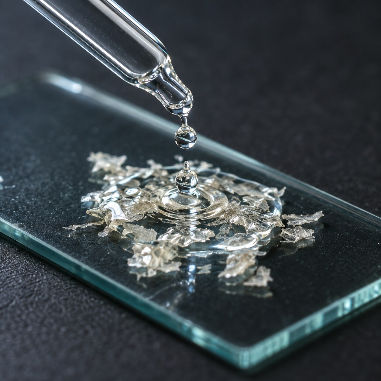
Figure 2. Potassium hydroxide is applied directly onto the collected skin, nail, or hair sample on the slide, where it will spend the next several minutes selectively breaking down keratin.
Some labs add a second ingredient to this same droplet: a blue or black chitin-binding stain, most commonly chlorazol black E or a fungal-specific fluorescent brightener like calcofluor white. These stains bind preferentially to chitin, meaning they latch onto fungal cell walls far more than onto anything else left in the sample. Under an ordinary light microscope, a chlorazol-black-stained fungal element appears distinctly blue-gray against the pale, dissolving background. Under a fluorescence microscope, calcofluor white causes fungal walls to glow a vivid blue-white when exposed to ultraviolet light — a striking, almost unmistakable signal that dramatically reduces how often a technician can miss a small or sparse fungal element sitting in a busy, cell-debris-filled field of view. Plain KOH without a stain still works and is still the most common version performed in a typical clinic, but the stained version is meaningfully more sensitive, particularly in the hands of someone reading fewer of these slides in a given week.
The concentration of the KOH solution itself is a deliberate balance. Too weak, and the keratin won't dissolve fast enough to give a usable view within a reasonable window. Too strong, and the solution can start forming crystalline artifacts as it evaporates slightly at the edges of the coverslip — tiny needle-like or granular structures that inexperienced readers sometimes mistake for fungal hyphae, a recognized source of false positives. This is one of several reasons the test, despite its apparent simplicity, genuinely benefits from being read by someone with real microscopy experience rather than treated as a fully mechanical, error-proof process.
What the Technician Is Actually Looking For Under the Microscope
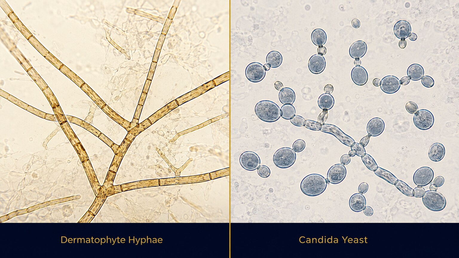
Figure 3. Under the microscope, dermatophyte fungi appear as long, branching, thread-like hyphae divided by internal walls called septa, while Candida yeast appears as small round or oval cells, often caught mid-bud.
Once the keratin has cleared enough to see through, the technician scans the slide methodically, moving the field of view in a grid pattern across the entire coverslip area rather than just glancing at the center — fungal elements are often sparse and unevenly distributed, so a careless scan is one of the most common reasons a true infection gets missed. What they're specifically hunting for falls into two broad visual categories, and which one they find (or don't find) carries real diagnostic weight.
The first category is hyphae — long, thread-like structures that branch off from each other at angles, often divided at intervals by thin internal cross-walls called septa. This branching, septate, thread-like appearance is the classic signature of dermatophytes, the group of fungi responsible for ringworm, athlete's foot, jock itch, and most nail fungus infections. A technician who spots true septate hyphae crossing multiple adjacent skin cells, especially ones that appear to be actively growing through and along cell boundaries rather than just sitting on top of debris, is looking at a strong positive.
The second category is yeast forms — small, round or oval cells, frequently seen in the act of budding, where a smaller daughter cell appears to be pinching off from a slightly larger parent cell like a tiny bubble forming on the side of a balloon. This appearance points toward Candida species rather than a dermatophyte, and it often comes paired with pseudohyphae — chains of elongated yeast cells that look almost, but not quite, like true branching hyphae, strung together end to end rather than branching at true right angles. The distinction matters clinically, because dermatophyte infections and Candida infections, while they can look similar on skin, sometimes respond to different antifungal medications and different treatment durations.
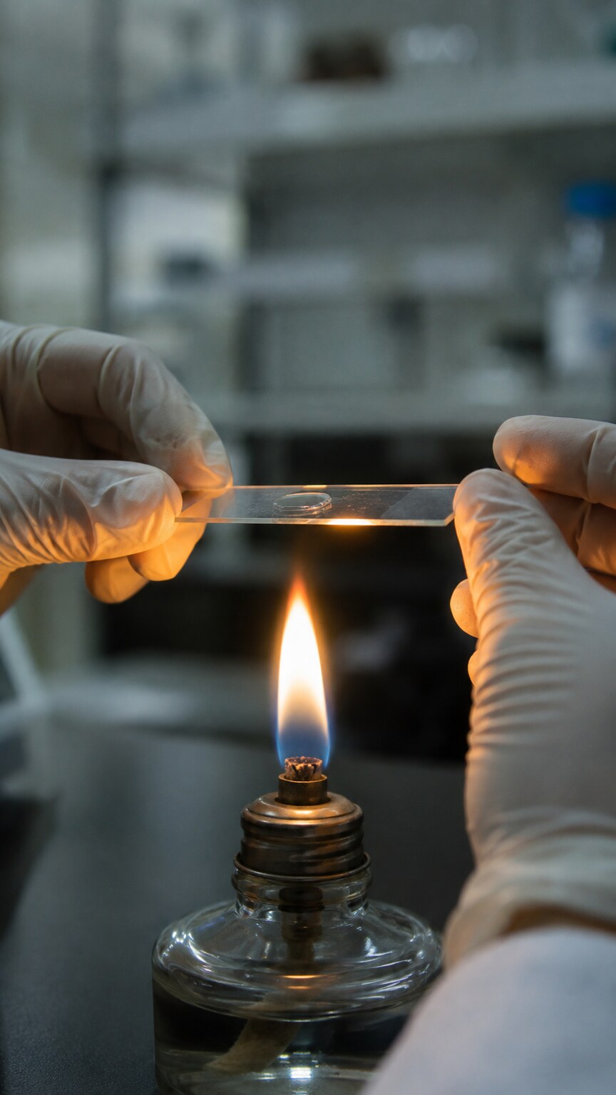
Figure 4. Gently warming the slide speeds up how quickly potassium hydroxide breaks down keratin, shortening the wait from roughly thirty minutes down to just a few.
Reading a KOH prep well is a genuine skill, not a box-checking exercise, and this is one of the test's most underappreciated limitations. Studies comparing KOH prep interpretation across different examiners have found meaningful variation in sensitivity depending on the reader's experience — an experienced dermatologist or trained lab technician who reads dozens of these slides a month will reliably catch fungal elements that a less experienced reader, looking at the exact same physical slide, would miss entirely. Air bubbles trapped under the coverslip, fibers from clothing or gauze that contaminated the sample, cell wall fragments, and even certain crystallized artifacts from the KOH solution itself can all superficially resemble hyphae to an untrained or rushed eye, and distinguishing real fungal structures from these mimics is exactly the kind of pattern recognition that comes from repetition, not instruction alone.
The condition of the sample also affects what's visible. A scraping that's too thick, with several overlapping layers of skin cells stacked on top of each other, can trap fungal elements in a dense, poorly-cleared region where they're nearly impossible to distinguish from surrounding debris, even after the keratin has had time to dissolve. This is why a well-performed test uses a thin, even scraping rather than a thick clump — more sample isn't automatically better if it's too dense to see through.
What a Positive Result Can — and Can't — Tell You
A positive KOH prep is a genuinely useful, actionable finding, but it's worth being precise about exactly what it confirms and what it leaves open. What it confirms, with reasonable confidence, is that fungal material — a real organism's cell walls, not a lookalike artifact — is physically present in the sample taken from that specific site, at that specific moment. In someone with a scaly, itchy, ring-shaped rash or a thickened, discolored nail, a positive KOH prep is usually enough on its own for a clinician to start antifungal treatment immediately, without waiting on anything further.
What a positive result does not reliably tell you is which specific species of fungus is responsible. A KOH prep can distinguish the broad category — septate branching hyphae suggesting a dermatophyte, versus budding yeast and pseudohyphae suggesting a Candida species — but it cannot identify whether a dermatophyte infection is specifically Trichophyton rubrum, Trichophyton mentagrophytes, Microsporum canis, or one of several dozen other dermatophyte species that all look essentially identical as thin branching threads under a microscope. For most everyday skin and nail infections, this species-level distinction doesn't change first-line treatment, which is part of why the KOH prep is so widely used as a fast, practical first step rather than something that always needs to be followed by more testing. But in cases that don't respond to standard treatment, involve an unusual location, or occur in someone who is immunocompromised, that missing species-level detail becomes clinically important, and a fungal culture or molecular test becomes the next step specifically because the KOH prep was never designed to provide it.
Why a KOH Prep Can Come Back Negative Even With a Real Infection
A negative KOH prep is considerably less definitive than a positive one, and this asymmetry trips a lot of people up. Sensitivity estimates for KOH preps performed and read under typical real-world conditions range widely in published research, commonly cited somewhere between roughly 40% and 90% depending on the body site, the skill of the person performing and reading it, and how the sample was collected — meaning a meaningful fraction of true fungal infections can produce a falsely negative result on any single test. Several practical factors drive this gap.
Sample site error is the single biggest one. As mentioned earlier, scraping from the center of a lesion instead of its active, expanding edge is one of the most common technical mistakes, and it alone can turn a genuinely fungal infection into a falsely negative slide. Recent use of an antifungal cream, powder, or even certain moisturizers before the sample was taken can also suppress or physically wash away enough fungal material to drop it below what's visible under the microscope, even if the underlying infection hasn't actually cleared — this is one of the most frequent reasons a clinician will specifically ask, before scraping, whether anything has been applied to the area recently. Nail samples add their own layer of difficulty: fungal elements in a thickened, damaged nail are often unevenly distributed through the nail plate, concentrated in certain layers or pockets rather than spread evenly throughout, so a scraping that happens to miss that specific pocket can come back clean despite a real infection sitting just beneath or beside the sampled spot.
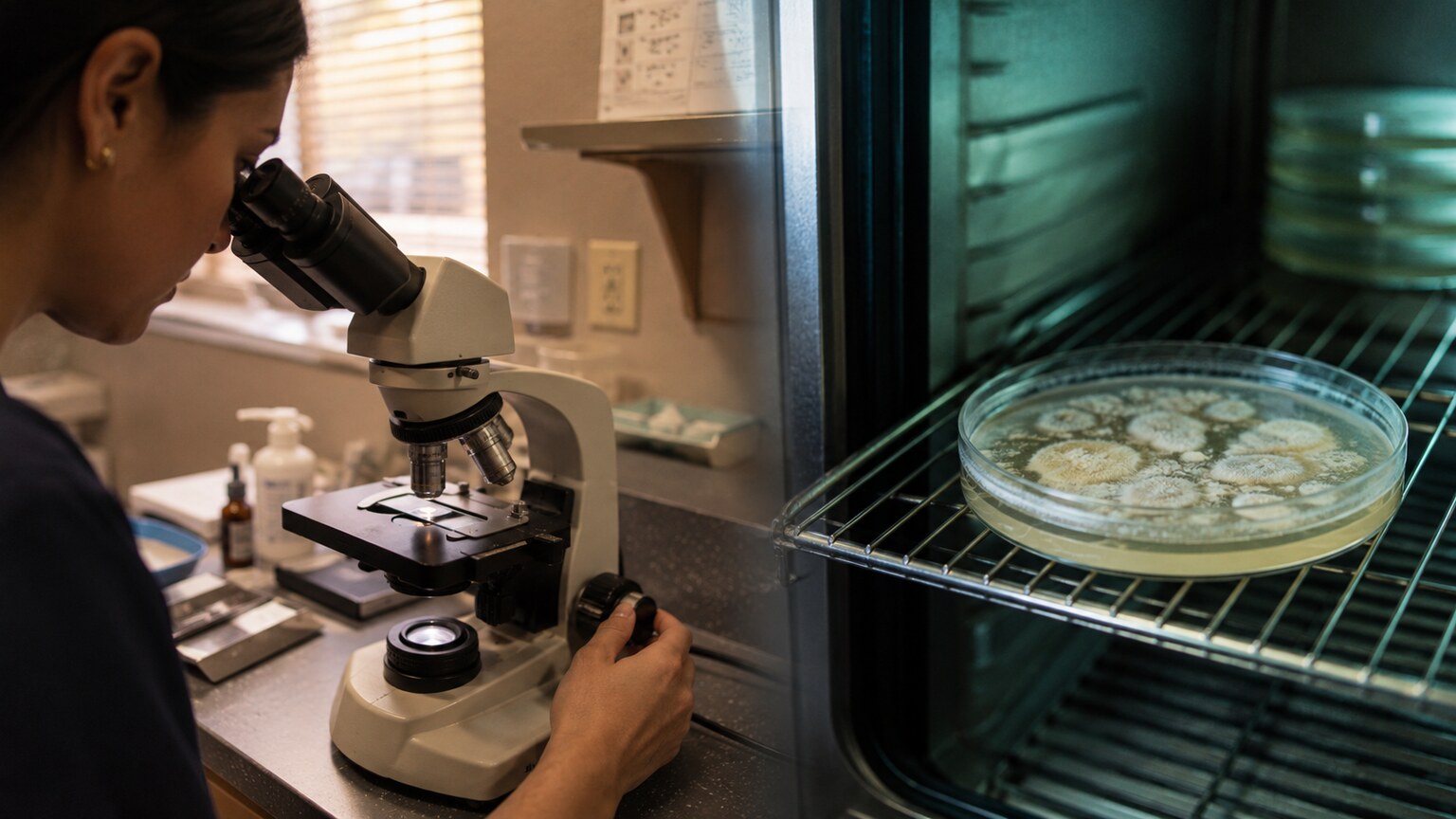
Figure 5. A KOH prep is read in minutes at the point of care, while a fungal culture is incubated for one to several weeks before a species-level identification is possible.
Because of this real gap between test sensitivity and biological reality, a clinician who strongly suspects a fungal infection despite a negative KOH prep — based on the appearance, location, and history of the rash or nail change — will often either repeat the scraping from a different or better spot, or move straight to sending a sample for fungal culture, which grows the organism over one to several weeks and can catch infections a single KOH prep missed, precisely because culture doesn't depend on a small enough population of fungal cells landing directly under the coverslip at the moment of reading. A single negative KOH prep is a data point, not a final verdict, especially when the clinical picture strongly suggests fungus.
KOH Prep vs. Fungal Culture vs. Molecular Testing
The KOH prep's real role in modern practice is best understood next to the other tools available for the same basic question, because each one trades speed for detail in a different way. The KOH prep itself is fast — minutes, not days — cheap, and can be performed and interpreted in the exam room without sending anything out, which makes it the default first step for a classic-looking rash, scaly patch, or discolored nail where the clinical picture is already fairly convincing. Its tradeoff is exactly what's been laid out above: no species identification, and a real chance of a false negative depending on technique and sample site.
Fungal culture takes the opposite tradeoff. A portion of the sample is placed onto a specialized growth medium and incubated, typically at room temperature, for anywhere from one to several weeks, giving any fungal organisms present time to multiply into a visible colony that can then be identified down to the species level based on its growth pattern, colony appearance, and microscopic structures. This extra detail matters most when first-line treatment has already failed, when the infection is in an unusual location, or when the patient's immune status means getting the exact organism right actually changes the treatment plan. The tradeoff is the wait — a culture-based diagnosis and treatment start can take weeks longer than a same-day KOH prep, which is a real cost when someone is uncomfortable or the infection is spreading.
Newer molecular tests, based on PCR technology that detects a fungus's genetic material directly rather than waiting for it to grow, sit in between the two on the speed-versus-detail spectrum — often returning species-level results within a day or two rather than weeks, with sensitivity that in several published comparisons exceeds both KOH prep and traditional culture. These tests are becoming more available, particularly for nail fungus, but they're generally more expensive and not yet universally offered in every clinic or covered by every insurance plan the way a simple in-office KOH prep is, which is part of why the decades-old KOH prep remains the practical first step in the vast majority of everyday cases despite these newer, more detailed alternatives existing.
Where Samples Actually Get Taken From
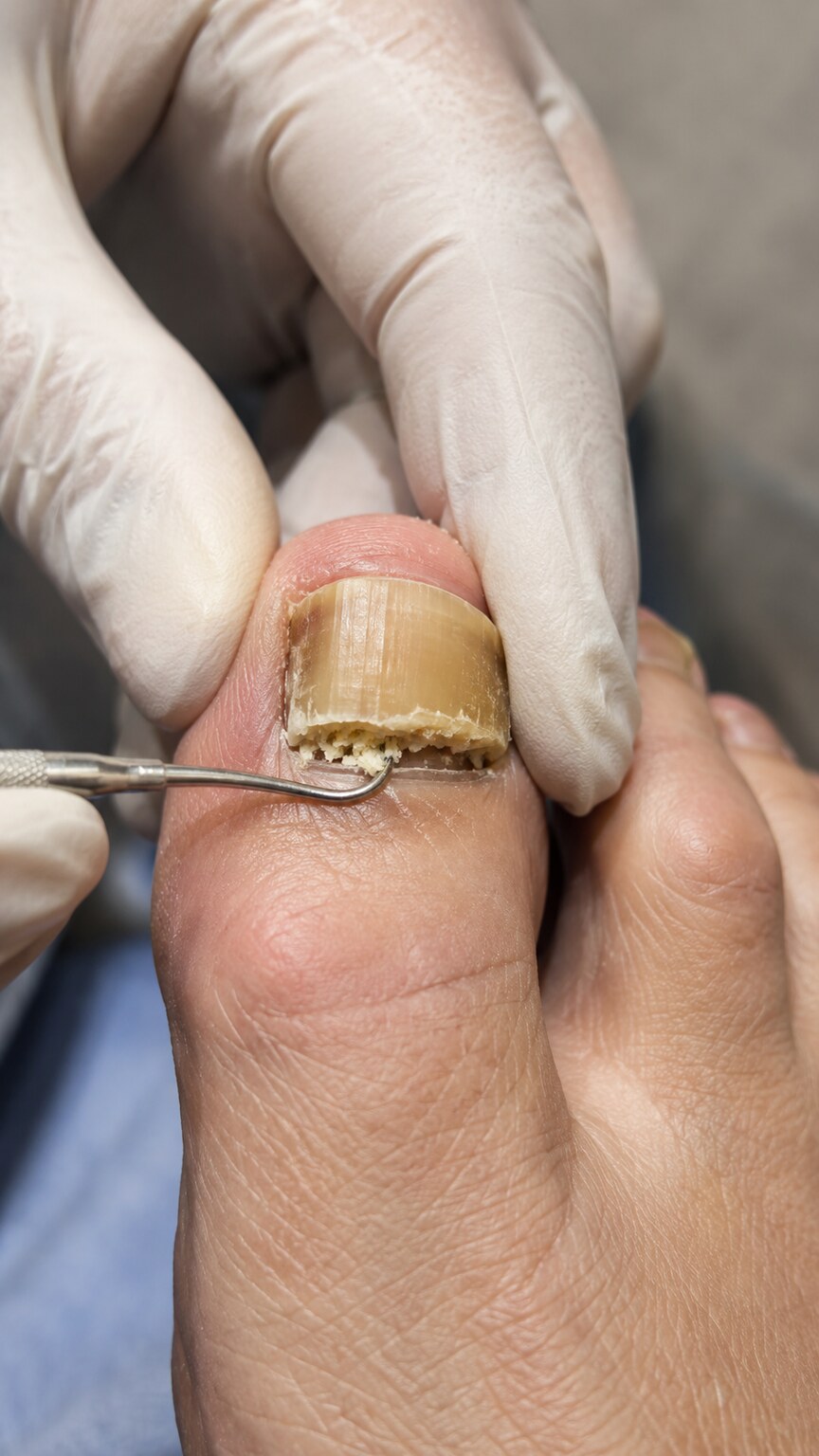
Figure 6. For suspected nail fungus, the sample is typically taken from beneath the nail plate near the border between healthy and affected tissue, where fungal density tends to be highest.
Exactly where and how the sample is collected changes depending on which body site is involved, and this is worth understanding because it explains why the same test can feel different depending on what's being checked. For a skin rash, the technique described earlier applies: a gentle scrape along the active, scaling border of the lesion, since that's where the fungus is actively invading fresh tissue.
For a suspected nail infection, the technique shifts. Rather than scraping the visible top surface of the nail, which is often mostly dead, compacted keratin with relatively little live fungal material, a clinician will typically clip or scrape debris from underneath the nail plate, at the border between clearly affected, discolored, crumbling nail and the nearby healthier-looking tissue. This subungual debris — the soft, often powdery material that builds up beneath a fungal-affected nail — tends to carry a much higher concentration of fungal elements than the hard nail plate itself, which is why simply clipping the visible white or yellow part of the nail and sending that alone often yields a disappointing, falsely negative result.
For suspected scalp involvement, particularly in children with a scaly, sometimes bald patch, sample collection often includes plucking a few affected hairs directly, in addition to scraping scale from the scalp surface, since certain dermatophytes that cause scalp ringworm grow specifically inside or around the hair shaft itself rather than only on the surrounding skin — a detail that would be completely missed if only surface scale were collected.
What Happens After the Result Comes Back
A positive KOH prep, in most everyday cases, is enough on its own to start treatment: a topical antifungal cream for a straightforward skin rash, or an oral antifungal medication for a nail infection, scalp involvement, or a widespread or stubborn skin infection, since topical treatments often can't penetrate deeply enough to clear an infection embedded in nail tissue or hair follicles. The specific medication chosen doesn't usually depend on knowing the exact species, since most common dermatophytes and Candida species respond to the same first-line drug classes.
A negative result, especially when the clinical picture still looks convincingly fungal, typically leads to one of three paths: a repeat KOH prep with a more careful or differently-located sample, a fungal culture sent out for a more sensitive, species-level answer, or — if the appearance was never entirely convincing for fungus to begin with — consideration of other explanations entirely, since several non-fungal skin conditions, including eczema, psoriasis, and certain bacterial infections, can superficially resemble a fungal rash closely enough to prompt the original test in the first place. A negative KOH prep, in that sense, does real diagnostic work even when it isn't the final answer — it helps rule fungus in or out as the clinician works through what's actually going on.
How the KOH Prep Became the Default First Step
It's worth pausing on why a technique this simple has survived, largely unchanged, as a frontline diagnostic tool for well over a century, in an era of genomic sequencing and automated lab analyzers. Potassium hydroxide preparations of skin scrapings were already being described in dermatology literature in the late 1800s, and the core method — scrape, dissolve, look — hasn't fundamentally changed since, because the underlying chemistry it exploits hasn't changed either: human keratin is still built the same way, and fungal chitin is still just as resistant to alkaline digestion as it always was. What has changed is everything around the core technique — better stains, fluorescence microscopy, standardized KOH concentrations, and clearer training on where and how to sample — but the basic principle that made the test useful in the 1890s is the exact same principle making it useful in a clinic today.
Part of what has kept it in continuous use is cost and access. A fungal culture requires incubators, specialized growth media, and technicians trained to identify colony morphology — infrastructure that isn't available in every primary care office, and that adds real cost even where it is. A KOH prep needs a microscope, a bottle of solution, and glass slides, all of which sit on a shelf in most dermatology offices and many general practices already. In parts of the world with less lab infrastructure, the KOH prep is sometimes the only fungal diagnostic available at all, which is part of why global health and tropical medicine literature still treats it as an essential technique worth teaching well, not a relic waiting to be replaced.
Common Mistakes That Quietly Skew the Result
Beyond sample-site error, a handful of smaller technical missteps show up repeatedly in the literature on KOH prep reliability, and knowing about them helps explain why two people with what looks like the same rash can walk away with different test experiences. Using a KOH concentration that's too dilute is one: some clinics prepare their own solution rather than buying a pre-mixed reagent, and if the concentration drifts too low, keratin dissolution can take far longer than the few minutes a busy clinic visit allows, tempting a rushed reader to call the slide negative before the keratin has actually cleared enough to see through.
Coverslip technique matters more than it sounds like it should. Pressing the coverslip down too hard can shatter or displace larger fungal structures, scattering fragments across a wider area and making them harder to recognize as connected, continuous hyphae. Too little pressure, on the other hand, leaves air bubbles trapped under the glass, and air bubbles are a notorious source of false-positive misreads — under low magnification, the curved edge of a trapped bubble can superficially resemble a rounded yeast cell or a fiber can mimic a hyphal strand, especially to someone reading quickly between other patients.
Contamination is another quiet source of error. Cotton or synthetic fibers from clothing, gauze, or even the cotton tip of a swab used to clean the area beforehand can end up on the slide and, at a glance, look enough like a fungal hyphae to confuse an inexperienced reader — the giveaway is usually that fibers don't branch the way true hyphae do, and they typically don't cross cell boundaries the way an invading fungus does, but recognizing that distinction reliably takes practice. This is one more reason experienced readers consistently outperform less experienced ones on identical slides, and why some larger practices route KOH preps to a specific staff member who reads the majority of them, rather than spreading the task evenly across whoever happens to be free.
What a Positive Slide Actually Looks Like to the Naked Eye Through the Scope
It helps to have a concrete mental picture of what the moment of a positive read actually looks like, since most descriptions stay abstract. Under low magnification, roughly 10x, the field of view generally looks like a pale, textured landscape — the dissolving remnants of skin cells forming an uneven, semi-transparent terrain, sometimes still showing faint cell-wall outlines like a ghost image of the tissue that used to be there. Scanning at this magnification is mostly about spotting anything that looks structurally different from that background: a long, unnaturally straight or gently curving line crossing multiple cells, or a cluster of small round shapes sitting together in a way loose cell debris typically doesn't.
Once something suspicious is spotted, the reader switches to higher magnification, typically 40x, to confirm what they're looking at. At this level, true dermatophyte hyphae resolve into clearly defined, roughly parallel-walled tubes, often with visible internal septa spaced along their length, sometimes branching off at an angle into a second thread. True hyphae also tend to show a subtle but consistent width along their length and cross over cell boundaries rather than stopping abruptly at the edge of one cell — a real biological structure growing through tissue, not a random fragment sitting on top of it. Budding yeast cells, at this same magnification, present unmistakably once a reader knows what to look for: a rounded cell with a smaller, clearly separate rounded projection budding from one side, connected by a visibly narrower neck — genuinely distinct from a stray dust particle or debris fragment, which almost never shows that specific budding geometry.
Frequently Asked Questions
Does a KOH prep hurt?
No — the scraping involved is superficial, taken from the outermost, already-dead layer of skin, nail, or hair, and most people describe it as a light scratching sensation at most. It's generally well tolerated even in young children, which is part of why it remains a practical first-line test in pediatric dermatology.
Can a KOH prep tell the difference between athlete's foot and a bacterial infection?
Yes, in the sense that a KOH prep specifically looks for fungal cell walls and won't show anything characteristic if the problem is bacterial rather than fungal. However, a negative KOH prep doesn't automatically confirm a bacterial cause — it only rules fungus in or out; a clinician still has to consider the full clinical picture, and sometimes additional bacterial cultures, to reach a bacterial diagnosis.
Why did my doctor order both a KOH prep and a fungal culture at the same time?
This is common when a clinician wants both a fast preliminary answer and a more detailed, species-level result. The KOH prep can confirm fungus is present within minutes and support starting treatment right away, while the culture sample, taken from the same scraping, continues incubating in the background to identify the exact species over the following one to several weeks — useful if the initial treatment doesn't work as expected.
If my KOH prep is negative, does that mean I definitely don't have a fungal infection?
Not necessarily. Because KOH prep sensitivity depends heavily on sample site, technique, and reader experience, a real fungal infection can still produce a negative result, particularly with nail samples or if antifungal products were used recently. If symptoms strongly suggest fungus despite a negative result, a repeat test or a fungal culture is a reasonable next step.
Should I stop using an antifungal cream before getting a KOH prep done?
Generally yes, if it's practical to wait. Antifungal products can suppress or physically remove enough fungal material from the surface of a lesion to cause a falsely negative result, even when the underlying infection is still present. Many clinicians recommend stopping topical treatment for at least a few days to a week before sampling, when the situation allows for that delay.
Can a KOH prep be done on a sample I collect myself at home?
It's technically possible to collect skin scale or nail debris at home into a clean, dry container, but reading the slide still requires a microscope and trained interpretation, so the sample ultimately needs to reach a clinic or lab either way. Some newer at-home fungal test kits use different methods entirely, such as mailing a sample for PCR-based analysis, rather than replicating the KOH microscopy step itself.
How Results Get Communicated Back
Because the KOH prep is usually read right in the clinic rather than sent to an outside lab, the way a result gets communicated often looks different from most other tests people are used to waiting on. Rather than a formal printed lab report arriving days later, the answer is frequently given verbally, in the same visit, sometimes with the clinician describing what they saw under the scope in plain terms — "I can see the branching threads, that confirms it's a fungal infection" — before treatment is even decided on. When a KOH prep is documented in the chart or an after-visit summary, it's typically recorded simply as positive or negative for fungal elements, sometimes with a brief note on whether hyphae, yeast, or both were seen, rather than a detailed report with reference ranges the way a blood test result would be presented. That simplicity is part of the point: the test exists to answer one narrow, practical question quickly, not to generate a document meant for extended independent interpretation later.
Conclusion
A KOH prep is, at its core, a chemistry trick applied to a microscope slide: dissolve everything human, leave the fungus standing. That simplicity is exactly what makes it so useful — a same-day, in-office answer to the specific question of whether fungal cells are physically present in a sample, without the wait, cost, or complexity of sending something out to a lab. But that same simplicity is also its boundary. It can point toward a dermatophyte or toward Candida based on shape alone, and it can hand a clinician enough confidence to start treatment immediately, but it was never built to name a species, and it depends heavily on exactly where and how the sample was taken. Understanding that boundary — what the test is actually built to answer, and what it was never designed to answer — is what turns a one-line lab result into something you can actually make sense of.
Still Not Sure What Your Results Mean?
Upload your labs and get a complete, visual, plain-language interpretation of every biomarker — delivered to your inbox in under 15 minutes.
Get My ReportThis article is for educational purposes only and does not constitute medical advice. Always consult your healthcare provider regarding your specific lab results.