
Why Would a Doctor Order a Fungal Blood Test Instead of a Skin Scraping?
A skin scraping and a fungal blood test both genuinely hunt for the exact same broad kind of organism — a fungus — but they're each specifically built to catch it in two anatomical places that could hardly be more different from one another. A skin scraping works reliably because the specific fungus causing something like athlete's foot or a stubborn, persistent nail infection is genuinely living right there, on the surface, in dead skin and keratin a simple blade can physically lift off in mere seconds. A fungal blood test exists because some infections don't stay on the surface at all — the fungus gets past the skin entirely and starts circulating inside the bloodstream itself, in a location no scraping tool could conceivably ever reach, no matter how deep it cut into the tissue. Choosing correctly between these two available tests isn't ever really a matter of simple convenience or clinician preference; it's always a direct, deliberate reflection of exactly where in the body a doctor genuinely suspects the fungus actually is currently located, and getting that particular choice wrong isn't merely a minor inconvenience to sort out later — a genuine bloodstream fungal infection missed simply because someone only ever checked the skin can, quite literally, turn fatal within a matter of just a few short days.
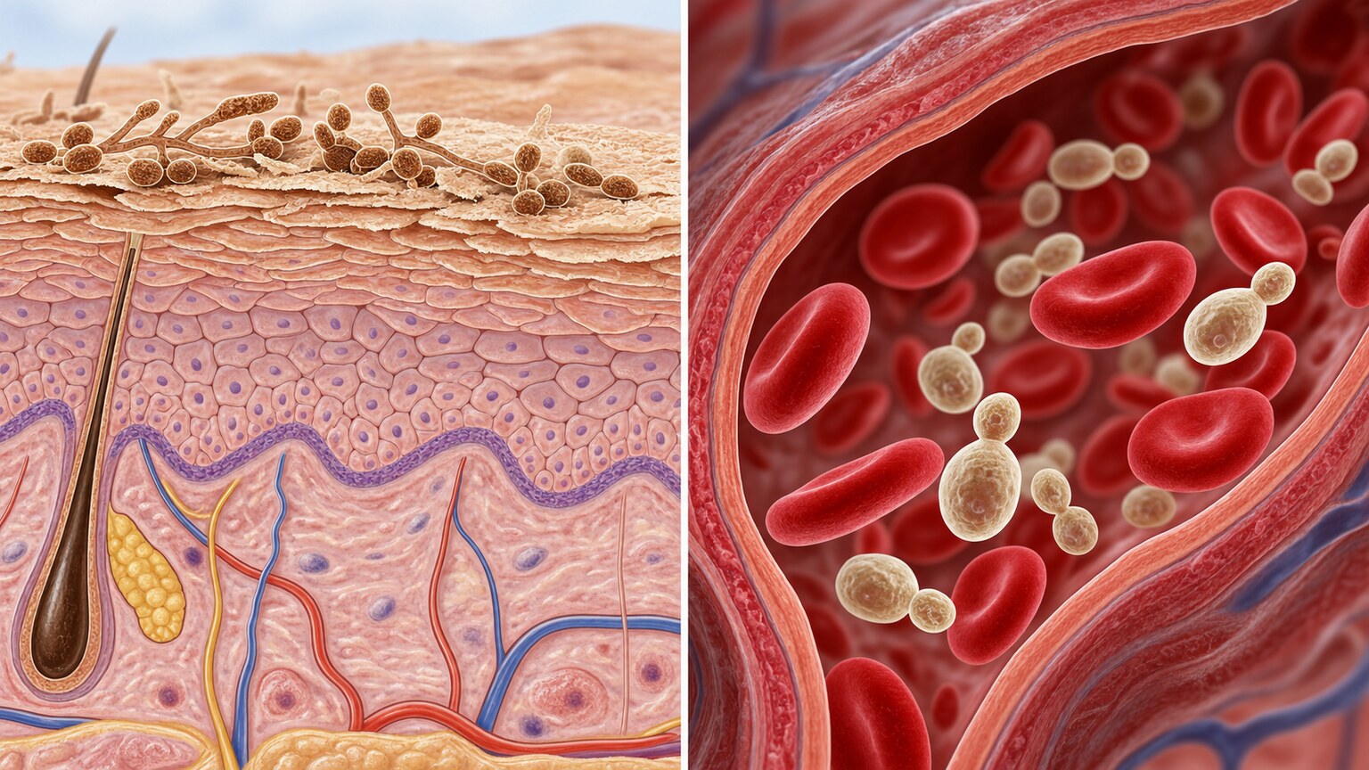
Figure 1. A skin scraping samples the outermost keratin layer where superficial fungi live; a bloodstream infection occupies an entirely different anatomical compartment that a surface sample can never reach.
Curious what the rest of your test results are telling you? Upload them and get a complete, plain-language breakdown in under 15 minutes.
Analyze My ResultsWhy a Skin Scraping Works at All
To understand why a blood test is sometimes necessary, it helps to understand exactly why a skin scraping works so well for the infections it's actually designed to catch. The fungi responsible for athlete's foot, ringworm, and most nail infections are called dermatophytes, and they have a very specific, limited diet: keratin, the tough structural protein that makes up the outermost layer of skin, hair, and nails. These fungi don't have the biological tools to invade living tissue or cross into blood vessels — they live entirely within dead, keratinized surface material, digesting it slowly as their sole food source. This is precisely why a scraping works: the fungus is sitting right there, physically accessible, in a layer of tissue a clinician can lift off with a blunt blade or edge of a glass slide in a matter of seconds, with essentially no meaningful discomfort involved and virtually no real risk to the patient at all.
Because dermatophytes are biologically restricted to this single, superficial compartment, a scraping isn't just a convenient shortcut — it's actually sampling the exact location where essentially the entire infection lives. There's no meaningful amount of the organism anywhere else in the body to miss. This is why, for the overwhelming majority of everyday fungal skin and nail complaints, a scraping examined under a microscope with potassium hydroxide, or sent for a standard fungal culture, is not just sufficient but genuinely the correct, complete answer — reaching for a blood test in this particular situation would be a genuinely meaningless, entirely wasted diagnostic step, since there's simply nothing at all circulating anywhere in the bloodstream for that test to actually find.
What Changes When the Infection Isn't on the Surface Anymore
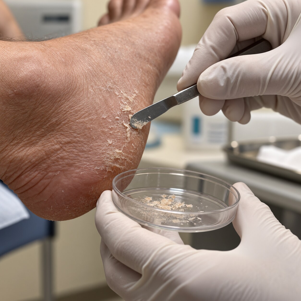
Figure 2. A skin scraping collects only the outermost keratin layer — an adequate and complete sample for a dermatophyte infection, since that's the only place the organism exists in that scenario.
A completely different category of fungal organism, most commonly species of Candida and Aspergillus, plays by different rules entirely. These fungi are naturally present in small, harmless numbers in places like the gut and on mucous membranes in most healthy people, kept in check by a functioning immune system and by competition from the body's normal bacterial population. Under the right — or rather, wrong — circumstances, these organisms can breach the barriers that normally contain them and enter the bloodstream directly, a condition called fungemia, or spread to invade internal organs entirely, called invasive fungal disease. Once a fungus is circulating in blood or embedded in deep tissue like the lungs, liver, or heart valves, there is no skin surface to scrape, because the infection was never on the skin to begin with.
This is the exact fork in the road this article is about. The moment a doctor suspects an infection has moved past the surface — into the bloodstream or into internal organs — a skin scraping stops being merely unhelpful and becomes actively the wrong tool for the job, capable of coming back completely clean while a genuinely dangerous, spreading infection continues untouched and undetected inside the body.
The Specific Clinical Signals That Shift Suspicion to the Bloodstream
Doctors don't reach for a fungal blood test based on a hunch — a specific combination of patient history and clinical presentation has to line up first. The single biggest risk factor is a significantly weakened immune system: patients undergoing chemotherapy, organ or bone marrow transplant recipients on immunosuppressive medication, people with poorly controlled HIV, and anyone with a very low neutrophil count (the specific white blood cell responsible for containing fungal overgrowth) are all at dramatically elevated risk of a fungus breaching its normal boundaries. In these patients, a fever that doesn't respond to standard antibiotics after several days is one of the most classic and important triggers for expanding the workup to include fungal blood testing, since a persistent fever unexplained by bacteria is a well-recognized signature of an underlying fungal bloodstream infection in this specific population.
A second major trigger is anyone with an indwelling medical device providing fungi a direct entry point past the skin's natural barrier — most importantly central venous catheters, the long-term IV lines used in hospitalized patients, particularly those in intensive care for extended periods. These catheters can become colonized by Candida species forming a protective biofilm on their surface, providing a direct, continuous bridge from the skin's surface bacteria and fungi straight into the bloodstream, bypassing the body's normal surface defenses entirely. Prolonged broad-spectrum antibiotic use is a third significant contributor, since these antibiotics wipe out the normal bacterial population that would otherwise compete with and suppress fungal overgrowth, giving Candida in particular an opportunity to multiply unchecked and eventually invade.
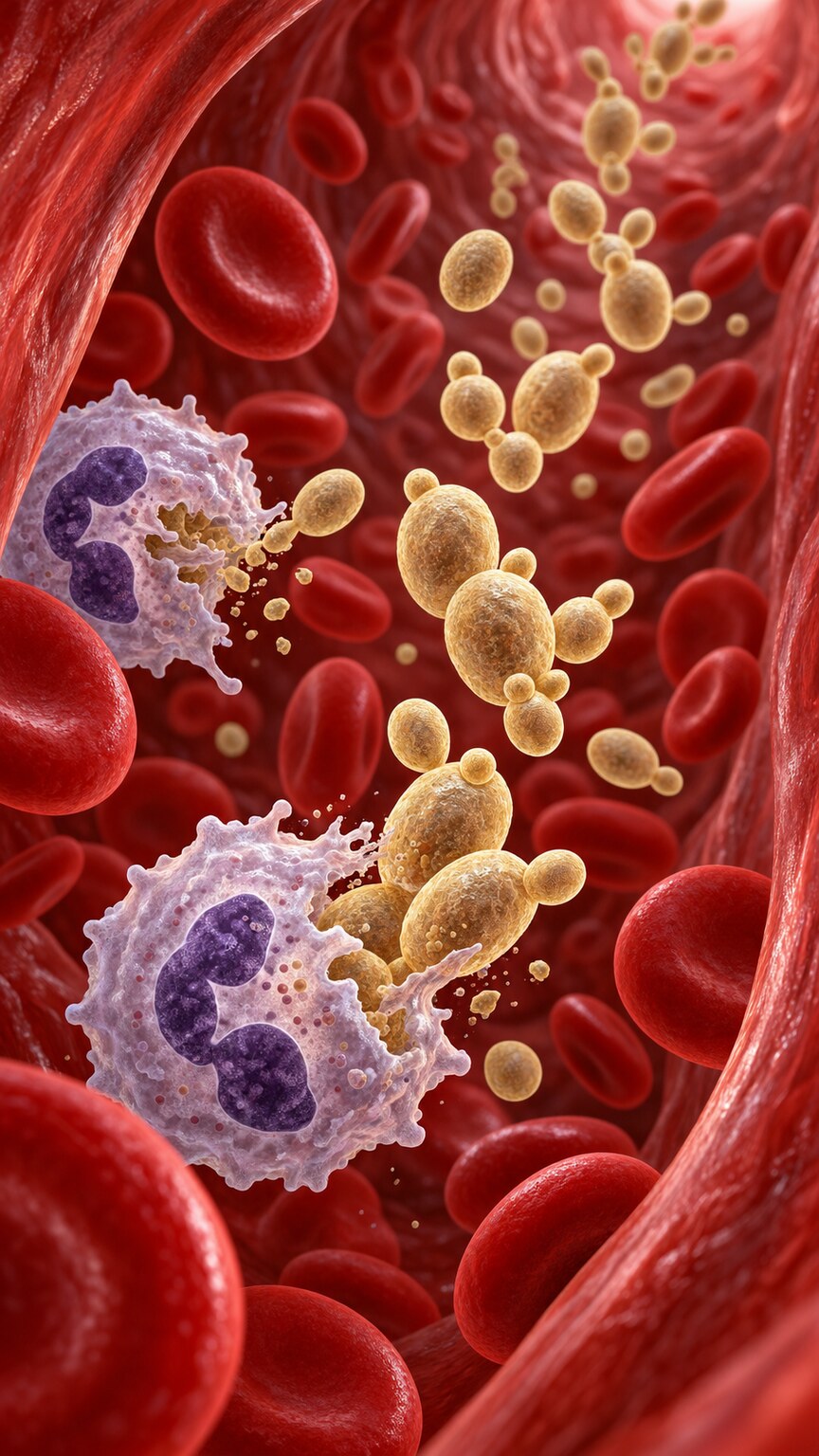
Figure 3. With too few neutrophils circulating to contain them, Candida cells that would normally be suppressed can multiply freely and spread through the bloodstream to distant organs.
Certain surgical and medical scenarios round out the picture: major abdominal surgery, particularly involving the gastrointestinal tract, can allow gut-resident Candida direct access to the abdominal cavity and bloodstream. Severe burns compromise the skin's barrier function over a large surface area, similarly allowing entry. Prolonged stays in an intensive care unit combine several of these risk factors simultaneously — central lines, broad-spectrum antibiotics, and often a weakened overall immune state — which is exactly why ICU patients represent one of the populations where clinicians maintain the highest ongoing suspicion for invasive fungal infection and the lowest threshold for ordering blood cultures alongside bacterial ones.
These risk factors rarely act alone in the patients who actually go on to develop a bloodstream infection — they tend to stack, and the more of them present at once, the higher the resulting risk becomes. A patient recovering from major abdominal surgery who also happens to be neutropenic from recent chemotherapy, for instance, carries meaningfully more risk than either factor would suggest in isolation, since the surgical breach in the gut lining and the weakened immune response needed to contain any resulting spread are both working against the same patient simultaneously. This is exactly why clinicians treat this list of risk factors as a cumulative picture to be weighed together, rather than as a simple checklist where any single item automatically triggers testing on its own.
How the Bloodstream Infection Actually Gets There
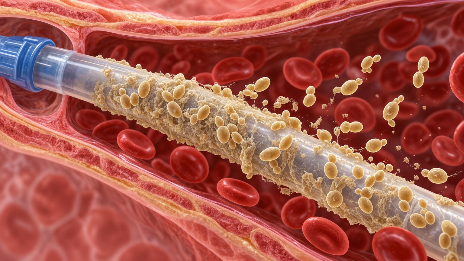
Figure 4. A biofilm of Candida cells coating the inner surface of a central line provides a direct, continuous route from the skin's surface into the bloodstream, bypassing the body's normal barriers entirely.
Understanding the actual mechanical route a fungus takes to reach the bloodstream helps explain why these specific risk factors matter so much clinically. Candida species normally live in modest numbers on skin and throughout the digestive tract as part of the body's ordinary microbial population, held in check by an intact immune system and by competition from bacteria. A central line, inserted directly through the skin into a large vein, physically breaches that surface barrier and provides a stable, moist surface fungi can adhere to and colonize, gradually building a biofilm — a structured, somewhat protected community of fungal cells — directly on the catheter's inner or outer surface. Cells shed from this biofilm enter the bloodstream continuously and directly, with no need to breach a skin barrier the catheter has already bypassed entirely.
In patients without a catheter, the gut itself becomes the more common entry route. When the intestinal lining is damaged — from chemotherapy, major surgery, or severe illness — combined with an immune system too weakened to contain the normal population of gut-resident Candida, the fungus can translocate directly across the compromised intestinal wall into the bloodstream, a process sometimes called gut translocation. Both routes explain why the underlying risk factors described in the previous section are so consistently associated with bloodstream fungal infection: they each represent a different physical breach in the barriers that would otherwise keep these organisms safely contained outside of circulation.
Why a Negative Skin Scraping Wouldn't Rule Anything Out Here
This is the central, practical point this entire article is built around: in a patient with suspected bloodstream or invasive fungal infection, a skin scraping would almost certainly come back completely negative, and that negative result would tell a doctor absolutely nothing useful about what's happening internally. The skin, in this scenario, was never involved in the first place — the infection took a different route entirely, through a catheter or through the gut, straight into the blood or into internal organs like the lungs, liver, spleen, or heart valves. Relying on a skin sample here wouldn't just be unhelpful; it would provide dangerously false reassurance in exactly the situation where speed and accuracy matter most, since untreated bloodstream candidiasis carries a mortality rate that published research has placed at somewhere between 25% and 40% even with treatment, rising considerably higher when treatment is delayed.
A fungal blood culture works by drawing blood directly into specialized culture bottles designed to support fungal growth, then incubating them under conditions that encourage any circulating fungal cells present to multiply to detectable levels — directly sampling the actual compartment where the suspected infection lives, rather than a location the organism was never present in to begin with. Newer, faster molecular and antigen-based blood tests, including one that detects a specific fungal cell-wall component called beta-D-glucan, have added an additional, often quicker layer of detection alongside traditional blood cultures, since traditional fungal blood cultures can still take several days to turn positive even when the organism is present — a meaningful delay in a situation where mortality risk climbs with every day treatment is withheld.
The Reverse Mistake: Ordering a Blood Test for an Ordinary Skin Infection
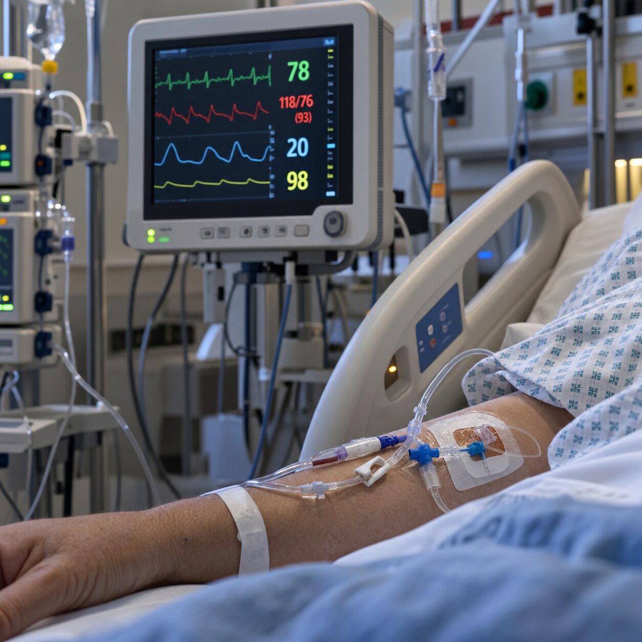
Figure 5. A persistent, unexplained fever in a hospitalized patient with a central line and no clear bacterial source is one of the most common triggers for expanding a workup to include fungal blood cultures.
It's just as important to understand the opposite error: ordering a fungal blood test on an otherwise healthy person with an ordinary case of athlete's foot or a fungal toenail is both clinically unnecessary and genuinely wasteful. Without any of the risk factors described earlier — no significant immune suppression, no central line, no recent major surgery or prolonged hospitalization, no unexplained fever — the biological likelihood of that same dermatophyte organism having crossed into the bloodstream is, for all practical purposes, essentially zero, since dermatophytes genuinely lack the specific biological machinery needed to actually invade living tissue in the first place at all. Ordering an invasive, more expensive blood test in this situation adds cost, a needless blood draw, and a delay in getting an actually useful answer, all without any realistic chance of finding anything a skin scraping wouldn't have already captured directly and far more simply.
This is exactly why the decision between these two tests isn't about which one is generally "better" or more thorough in some abstract sense — it's entirely about matching the test to the specific anatomical location a real clinical picture actually points toward. A more invasive, more expensive test isn't automatically the more rigorous choice; the right test is whichever one actually samples the location where the infection genuinely, biologically exists.
The Gray Zone: When a Skin Finding Prompts a Blood Test Too
Some clinical situations genuinely call for both tests rather than a strict either-or choice, and understanding this gray zone rounds out the full picture. A person with poorly controlled diabetes and a chronic, non-healing foot ulcer represents a good example: the visible wound itself might be sampled and cultured the way any skin or soft-tissue infection would be, but if that same patient also has a fever, appears systemically unwell, or shows signs the infection may be tracking into deeper tissue or bone, blood cultures are added specifically to rule out the infection having spread beyond the local wound into the bloodstream. Similarly, a severely immunocompromised patient who develops a new, unusual-looking skin lesion might have that lesion biopsied and cultured locally while blood cultures are drawn simultaneously, since in this specific population an unusual skin finding can occasionally represent the visible tip of a deeper, disseminated fungal infection rather than a purely localized one.
These combined-testing scenarios reinforce the same underlying logic this whole article has been built around: the test selection always follows the actual, biologically plausible location of the infection, and when that location is genuinely uncertain or when both a superficial and a systemic process are plausible simultaneously, sampling both compartments rather than guessing is the appropriate, careful approach.
How the Two Sample Types Are Actually Processed Differently
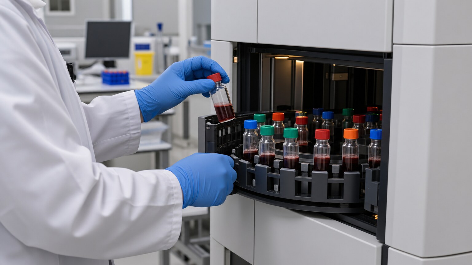
Figure 6. Fungal blood cultures are incubated in specialized automated systems that continuously monitor for signs of microbial growth, a very different process from the plate-and-microscope method used for a skin scraping.
Beyond the anatomical difference in where each sample comes from, the two tests are also handled quite differently once they reach the lab. A skin scraping is typically examined directly under a microscope after treatment with potassium hydroxide, which dissolves the surrounding skin cells and leaves fungal structures clearly visible, and a portion is often also placed onto a solid culture plate to grow out and confirm the specific organism over the following one to several weeks. A blood sample intended for fungal testing, by contrast, is drawn directly into specialized liquid culture bottles containing a growth medium formulated to support fungal proliferation, then loaded into an automated incubator system that continuously monitors for the chemical signs of active microbial growth, flagging a bottle as positive as soon as those signs appear, sometimes within one to several days depending on how many organisms were present in the original sample.
This difference in processing reflects the different stakes involved: a skin scraping's turnaround time of one to several weeks is perfectly acceptable for a slow-growing, non-urgent superficial infection, while the automated, continuously monitored blood culture system exists specifically because a bloodstream infection can turn fatal within days, and every hour saved in detection genuinely matters for a patient's real chance of survival.
What Happens After Each Test Comes Back
The path forward after each test also diverges sharply, reflecting the different urgency of what each one is actually checking for. A positive skin scraping or culture typically leads to a topical or oral antifungal medication, prescribed and taken on an outpatient basis over several weeks, with no particular time pressure since the infection, while uncomfortable, poses no immediate systemic danger. A positive fungal blood culture, on the other hand, typically triggers immediate intravenous antifungal treatment, often started even before the specific organism is fully identified, given how quickly a bloodstream infection can progress; identification of the exact species and its antifungal susceptibility follows over the next several days and may prompt a change in medication once that more detailed information is available. Any central line suspected as the source is generally removed as part of treatment, since leaving an infected catheter in place makes clearing the infection significantly more difficult, regardless of which antifungal medication is used.
A Simple Way to Think Through Which Test Applies
Bringing this entire framework down to something genuinely usable: the central question isn't "which test is more thorough" but "where does the clinical picture actually suggest this fungus is living." A localized, uncomplicated skin, nail, or scalp complaint in someone without significant risk factors points toward a skin scraping, since that's exactly where a dermatophyte infection would be found and the only place it could realistically exist. A persistent, unexplained fever in someone with a central line, recent major surgery, significant immune suppression, or prolonged hospitalization points toward blood cultures, since these are precisely the circumstances under which a fungus can breach its normal boundaries and enter circulation. And a situation combining both a visible skin or wound finding with systemic signs of illness reasonably calls for sampling both compartments rather than assuming one location necessarily rules out the other.
Which Fungal Species Show Up Most Often in Each Location
It's worth being specific about which organisms actually turn up in each of these two very different tests, since the contrast reinforces just how separate these two clinical worlds really are. Skin scrapings for ordinary superficial infections overwhelmingly grow dermatophytes from three main genera: Trichophyton, the most common cause of athlete's foot and most nail infections; Microsporum, frequently responsible for scalp ringworm, especially in children; and Epidermophyton, a less common but recognized cause of both skin and nail infections. These three genera account for the vast majority of everyday superficial fungal complaints seen in primary care and dermatology.
Fungal blood cultures, in sharp contrast, are dominated by an entirely different set of organisms. Candida albicans remains the single most commonly identified species in bloodstream infections, though several other Candida species, including some that are naturally more resistant to standard antifungal medications, have become increasingly common in recent decades, particularly in hospitalized patients who've previously received antifungal treatment. Aspergillus species, while less frequently isolated directly from blood cultures, are a leading cause of invasive fungal disease in the lungs and other organs in severely immunocompromised patients, often requiring specialized tissue or respiratory sampling rather than blood culture alone to confirm. The near-total lack of overlap between these two organism lists is itself a clear illustration of why the two tests exist as separate, purpose-built tools rather than interchangeable options for the same underlying question.
Why Fungal Bloodstream Infections Have Become More Common Over Time
Invasive fungal infections, once considered relatively uncommon, have become a steadily growing concern in modern hospital medicine, and understanding why helps explain why this specific test decision comes up more often today than it did decades ago. Advances in cancer treatment, organ transplantation, and intensive care medicine have meant that far more patients now survive conditions that once would have been fatal on their own — but many of these same life-saving treatments, from chemotherapy to immunosuppressive medication to prolonged ICU stays with central lines, directly create the exact conditions under which fungi can breach their normal boundaries. In effect, modern medicine's success at keeping critically ill patients alive longer has simultaneously expanded the population of people vulnerable to this specific category of infection, which is a large part of why fungal blood testing has become a more routine and more clinically important consideration than it was a generation ago.
The increasing use of broad-spectrum antibiotics across modern medicine compounds this trend further, since these medications, while essential for treating bacterial infections, simultaneously suppress the normal bacterial populations that would otherwise help keep fungal organisms like Candida in check throughout the body. This combination of a larger vulnerable patient population and more widespread antibiotic use has made invasive fungal infection a significantly more prominent concern in hospital-based medicine than it was in earlier decades, and it's part of why clinical guidelines have increasingly emphasized maintaining a lower threshold for considering fungal blood testing in exactly the higher-risk populations described earlier in this article.
The Role of Antifungal Susceptibility Testing Once an Organism Is Identified
Once a fungal blood culture turns positive and the specific organism is identified, the workup doesn't stop there — a further test called antifungal susceptibility testing is typically performed, checking exactly which antifungal medications the specific strain isolated will actually respond to. This step has become increasingly important as resistant Candida strains, capable of surviving exposure to one or more standard antifungal drug classes, have become more prevalent in hospital settings, particularly among patients who've received prior antifungal treatment. Skin scrapings for ordinary dermatophyte infections rarely require this same level of individualized susceptibility testing, since resistance to standard topical and oral antifungal treatments remains considerably less common and clinically significant in that population, another meaningful difference in how thoroughly each type of positive result gets worked up afterward.
This susceptibility step directly shapes the treatment decisions that follow a positive blood culture: a patient started on a particular antifungal medication based on initial clinical judgment may need to switch to a different drug entirely once susceptibility results reveal the specific strain is resistant to the original choice, a real-time adjustment that underscores why bloodstream fungal infections are managed with such active, ongoing monitoring rather than a single fixed treatment course decided at the outset and left unchanged.
What This Looks Like From the Patient's Side
For someone actually experiencing this situation rather than reading about it abstractly, it helps to know what each pathway generally feels like in practice. A skin scraping is quick, done in a routine outpatient visit, involves no needles, and results — whether from direct microscopy or a full culture — come back over a timeline of days to a few weeks, generally without any sense of urgency communicated by the care team, because there typically isn't any. A workup that includes fungal blood cultures, by contrast, usually happens in a hospital or more urgent care setting, is accompanied by other tests happening simultaneously (bacterial blood cultures, imaging, additional bloodwork), and is generally treated by the care team with a distinctly different sense of urgency — antifungal treatment may begin empirically before any culture result is even back, precisely because of how quickly this category of infection can become dangerous.
Recognizing this difference in tone and pacing can itself be a useful clue for a patient or family member trying to understand what's happening: if a care team is moving quickly, drawing multiple blood tests, and starting treatment before a definitive result is available, that pace of action reflects real, informed clinical urgency about a bloodstream process, not overreaction — exactly the opposite situation from a routine, unhurried skin scraping sent out for a nail or scalp complaint.
Special Considerations in Children and Newborns
This same test-selection logic applies to children, but a few population-specific details are worth understanding separately. Premature and critically ill newborns in neonatal intensive care represent one of the highest-risk pediatric populations for invasive fungal infection, since their immune systems are still developing, they frequently require central lines and prolonged hospitalization, and their skin barrier itself is less mature and more easily breached than an older child's or adult's. Because of this combination, neonatal intensive care units generally maintain a notably low threshold for fungal blood testing in a premature infant with unexplained instability, fever, or feeding intolerance, even before more classic risk factors would typically be considered.
Outside of the intensive care setting, healthy children are actually more prone than adults to certain superficial fungal infections, particularly scalp ringworm, simply due to close physical contact common in school and daycare settings and shared items like combs and hats. In this far more common pediatric scenario, a simple skin or scalp scraping remains the correct and sufficient test, following exactly the same logic that applies to adults with uncomplicated superficial infections — the organism responsible for ordinary childhood ringworm has no more ability to invade the bloodstream than it does in an adult.
How Imaging Complements Blood Testing in Suspected Invasive Disease
When invasive fungal disease is strongly suspected, blood cultures are frequently ordered alongside imaging studies rather than in isolation, since a positive blood culture confirms the organism is present in circulation but doesn't necessarily show where else in the body it may have already spread. A CT scan of the chest, for instance, can reveal characteristic patterns of fungal involvement in the lungs, particularly relevant for suspected Aspergillus infection in severely immunocompromised patients, sometimes even before blood tests turn positive. An echocardiogram may be ordered specifically to check the heart valves for fungal vegetations, a serious complication that can develop when a bloodstream infection seeds itself onto a heart valve, and an eye examination is often performed as part of the standard workup after a confirmed Candida bloodstream infection, since the fungus can seed the retina and cause vision-threatening damage without producing any symptoms early on.
This layered approach — blood testing combined with targeted imaging based on where the specific organism is known to commonly spread — reflects the broader point running through this entire article: once an infection has left the skin's surface and entered systemic circulation, a single test in isolation rarely tells the complete story, and a more comprehensive, multi-pronged workup becomes necessary specifically because the infection now has access to essentially anywhere in the body the bloodstream reaches.
Frequently Asked Questions
Can a skin fungus ever turn into a bloodstream infection on its own?
This is extremely rare with common dermatophytes, since they lack the biological ability to invade living tissue. Bloodstream fungal infections almost always involve different organisms, most commonly Candida, entering through a different route like a central line or the gut rather than spreading from an ordinary skin infection.
Why does a fungal blood culture take days when other blood tests come back same-day?
Fungi grow more slowly than bacteria, and a culture needs enough organisms to multiply to detectable levels before the automated system can flag it as positive. Newer antigen-based tests can sometimes provide a faster preliminary answer alongside the traditional culture.
Is a fungal blood test ever done just as a precaution, without symptoms?
In very high-risk patients, such as certain transplant recipients or those with prolonged severe neutropenia, some centers use proactive screening strategies. For most patients, though, blood testing is triggered by a specific clinical concern like unexplained fever rather than performed as routine precaution.
If my skin scraping was negative, could I still have a serious fungal infection elsewhere?
Yes, if your clinical picture includes risk factors like a central line, significant immune suppression, or unexplained fever. A negative skin scraping only rules out infection at the specific surface site sampled, not a separate, deeper infection with a different cause.
Why is a central line removed if a bloodstream fungal infection is found?
Fungi can form a protective biofilm on the catheter's surface that's difficult for antifungal medication to penetrate. Removing the line eliminates that ongoing source of infection and makes clearing it from the bloodstream considerably more likely.
Does having diabetes alone mean I need fungal blood testing for a skin infection?
Not automatically. Diabetes does raise general infection risk, but a routine fungal skin or nail infection in someone with well-controlled diabetes and no other risk factors is still typically evaluated with a skin scraping, not a blood test.
Are newborns tested differently than older children?
Yes. Premature or critically ill newborns in intensive care are considered high-risk for invasive fungal infection due to an immature immune system and skin barrier, and blood testing is often considered earlier and more readily than in an otherwise healthy older child.
Why would a doctor also order a CT scan or eye exam alongside blood cultures?
Once a fungus is confirmed in the bloodstream, it can spread to organs like the lungs, heart valves, or eyes. Imaging and exams check whether it has already seeded a distant site, since a positive blood culture alone doesn't reveal how far the infection has spread.
Conclusion
A skin scraping and a fungal blood test were never really competing options for the same underlying job in the first place — they're two purpose-built tools, each carefully designed to sample two entirely different anatomical compartments of the body, and the right choice always comes down to where the clinical picture genuinely suggests the fungus actually is. Dermatophytes responsible for ordinary, everyday skin and nail infections stay biologically confined entirely to dead surface keratin, which is exactly what makes a simple scraping both a genuinely sufficient and a completely correct diagnostic choice in that specific situation. Candida and similar organisms capable of breaching the body's normal barriers, most often through a central line or a compromised gut lining, require sampling the bloodstream directly, since a surface sample taken from skin that was never actually involved in the first place would provide nothing whatsoever but dangerously misleading, false reassurance at exactly the wrong moment. Understanding this distinction is what actually explains why your doctor reached for one test over the other — not caution for its own sake, but a deliberate, biologically grounded match between the specific test ordered and the one actual place in the body where that particular infection could realistically be found, given everything already known and documented about that patient's own individual risk factors and full clinical presentation at the time.
Still Not Sure What Your Results Mean?
Upload your labs and get a complete, visual, plain-language interpretation of every biomarker — delivered to your inbox in under 15 minutes.
Get My ReportThis article is for educational purposes only and does not constitute medical advice. Always consult your healthcare provider regarding your specific lab results.