
Can Blood Tests Detect Certain Parasitic Infections?
Yes — but which blood test, and what it can actually find, depends enormously on which parasite is being asked about. A handful of parasites live directly inside the bloodstream, which means a lab technician can put a drop of your blood under a microscope and, with the right stain and enough patience, literally watch the organism swimming among your red blood cells. Malaria works this way, and so do a few rarer infections like babesiosis and certain filarial worms. But most of the parasites people worry about — the ones that cause intestinal symptoms, or that get picked up after a trip abroad, or that show up as a strange finding on a stool test — never circulate freely in the blood at all. For those, a blood test can only detect the immune system's reaction to the parasite, not the parasite itself, and that reaction can take weeks to develop or, in some cases, persist for years after an infection has already been cured. Understanding which category your situation falls into changes everything about how to interpret a result, including what a "negative" actually means, and it's one of the more common gaps between what a lab report technically says and what a worried person assumes it says.
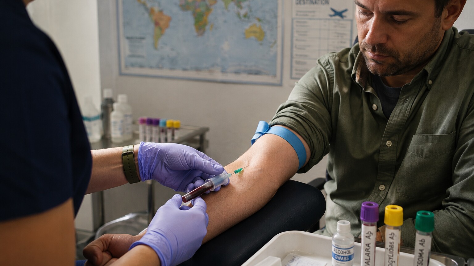
Figure 1. When someone returns from travel with fever, a blood draw is often the first step toward identifying a parasitic cause.
Curious what the rest of your bloodwork is telling you? Upload your results and get a complete, plain-language breakdown in under 15 minutes.
Analyze My ResultsThe Two Fundamentally Different Ways a Blood Test Can Find a Parasite
Before looking at any specific infection, it helps to understand that "a parasite blood test" isn't one single kind of test — it's a label covering two completely different strategies, and mixing them up is where most of the confusion starts. The first strategy is direct detection: the lab is trying to physically see or genetically identify the actual organism in a sample of your blood. This is only possible for parasites that spend part of their life cycle circulating freely in the bloodstream itself, rather than hiding inside organs, muscle tissue, or the intestines. The second strategy is indirect detection: instead of looking for the parasite, the lab looks for antibodies — proteins your immune system manufactures specifically to fight that parasite — or, less commonly, for fragments of the parasite's own proteins (antigens) that have leaked into circulation. Indirect testing works even when the parasite itself never enters the bloodstream, because the immune response to it does. The tradeoff is that indirect tests answer a slightly different question than people assume: they tell you whether your immune system has encountered the parasite, not necessarily whether the parasite is still there right now.
When Doctors Can Actually See the Parasite Under a Microscope
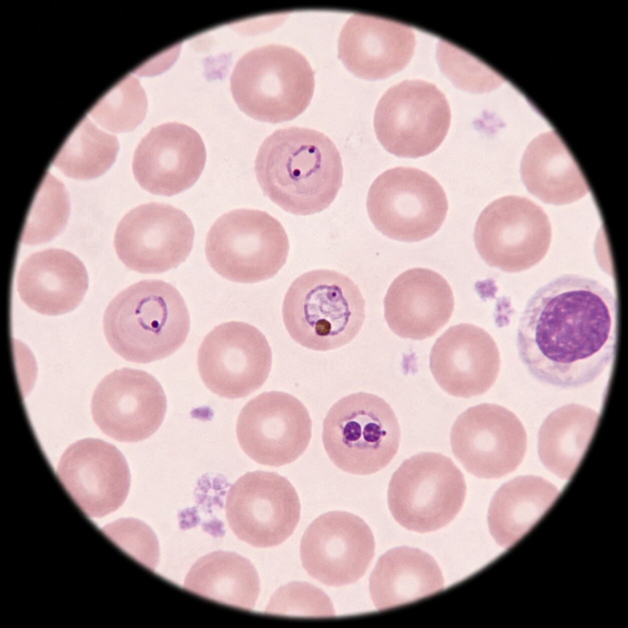
Figure 2. A thin blood smear lets a technician see the parasite's exact shape and stage inside individual red blood cells.
Malaria is the textbook example of direct visualization, and it's still the gold-standard method nearly a century after it was first described. A drop of your blood is smeared onto a glass slide two ways — a "thick" smear, which concentrates a larger volume of blood to make it easier to spot even a small number of parasites, and a "thin" smear, which spreads the blood into a single layer so the parasite's exact shape can be studied. Both are stained with a dye called Giemsa stain, which colors the parasite's internal structures a distinctive purple-pink against the pale background of the red blood cell, letting a trained technician identify not just that malaria is present, but which of the five species of Plasmodium is causing it and roughly what percentage of red blood cells are infected — a number that directly informs how urgently treatment is needed.
A small number of other infections are found the same direct way. Babesiosis, a tick-borne infection that in some regions is confused with Lyme disease because they share a tick vector, is also diagnosed by spotting the parasite inside red blood cells on a Giemsa-stained smear — under the microscope it can look strikingly similar to early-stage malaria, which is one reason travel and exposure history matters so much to the person interpreting the slide. Certain filarial worms, which cause lymphatic filariasis, release immature larval forms called microfilariae into the bloodstream, and these can sometimes be seen swimming across a blood smear in real time; because several filarial species release their larvae mainly at night to match the biting habits of the mosquitoes that spread them, the blood sample sometimes has to be drawn specifically after dark to catch them at their peak concentration — a detail that surprises most people the first time they hear it, since almost every other blood test in medicine can be drawn whenever is convenient.
Malaria diagnosis also illustrates why a single test result is rarely the end of the story with direct-visualization methods. Because the amount of parasite in the blood can rise and fall in cycles tied to the parasite's own reproductive rhythm inside red blood cells, a single blood smear drawn at the wrong moment can occasionally miss a low-level infection even though the organism is genuinely present. For this reason, standard practice when malaria is strongly suspected is to repeat the smear every twelve to twenty-four hours for up to three samples before concluding the result is truly negative, rather than accepting one look under the microscope as the final word. Rapid diagnostic tests, which detect malaria antigens rather than the organism itself and can produce a result in as little as fifteen to twenty minutes without a microscope, have become a valuable complement in settings where immediate answers matter more than the added detail a smear provides, though a positive rapid test is still typically confirmed with microscopy when possible, since the rapid format can occasionally continue reading positive for a period after the infection has actually been treated and cleared.
Why Most Intestinal and Tissue Parasites Never Show Up in Blood at All
The parasites people are most commonly screened for — the ones behind irritable digestion, unexplained weight loss, or a positive stool test after international travel — mostly belong to a different category entirely. Organisms like Giardia, Cryptosporidium, and the various species that cause amoebic infections live and reproduce inside the intestines, shedding cysts or eggs into stool rather than circulating through the bloodstream. Because they never leave the gut in appreciable numbers, no amount of blood testing will find the organism itself; a stool sample, examined directly under a microscope or tested with a stool antigen panel, remains the only way to detect them directly. This is a genuinely common source of confusion, since it seems intuitive that "a blood test checks everything," but a blood test can only find what is actually present in blood — an organism that never enters the bloodstream is, from blood's point of view, invisible.
Some tissue-dwelling parasites occupy a middle ground. Toxoplasma gondii, the organism behind toxoplasmosis, spends most of a chronic infection dormant inside cyst-like structures in muscle and brain tissue, essentially invisible to any blood test looking for the organism itself. Trichinella, acquired from undercooked meat, burrows into skeletal muscle. Schistosoma species mature inside blood vessels around the intestines or bladder but release their eggs into stool or urine rather than staying detectable in a general blood draw. For every one of these, direct visualization in a standard blood sample essentially doesn't work — which is exactly the gap that antibody testing exists to fill.
It helps to think of it in terms of real estate rather than biology alone: a blood test can only ever report on what's actually passing through the bloodstream at the moment the sample is drawn. A parasite that spends its entire adult life anchored inside a muscle fiber, curled inside a cyst in brain tissue, or attached to the wall of the small intestine simply isn't traveling through the vein a needle happens to access, no matter how sophisticated the laboratory equipment analyzing that sample is. This is also why doctors sometimes order imaging — an ultrasound, CT scan, or MRI — alongside or instead of blood work when a tissue-dwelling parasite like Echinococcus, which forms slow-growing cysts in the liver or lungs, is suspected; the cyst itself can sometimes be seen directly on imaging even when serology results are ambiguous, giving a second, independent line of evidence that doesn't depend on the immune system's antibody response at all, and that can be especially reassuring in cases where the antibody picture alone is genuinely hard to read.
Antibody and Antigen Testing — the Indirect Route
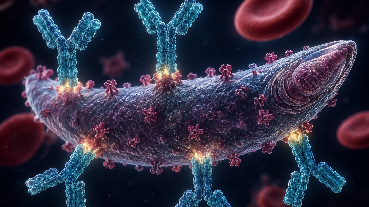
Figure 3. Antibody tests don't detect the parasite directly — they detect the immune system's Y-shaped weapons built specifically to fight it.
Serology, the branch of testing that looks for antibodies in blood, is how most tissue-dwelling and gut-dwelling parasites end up being confirmed even though the organism itself can't be seen. When your immune system encounters Toxoplasma, Strongyloides, Trichinella, Schistosoma, or the trypanosome that causes Chagas disease, it manufactures antibodies shaped specifically to recognize that particular parasite's surface proteins, the way a key is cut to fit one specific lock. A serology test essentially checks whether your blood contains keys that fit the lock of a given parasite. Two main classes of antibody are usually measured: IgM, which tends to appear first and signals a recent or active infection, and IgG, which develops later, sticks around for a much longer time, and in many parasitic infections can remain detectable for years or even for life, long after the parasite itself has been eliminated or gone dormant. This is precisely why a positive IgG result on its own is often described by clinicians as evidence of exposure rather than proof of an active, ongoing infection — the antibody is a historical record of having met the parasite at some point, not necessarily a live status update.
Strongyloides is a particularly instructive example of why this matters clinically. This intestinal roundworm has an unusual ability to complete an internal life cycle entirely inside its host, quietly reinfecting the same person for decades without ever causing dramatic symptoms — something known as autoinfection. Because the worm burden in the gut can be too low and too intermittent for a stool exam to reliably catch, serology has become the preferred screening tool, especially before someone starts an immunosuppressive medication, since a dormant Strongyloides infection can turn into a severe, sometimes fatal, hyperinfection if the immune system that had been holding it in check is suppressed. Chagas disease follows a similar pattern: the acute phase is often mild enough to go unnoticed, but the chronic phase can persist silently for 10 to 30 years before causing heart or digestive complications, and it's almost always the chronic-phase antibody test — not a hunt for the trypanosome itself — that eventually uncovers it.
Toxoplasmosis and Pregnancy — Why Antibody Timing Matters So Much
Nowhere does the timing of antibody testing matter more than in pregnancy screening for toxoplasmosis. Most adults who carry Toxoplasma gondii antibodies were infected years or decades earlier, usually with no memory of ever being sick, and the parasite's dormant cysts pose essentially no ongoing risk to a healthy adult immune system. The concern shifts dramatically, however, when a person becomes infected for the first time during pregnancy, because the parasite can cross the placenta and, depending on the trimester, cause serious harm to a developing fetus — ranging from vision problems to more severe neurological effects. This is exactly why obstetric guidelines in many countries focus so heavily on distinguishing a brand-new infection from old, harmless immunity, rather than simply reporting whether antibodies are present at all.
In practice, this means a pregnant patient with a positive IgG and a negative IgM is usually reassured that her exposure happened long before conception and poses no threat to the pregnancy, while a positive IgM triggers a more careful workup, since IgM alone can sometimes linger for months after an infection or, less commonly, produce a false-positive result. When the picture is ambiguous, labs can turn to a specialized test called IgG avidity, which measures how tightly the antibodies bind to the parasite; antibodies produced early in a fresh infection bind loosely (low avidity), while antibodies from an old, mature immune response bind tightly (high avidity) — in effect giving the lab a rough timestamp on when the exposure most likely occurred, months after the fact, just from the physical characteristics of the antibody itself.
Schistosomiasis — When the Worm Itself Never Comes Near a Blood Test
Schistosomiasis offers one of the clearest illustrations of how a parasite's specific anatomy dictates the entire testing strategy built around it. Adult Schistosoma worms actually do live inside blood vessels — specifically the veins draining the intestines or bladder, depending on the species — which might suggest a blood smear should be able to find them the way it finds malaria. In reality, the adult worms are far too large and too few in number to ever appear floating freely in a standard blood draw the way single-celled malaria parasites do; a blood smear was never built to spot something the size and shape of a thin worm nestled inside a specific vein. Instead, the worms release large numbers of microscopic eggs that travel to the intestines or bladder to be shed in stool or urine, which is why direct diagnosis in endemic settings still relies on finding those eggs under a microscope in the appropriate body fluid rather than in blood at all.
For someone testing outside an endemic region — for instance, a returning traveler with a low egg burden that a stool or urine exam might miss entirely — antibody testing becomes the more sensitive option, since it doesn't depend on catching eggs in the right sample at the right moment. The tradeoff mirrors what's true of every serology test: a positive antibody result confirms exposure to Schistosoma at some point, and needs to be paired with symptoms, geography, and sometimes a follow-up egg count to determine whether treatment is currently needed, versus reflecting a past infection that has already resolved.
The Window Period Problem — Why a Negative Result Doesn't Always Mean "No Infection"
Antibody tests share a limitation with every other kind of immune-response testing in medicine: they can only detect a response that has already had time to develop. After a new infection, it typically takes somewhere between one and several weeks for the immune system to produce enough antibody to register on a standard test — a stretch of time referred to as the window period — the same underlying concept used in testing for viral infections, applied here to a very different class of organism. Someone tested three days after a mosquito bite that transmitted a filarial parasite, for example, may test antibody-negative simply because their immune system hasn't caught up yet, not because no infection is present. This is one of the main reasons a doctor evaluating a possible parasitic infection will often ask pointed questions about exactly when and where exposure might have happened — the timing tells them whether a current negative result is trustworthy yet, or whether it's simply too early to know and a repeat test in a few weeks is warranted.
The opposite problem — a positive result that persists long after the infection is gone — is just as important to understand. Because IgG antibodies can remain detectable for years, someone who was successfully treated for toxoplasmosis a decade ago may still show a positive IgG result today, even though there's no active infection left to treat. Clinicians typically resolve this ambiguity by looking at the pattern together: a positive IgM alongside a positive IgG generally points toward a more recent infection, a positive IgG with a negative IgM more often reflects old, resolved exposure, and in genuinely ambiguous cases, a rising antibody level measured across two samples drawn several weeks apart — called titers — can reveal whether the immune response is actively climbing (suggesting a current infection) or has already plateaued (suggesting past exposure).
The Quiet Clue Hiding in a Routine Complete Blood Count
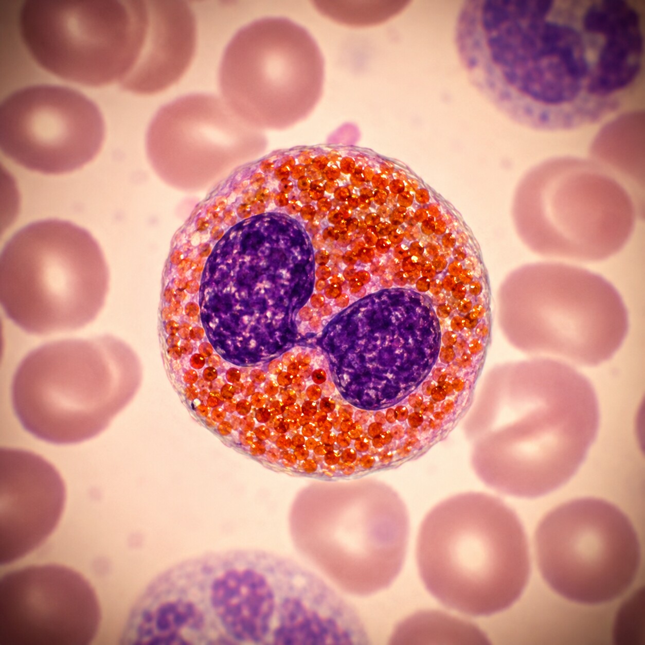
Figure 4. Eosinophils, identifiable by their bright orange-red granules, are the immune cells most closely tied to parasitic infection.
Long before anyone orders a dedicated parasite test, a routine complete blood count — one of the most common blood panels ordered in medicine — can drop a hint that something is worth investigating further. Among the five types of white blood cells your immune system uses, one type called eosinophils specializes specifically in fighting parasites, particularly the larger, multicellular ones like worms, in a way that other white blood cells generally don't. Eosinophils release toxic proteins aimed at organisms too large to be swallowed whole the way bacteria are, and when a parasitic worm is present anywhere in the body, the bone marrow frequently ramps up eosinophil production in response, causing the eosinophil count on a CBC to climb above the normal range — a finding called eosinophilia. Because this pattern shows up on such a routine, inexpensive test, an elevated eosinophil count found for an entirely unrelated reason is one of the more common ways a hidden parasitic infection first comes to a doctor's attention, prompting more specific serology or stool testing that wouldn't otherwise have been ordered.
It's worth being clear about the limits of this clue, though. Eosinophilia is a nonspecific signal — allergies, asthma, certain medications, and a handful of unrelated conditions can raise eosinophils too, and, confusingly, some parasitic infections don't reliably raise them at all, particularly protozoan infections like Giardia or Toxoplasma, which tend to trigger a different arm of the immune system entirely. A normal eosinophil count is therefore reassuring but not proof of absence, and an elevated one is a reason to look further, not a diagnosis on its own. The real value of the number is as a trigger for the right follow-up test, not as a final answer by itself.
PCR and Molecular Testing — Finding the Parasite's Own DNA
Over the past two decades, a third detection method has become increasingly available for parasitic infections: polymerase chain reaction (PCR) testing, which searches a blood sample directly for fragments of a parasite's genetic material rather than looking for the organism itself under a microscope or waiting for an antibody response. Because PCR can amplify even a tiny amount of parasite DNA into a quantity large enough to detect, it can pick up infections with a parasite burden too low for a blood smear to catch by eye, and because it's detecting the organism's genetic fingerprint directly, it isn't subject to the days-to-weeks delay of an antibody response — making it especially useful very early in an infection, when serology might still be falsely negative. PCR has become particularly valuable for malaria in cases with a very low parasite count, for distinguishing between look-alike species like Plasmodium and Babesia when a smear result is ambiguous, and for detecting Chagas disease during its brief acute phase, before chronic-phase antibodies have had time to fully develop.
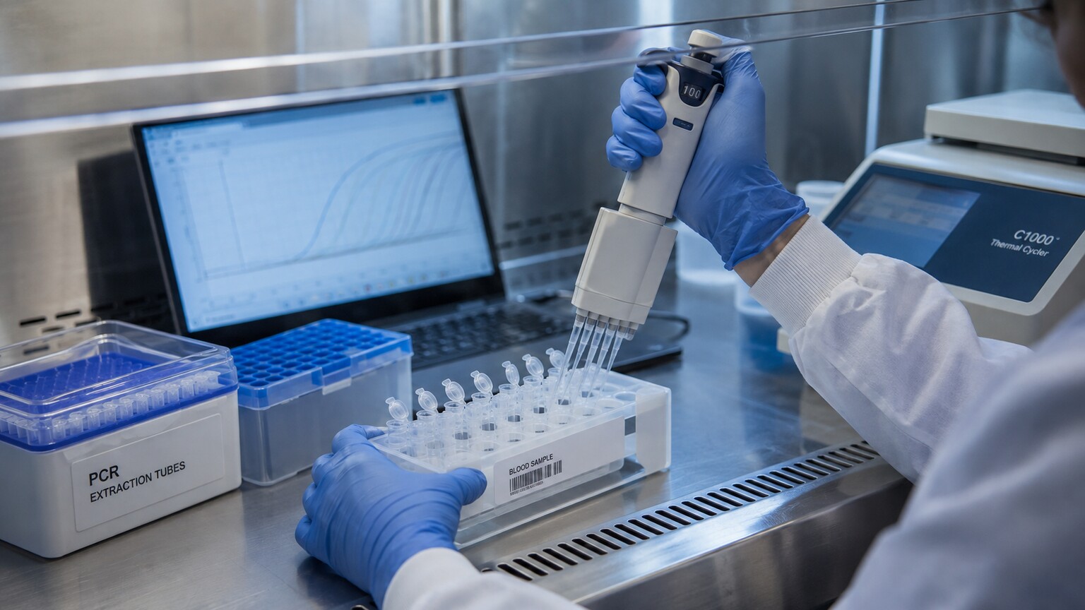
Figure 5. Molecular testing amplifies trace amounts of parasite DNA from a blood sample, catching infections a microscope alone might miss.
PCR isn't a universal replacement for the older methods, though — it's typically more expensive, requires specialized equipment that isn't available in every lab or every country, and for a handful of parasites it hasn't outperformed a skilled technician's eye on a well-prepared blood smear, which remains fast, cheap, and, in malaria in particular, still capable of delivering something PCR generally can't in an emergency setting: an immediate estimate of how severe the infection is, based directly on what percentage of red blood cells are visibly infected. In practice, most clinical labs use these methods as complements rather than substitutes, reaching for PCR specifically when microscopy or serology alone leaves real uncertainty.
Chagas Disease and Blood Donation — How Serology Quietly Protects the Blood Supply
One of the largest-scale, most consequential uses of parasite serology has nothing to do with a sick patient walking into a clinic at all — it happens behind the scenes, every time someone donates blood. Chagas disease, caused by the parasite Trypanosoma cruzi and historically concentrated in parts of Latin America, can be transmitted not only through the bite of the triatomine insect that normally spreads it, but also through blood transfusion and organ transplantation, since the trypanosome can circulate at low levels in an infected person's blood for years or decades without ever producing noticeable symptoms. Because millions of people who were exposed decades ago and never developed symptoms are otherwise perfectly eligible blood donors, national blood supplies in countries with any meaningful Chagas prevalence routinely screen every donation for Trypanosoma cruzi antibodies, catching silent chronic infections that the donor themselves may have had no idea they were carrying.
This screening program is a useful case study in how much weight a single antibody test can carry once it's applied at scale: a positive result on a donor screen doesn't necessarily mean that individual is in any immediate danger, since many people live full lives with chronic Chagas infection and never develop the heart or digestive complications that can eventually appear. But it does mean their blood is no longer used for transfusion, and it opens the door to a conversation about further evaluation and, in some cases, treatment — all triggered by a serology test that was never ordered because anyone suspected an active illness, but because population-level screening is simply the only practical way to catch an infection this good at staying hidden.
What Determines Which Test Your Doctor Actually Orders
Given how many different testing strategies exist, the specific test ordered comes down almost entirely to two pieces of information: where you've been, and what your body is doing. Recent travel to a malaria-endemic region with a fever is one of the few genuine medical urgencies in parasitology, since malaria can turn severe within days, so a blood smear is typically ordered immediately rather than waiting on results that take longer to process. A history of eating undercooked pork or wild game points toward Trichinella serology. Freshwater exposure in a region where schistosomiasis is present points toward that specific antibody panel. Chronic, vague gastrointestinal symptoms after travel more often lead to stool testing first, with blood-based serology reserved for parasites, like Strongyloides, that are notoriously difficult to catch in stool. An incidentally elevated eosinophil count on an unrelated blood panel, with no clear travel history at all, often prompts a broader serology screen to check for common possibilities rather than one targeted test. None of this is guesswork on the clinician's part — it reflects the underlying biology of where each parasite actually lives in the body, and matching the right tool to that biology is what makes the difference between a test that can find the answer and one that was never going to.
Cost and turnaround time factor into these decisions too, in ways that are easy to overlook from the patient side of the conversation. A blood smear can be read within the hour by a trained technician with nothing more than a microscope, glass slides, and Giemsa stain — equipment and expertise available in nearly every hospital lab in the world, which is part of why it has remained the frontline tool for malaria for so long despite newer alternatives existing. Serology panels typically take longer, often a day or more, since many rely on batches of samples run together for efficiency rather than one-at-a-time processing. PCR, while fast once a sample reaches a lab equipped to run it, often has to be sent out to a reference laboratory or major medical center, adding days to the turnaround in smaller communities. A clinician weighing these tradeoffs against how urgently an answer is needed — same-day for a possible malaria case with a high fever, versus a more relaxed timeline for a chronic, stable-seeming case of suspected old exposure — is quietly part of what determines which specific test lands on the order form.
Frequently Asked Questions
If my blood test for parasites came back negative, does that rule out an infection?
Not necessarily, and the answer depends entirely on which parasite was being tested for and by which method. A negative blood smear for malaria drawn during an active infection is genuinely reassuring, since the organism is either present in the blood or it isn't. A negative antibody test, on the other hand, can simply mean the test was drawn too early, before the immune system had time to respond — and for parasites that live in the gut or in tissue rather than in blood, a blood test was never going to detect the organism directly in the first place, regardless of timing.
Why did my doctor order both a blood test and a stool test?
Because they're checking two different things. A stool test looks directly for organisms or their eggs in the intestines, which is where most gut-dwelling parasites actually live and can be found. A blood test, in this context, is usually checking for the immune system's antibody response or for a clue like elevated eosinophils — useful supporting evidence, but not a replacement for looking at stool when the parasite in question lives in the gut.
Can a routine blood panel accidentally reveal a parasitic infection I didn't know I had?
Yes, this happens fairly often. An elevated eosinophil count on a standard complete blood count, ordered for an entirely unrelated reason, is one of the more common ways a silent parasitic infection first gets noticed, prompting a doctor to order more specific follow-up testing. It's an indirect clue rather than a diagnosis on its own, but it's frequently the first thread that gets pulled.
If I already had a positive antibody test years ago, will every future blood test show positive too?
Often, yes — this is one of the most common points of confusion in parasite serology. IgG antibodies against many parasites, including Toxoplasma and Trypanosoma cruzi, can remain detectable for years or for life, even long after any active infection has resolved or been successfully treated. A positive IgG on a future test usually reflects that old exposure rather than a new or ongoing infection, which is why clinicians interpret these results alongside IgM status, symptoms, and timing rather than treating a positive IgG alone as evidence of current illness.
How soon after possible exposure should a parasite blood test be done?
It depends on the parasite and the test type. Direct methods like a malaria blood smear can detect the organism as soon as it's circulating in measurable numbers, sometimes within days of symptom onset. Antibody-based tests generally need one to several weeks after exposure before the immune system has produced enough antibody to register reliably, so testing too early can produce a falsely reassuring negative result. When there's a real concern and the timing is uncertain, a doctor may recommend an initial test followed by a repeat test several weeks later to confirm the result either way.
Conclusion
Whether a blood test can catch a parasitic infection isn't a yes-or-no question so much as a "which parasite, and what kind of test" question. A small group of blood-dwelling organisms, malaria chief among them, can be seen directly under a microscope or detected by PCR, giving fast and highly specific answers. Everything else relies on catching the immune system's footprint — antibodies that take time to rise and, once risen, can linger for years — or on an indirect clue like an elevated eosinophil count nudging a doctor toward the right follow-up test. None of these tools is wrong; they're simply built for different biology, and the most common source of confusion is expecting one blood draw to answer a question that actually requires knowing where, in the body, a particular parasite chooses to live. Anyone with recent travel, unexplained gastrointestinal symptoms, or an unusual result on a routine blood panel has good reason to bring that specific context to a healthcare provider, since it's the detail that tells them which test will actually work.
It's also worth remembering that a single blood test, of any kind, is rarely meant to stand entirely on its own. Clinicians reading a serology result are almost always weighing it against the same three things at once: the biological plausibility of exposure given where you've actually been and what you've actually eaten or been bitten by, the timeline of your symptoms relative to how quickly that particular parasite tends to cause them, and, where available, a second data point — a repeat titer, an eosinophil count, a stool exam, or an imaging study — that either supports or complicates the first result. A lab value in isolation, stripped of that context, can be genuinely difficult to interpret correctly even for an experienced clinician, which is exactly why the conversation about travel history and symptom timing that can feel repetitive during a doctor's visit is doing real diagnostic work behind the scenes, not just filling out a form, and it's part of why bringing a clear, honest account of that history to an appointment tends to get to the right answer faster than the test result alone ever could.
Still Not Sure What Your Results Mean?
Upload your labs and get a complete, visual, plain-language interpretation of every biomarker — delivered to your inbox in under 15 minutes.
Get My ReportThis article is for educational purposes only and does not constitute medical advice. Always consult your healthcare provider regarding your specific lab results.