
Understanding How Parasites Are Identified in Lab Testing
Handing over a stool sample feels like a strange, low-tech, almost old-fashioned step in an otherwise high-tech modern medical system, and it's easy to picture what happens next as simple: someone smears a bit on a slide and looks at it under a microscope. The real process is considerably more deliberate and technically involved than that mental picture suggests, and understanding it actually explains a lot about why parasite testing sometimes takes several days rather than hours, why your doctor might ask for more than one sample collected on different days, and why a lab report can list a specific method by name right next to your result. This article walks through the actual multi-step identification process — concentration, staining, microscopic identification, and increasingly, molecular and antigen-based methods — and why each step exists to solve a specific technical problem the one before it couldn't solve on its own, from start to finish exactly as a real sample moves through it.
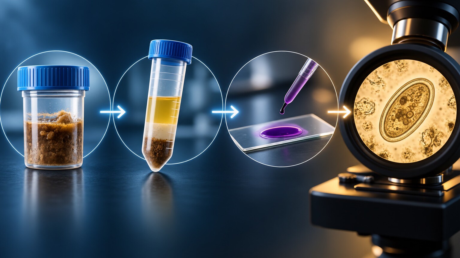
Figure 1. Parasite identification is a sequential laboratory workflow — concentration, preservation, staining, and microscopic or molecular analysis — not a single glance under a microscope.
Why a Raw Sample Alone Usually Isn't Enough
A direct wet mount — placing a tiny, unprocessed smear of stool directly on a slide with a drop of saline and looking at it immediately under a microscope — is the fastest possible method, and it's genuinely useful for one specific job: catching motile trophozoites, the actively moving, feeding form some protozoa take, since that movement is easiest to see in a fresh, unprocessed sample before the organism settles or dies. But a direct wet mount examines only a tiny fraction of a gram of stool, and most parasite eggs, cysts, and larvae are shed in relatively low numbers, especially early in an infection or with a light organism burden. Looking at an unconcentrated sample is a little like trying to find a handful of specific grains of sand scattered across an entire beach by only checking one small patch — the organism can genuinely be present in the sample and still go completely unseen simply because the technician's microscope field never happened to land on it.
There's a second, separate reason a direct wet mount alone falls short, beyond simple rarity: stool naturally contains an enormous amount of undigested plant fiber, bacteria, yeast, and other debris that visually competes with any parasite structures present, making a raw, unprocessed sample genuinely cluttered to examine even when an organism is sitting right there in the field of view. A direct wet mount does still have one genuine, meaningful advantage no downstream processing step can fully replicate, however, which is exactly why it's typically performed first, before any of the other methods described below, rather than skipped in favor of them: it's the only method that can catch a trophozoite's characteristic movement — a gliding or flowing motion in some species, a more jerky, directional movement in others — while the organism is still alive, since every later step in this process, by design, either kills or otherwise chemically alters the organism to prepare it for a different, more detailed kind of examination further down the line.
Concentration: Making Rare Organisms Easier to Find
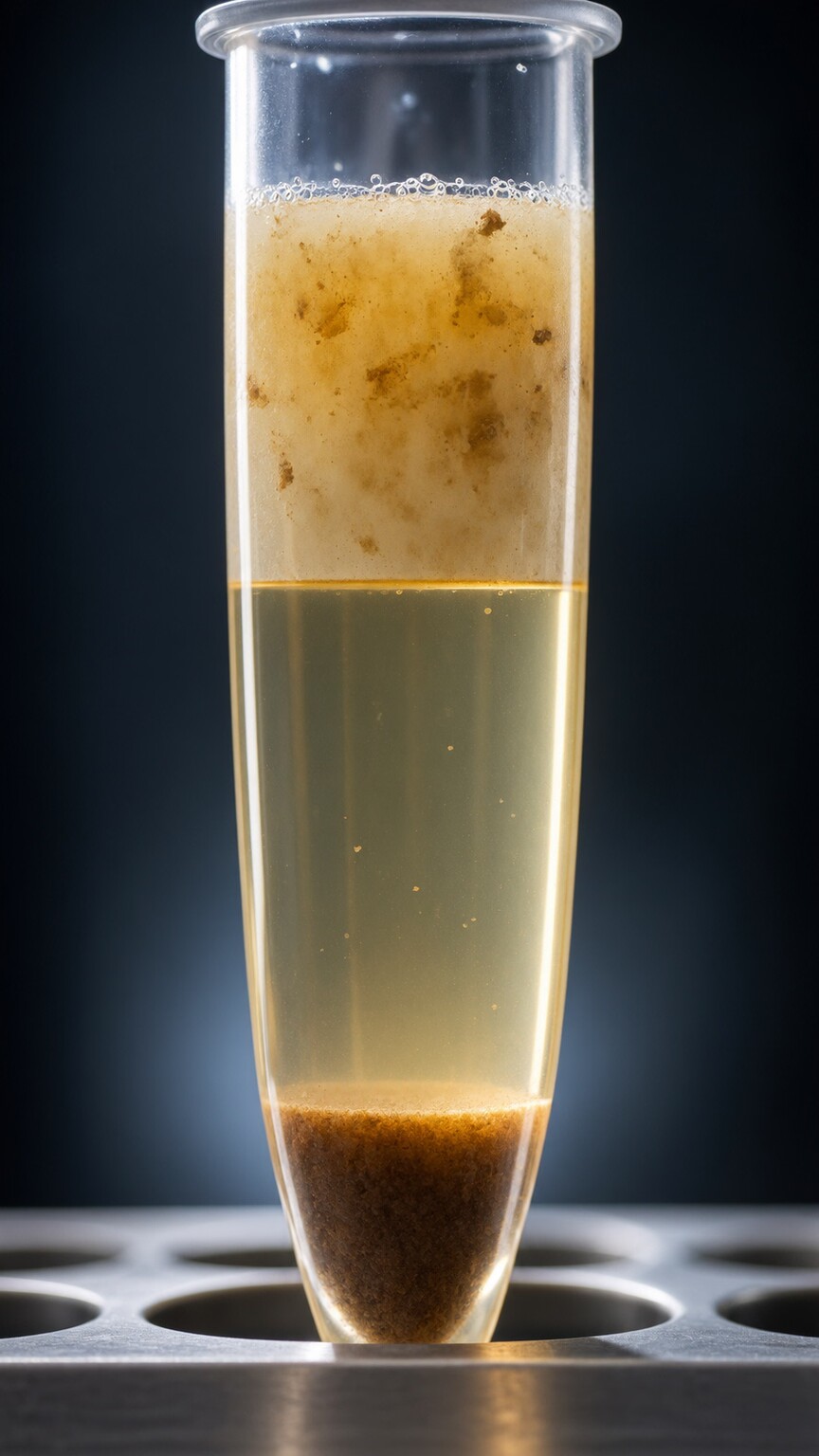
Figure 2. Formalin-ethyl acetate concentration separates a stool sample into distinct layers by density, pulling heavier parasite eggs and cysts into a small sediment pellet that's far easier to examine than raw stool.
This is exactly the specific, well-documented, longstanding technical problem that concentration techniques, described in detail next, were originally developed and refined over decades to solve. The most widely used method, formalin-ethyl acetate sedimentation, works by mixing the stool sample with a formalin solution (which preserves and kills the organisms present, stopping the sample from changing before it can be examined) and ethyl acetate (a solvent that dissolves fecal fat and debris). The mixture is then spun in a centrifuge at high speed, and because different components of the mixture have different densities, they separate into distinct layers: a top layer of dissolved fat and debris, a middle layer of formalin solution, and a small, dense sediment pellet at the very bottom containing the heavier material — including any parasite eggs, cysts, or larvae present, since these structures are generally denser than the surrounding organic debris.
The technician then discards the upper layers and examines only that concentrated sediment pellet, which contains organisms from a proportionally much larger amount of original stool than a single unconcentrated smear ever could. This single step is why a properly performed lab test can detect organisms present in numbers far too low for a direct wet mount to ever catch, and it's a major part of why the O&P (ova and parasite) exam remains a meaningfully more sensitive method than simply looking at raw stool, even though both ultimately rely on the same microscope and the same trained eye.
An older concentration technique, zinc sulfate flotation, works through the opposite physical principle and is occasionally still used for specific purposes: instead of spinning heavier material down to the bottom, it uses a solution dense enough that parasite eggs and cysts, being lighter than the solution itself, float upward to the surface, where they can be collected on a wire loop or a coverslip placed directly on top. Flotation techniques tend to produce a cleaner, less debris-cluttered final preparation than sedimentation, making some organisms easier to spot clearly, but they carry a real tradeoff: certain heavier or operculated (hinged-lid) eggs, including some tapeworm and fluke eggs, don't float reliably in the solution and can be missed entirely by a flotation-only approach, distorting collapsed by the solution's high salt concentration rather than staying at the surface. This is exactly why formalin-ethyl acetate sedimentation, which reliably captures organisms across a broader range of size and density, has become the more universally used default method in most modern clinical labs, with flotation reserved for specific situations or research settings where its particular strengths outweigh this blind spot.
Trying to make sense of a stool or blood lab report on your own? Upload your results and get a complete, plain-language breakdown in under 15 minutes.
Analyze My ResultsStaining: Turning a Transparent Organism Into a Visible One
Concentration solves the problem of finding rare organisms, but it doesn't solve a second, separate problem: many protozoa, especially their internal structures, are nearly transparent and extremely difficult to distinguish from surrounding debris or from each other under plain light. This is where a permanent stained smear comes in — a separate portion of the sample, fixed onto a slide with a chemical preservative and then stained, most commonly with a trichrome stain, a dye combination that colors different cellular structures in contrasting shades (typically greenish-blue cytoplasm against a more purplish-red nucleus).
This staining step is specifically what allows a technician to identify a protozoan down to the exact species level, since correctly and confidently distinguishing between closely related organisms — some entirely harmless, some genuinely clinically significant — often comes down to subtle internal features: the exact number and arrangement of nuclei, the size and position of a structure called the karyosome inside the nucleus, or the precise shape of chromatoid bodies (rod-like internal structures some cysts contain). Without staining, many of these distinguishing features are essentially invisible, which is exactly why a lab report distinguishing "Entamoeba histolytica" (a species that can cause genuine invasive disease) from the closely related, generally harmless "Entamoeba dispar" — two organisms that look nearly identical unstained — often requires this specific staining step, or in many modern labs, a molecular test entirely, discussed further below.
Trichrome isn't the only staining option available, and which specific one a lab actually reaches for depends partly on exactly what it's trying to see in that particular sample. An alternative, iron-hematoxylin staining, produces even sharper contrast for certain fine internal structures and is sometimes preferred specifically for identifying particular protozoan species whose distinguishing features are subtle even under trichrome, though it takes longer to perform and involves more preparation steps, which is part of why trichrome remains the more commonly used default in routine clinical practice. A separate, specialized stain, modified acid-fast staining, is used specifically for a different group of organisms — Cryptosporidium and Cyclospora — which don't take up trichrome stain well at all due to a chemically resistant outer wall, but do reliably absorb this particular acid-fast dye, turning a distinctive pink-red against a blue-green background. Without this specific stain, these two particular organisms can be genuinely difficult to spot with any of the other methods described in this article, which is exactly why a lab suspecting either one specifically requests this targeted staining approach rather than relying on trichrome alone.
The Trained Eye: What Identification Actually Requires
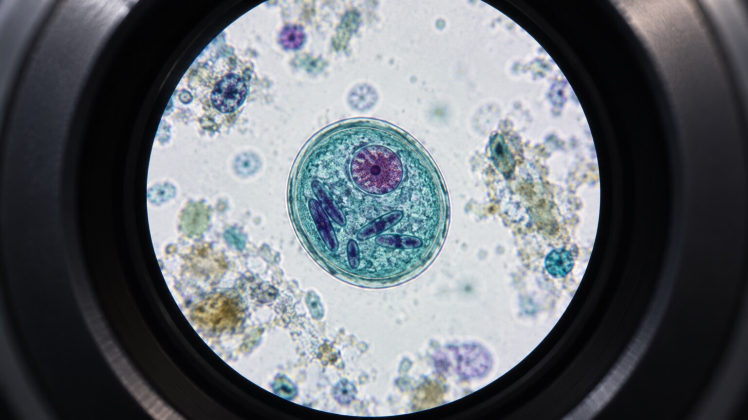
Figure 3. Correct species identification often hinges on subtle internal morphological details — nuclear count, karyosome position, and chromatoid body shape — that only become clearly visible after staining.
Reading a stained slide accurately is a genuinely specialized clinical skill, not a routine task any lab worker can pick up casually. Clinical microbiologists and medical laboratory scientists trained in parasitology spend considerable time learning to distinguish dozens of morphologically similar organisms from each other and from non-parasitic look-alikes — plant material, yeast cells, and pollen grains commonly found in stool can all superficially resemble parasite eggs or cysts to an untrained eye, and misidentifying one of these harmless look-alikes as a parasite (or vice versa) is a recognized source of laboratory error. This is exactly why proficiency testing — where labs are periodically sent unknown samples specifically to verify their technicians can identify organisms correctly — is a required, ongoing part of maintaining laboratory accreditation for parasitology testing, rather than a one-time qualification.
A significant part of this specialized training involves learning what a parasite structure is not, which turns out to be just as important as learning what it is. Stool naturally contains a wide range of plant material from a normal diet — undigested pollen grains, plant hair cells, starch granules, and fungal spores — several of which can superficially resemble a parasite egg or cyst in size and rough shape at a quick glance, particularly to someone newer to the field. Distinguishing a genuine helminth egg from a look-alike plant structure generally comes down to a combination of precise size measurement (parasite eggs and cysts fall within well-established, narrow size ranges, so a structure just outside that range is a meaningful red flag against a parasite identification), shell or wall texture under higher magnification, and internal content, since a true egg typically contains a developing embryo or a characteristic internal structure that a plant fragment simply won't show. This is exactly the kind of judgment call that separates a properly trained parasitology technician from someone simply looking at a slide, and it's a large part of why proficiency testing programs specifically include deliberately ambiguous or look-alike samples, not just clear-cut positive and negative controls.
Air bubbles trapped under the coverslip during slide preparation present a similarly deceptive, if more mundane, pitfall worth mentioning: a spherical air bubble, viewed at the wrong angle or focal depth, can occasionally mimic the rough size and rounded outline of a protozoan cyst closely enough to give an inexperienced observer pause, which is exactly why adjusting the microscope's fine focus to examine a suspicious structure from multiple depths — something a genuine three-dimensional organism will look subtly different at, while a flat air bubble largely won't — is one of the very first practical habits taught in parasitology training, alongside the size-measurement and internal-content checks described above.
Measurement itself is a more central part of the identification process than most people would guess. A calibrated micrometer scale built directly into the microscope's eyepiece lets a technician measure a structure's exact dimensions in micrometers while it's still under direct observation, and this measurement is frequently the single deciding factor between two otherwise similar-looking possibilities. Several helminth eggs, for example, look broadly similar in overall shape to an untrained eye but fall into distinctly different, non-overlapping size ranges specific to their species — meaning a technician who skips this measurement step, or measures imprecisely, risks a genuine misidentification even while looking directly at a textbook-clear structure, simply by relying on shape and general impression alone rather than confirming the specific size range. This calibrated eyepiece scale itself has to be verified against a known reference standard on a regular schedule as part of routine lab quality control, since even a subtle drift in its calibration would silently skew every single size-based measurement made with it until caught.
Molecular Testing: Finding the Organism's Genetic Signature Directly

Figure 4. Multiplex PCR testing detects small fragments of a parasite's own DNA directly, sidestepping the need for a visually intact, recognizable organism to be present in the sample at all.
An entirely different approach, now used increasingly often either alongside or instead of microscopy, is molecular testing — most commonly a multiplex PCR (polymerase chain reaction) panel. Rather than relying on a technician visually recognizing an intact organism's shape, PCR looks for a specific, small fragment of genetic material — DNA or RNA — unique to a given parasite species, and then chemically copies that fragment millions of times over in repeated cycles until there's enough of it to reliably detect, even if the original amount present in the sample was vanishingly small.
This genetic-signature approach solves several of microscopy's inherent limitations at once: it doesn't require the organism to be intact, alive, or in a particular life-cycle stage, and one panel can simultaneously screen for a dozen or more different parasitic, bacterial, and viral causes of gastrointestinal illness from a single stool sample submitted just once. Its main tradeoffs are cost and availability — multiplex PCR panels remain more expensive, require specialized laboratory equipment, and are less universally available than a standard O&P exam — and the fact that a PCR panel only tests for the specific organisms its particular panel was designed to detect, meaning an unusual or unexpected parasite outside that panel's target list could still be missed even on an otherwise thorough molecular test.
It's worth understanding the actual chemistry behind why PCR is capable of detecting such a tiny starting amount of genetic material, since the mechanism itself explains both the method's power and one of its more debated limitations. PCR works in repeated cycles, each one roughly doubling the number of copies of the target DNA fragment present — starting from just a handful of original copies, after around 30 to 40 cycles (a process that takes a few hours total), the reaction can generate billions of copies of that same fragment, more than enough for standard detection equipment to reliably register a positive signal. This exponential doubling is what allows PCR to find a genuine needle in a haystack that traditional microscopy would need many, many more organisms present to ever spot directly.
This same extreme sensitivity is also the source of a genuine, actively discussed limitation: PCR detects genetic material, not necessarily a live, actively infecting organism, meaning it can occasionally pick up DNA from an organism that's already dead, cleared, or present in such a small, clinically insignificant quantity that it wouldn't actually be causing any disease. Distinguishing a clinically meaningful positive from this kind of low-level detection is an active area of ongoing research and refinement in molecular parasitology, and it's part of why some clinicians interpret a PCR result together with the patient's actual symptoms and history rather than treating any positive molecular finding as automatically equivalent to active, clinically significant infection requiring treatment.
Quantitative PCR, a variant that reports not just presence or absence but an estimated relative amount of genetic material detected, has emerged partly as a response to this exact limitation, since a very low quantity result carries different clinical weight than a strongly positive one and gives a clinician more nuanced information to weigh alongside the patient's actual presentation. This more granular reporting isn't universally available on every panel yet, but it represents the direction the field is generally moving as molecular methods continue to mature and labs work to make results more clinically actionable rather than simply more sensitive.
Antigen Testing: A Fast, Targeted Middle Ground
A third method, antigen testing, sits conceptually between traditional microscopy and full molecular testing. Rather than looking for the organism itself or its genetic material, an antigen test uses lab-made antibodies designed to bind specifically to a protein found on a particular parasite's surface — conceptually similar to how a home pregnancy test's antibodies bind to a specific hormone. This approach is commonly used as a fast, targeted test for two specific, commonly encountered organisms, Giardia and Cryptosporidium, both of which can be genuinely difficult to spot reliably and consistently under a microscope even in a properly concentrated, well-stained sample, since their cysts are quite small and can easily resemble ordinary debris.
Antigen testing typically returns a result faster than a full microscopic workup and doesn't require the same level of specialized morphological training to interpret, which makes it a practical option in settings without ready access to an experienced parasitology microscopist. Its limitation is narrowness: an antigen test only checks for the one or two specific organisms it was designed to detect, so a negative antigen result says nothing about the dozens of other parasites a broader O&P exam or PCR panel would have screened for at the same time.
Several formats of antigen testing exist, and understanding the difference clarifies why one lab's turnaround time might be dramatically faster than another's for what looks like the same test on paper. An ELISA (enzyme-linked immunosorbent assay) format runs samples in a specialized plate reader, typically processing a batch of samples together, and offers slightly higher sensitivity than the alternative rapid-format tests, but usually requires being run at specific times during a lab's daily workflow rather than the instant a sample arrives. A lateral flow immunochromatographic assay — visually similar in concept to a home rapid COVID or pregnancy test, with a sample applied to one end of a test strip and a colored line appearing if the target antigen is present — trades a small amount of sensitivity for genuine speed, often producing a result within 15 to 30 minutes and requiring no specialized batch-processing equipment, which is exactly why it's the more common choice in urgent-care or point-of-care settings where a same-visit answer meaningfully changes what happens next for the patient.
Helminths vs. Protozoa: Why Identification Strategy Differs by Organism Type
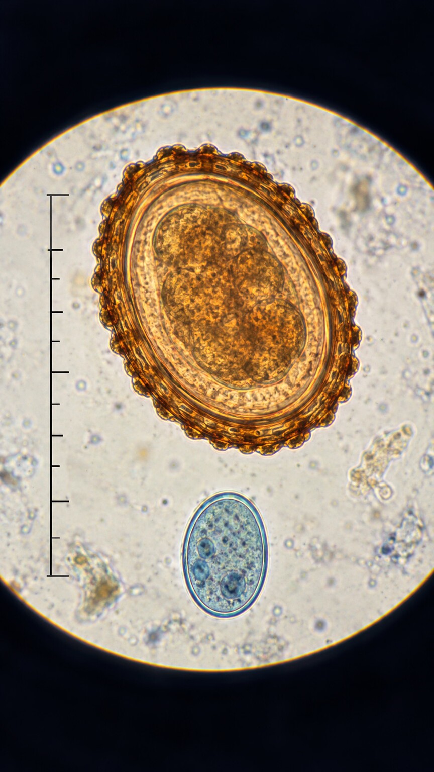
Figure 6. Helminth eggs are generally large and structurally distinctive enough to identify by shape alone, while protozoan cysts are smaller, less visually distinct, and typically require staining for a confident species-level call.
The two broad categories of intestinal parasites — helminths (worms) and protozoa (single-celled organisms) — genuinely differ enough in size and structure that the identification process leans on different strengths for each. Helminth eggs are relatively large, often with a distinctive shell shape, surface texture, or size that alone allows a trained technician to identify the genus, and sometimes species, from concentration and direct microscopy without necessarily needing a permanent stain at all — a roundworm egg's thick, mammillated (bumpy) outer shell, for instance, looks distinctly different from a whipworm egg's barrel shape with clear polar plugs at each end, and both look nothing like a tapeworm egg's thin, near-spherical shell.
Protozoa present the opposite challenge: they're single cells, often only a few micrometers across, and many different species share a broadly similar round or oval shape at a casual glance. This is exactly why the permanent stained smear described earlier carries so much more of the identification burden for protozoa specifically — the precise, fine internal detail a proper stain reveals (exact nuclear count, karyosome position, chromatoid body shape) is frequently the only reliable way to tell one protozoan species from another, whereas a helminth egg's overall size and shell structure alone are often sufficient. This distinction is part of why a lab report sometimes identifies a worm egg down to species with confidence from concentration alone, while a protozoan finding might explicitly note it required the stained smear, or a follow-up antigen or molecular test, to confirm.
Larvae, a third distinct form some helminths take at a specific point in their life cycle, present their own particular identification wrinkle worth mentioning here. Unlike an egg, which is generally a static, inert structure whose shape and shell alone provide most of the identifying information, a larva is an actively developing form with its own set of distinguishing features — the shape and proportions of its internal structures, its overall length and the ratio between different body segments, and in some cases, subtle differences visible in its motility pattern that echo the trophozoite-movement clue described earlier for protozoa. Because certain hookworm and threadworm larvae look genuinely similar to each other at a casual glance, correctly telling them apart sometimes requires a specific technique called a Baermann or agar plate culture, which allows larvae to actively migrate out of a stool sample and into a collection medium over a period of hours, both concentrating them and, in the agar plate variant, leaving visible migration tracks in the agar itself that add yet another piece of identifying information beyond the larva's static appearance alone.
Why Doctors Often Ask for More Than One Sample
Understanding this whole process explains a detail many patients find frustrating without context: why a doctor sometimes asks for stool samples collected on three separate days rather than accepting just one. Many parasites don't shed eggs, cysts, or larvae into stool continuously and evenly — shedding can be intermittent, heavier on some days than others, depending on the organism's specific life cycle. A single sample, even one processed through every method described above, only captures a snapshot of whatever happened to be shed on that particular day. Collecting samples across multiple days meaningfully increases the overall chance of catching the organism during a window when it's actually present in detectable numbers, which is a completely different, and complementary, strategy from any single identification technique — it's addressing timing, not sensitivity of the test method itself.
Travel history and specific exposure risks factor into this same decision in a way worth understanding, since they change how aggressively a doctor pursues repeat testing after an initial negative result. Someone with a strong, specific exposure — recent travel to a region where a particular parasite is common, a known contaminated water source, or close contact with a diagnosed case — carries a higher pre-test likelihood of true infection, which is exactly the situation where a single negative result is least reassuring and most likely to prompt a doctor to request additional samples or move directly to a more sensitive method like PCR, rather than accepting the first negative at face value. Someone being tested with no particular exposure history or risk factor, by contrast, starts from a lower baseline likelihood, meaning a single well-processed negative carries more genuine reassurance in that specific context — the same negative result, in other words, means something different depending on the pre-test probability a doctor is weighing it against, which is a broader principle in diagnostic testing generally, not unique to parasitology.
A Worked Example: Following One Sample Through the Full Process
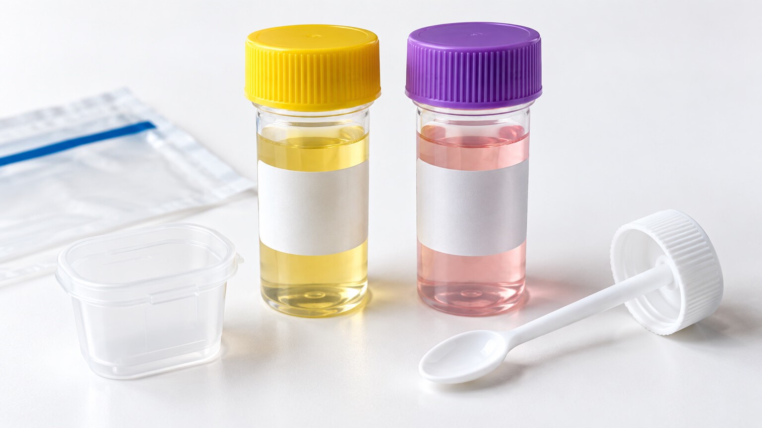
Figure 5. A standard O&P collection kit typically includes two separate preservative vials — one for concentration, one for the permanent stained smear — since each downstream method requires the sample preserved differently.
Consider a patient recently returned from international travel with persistent diarrhea, whose doctor orders a standard O&P exam. At home, the patient collects a sample into two provided preservative vials — one containing formalin for the concentration step, one containing a different fixative specifically for the permanent stained smear — since each downstream method requires the specimen preserved in a slightly different way to work correctly. At the lab, the formalin-preserved portion goes through the ethyl acetate concentration and centrifugation process described earlier, and the resulting sediment is examined under direct microscopy — this catches nothing definitive on the first pass, since only a handful of ambiguous, debris-obscured structures are visible.
The second vial's sample is then prepared as a permanent trichrome-stained smear, and under this stain, the technician identifies a protozoan cyst with a specific internal nuclear pattern consistent with Giardia — but because Giardia cysts can be genuinely tricky to distinguish with certainty from morphologically similar structures even when stained well, the lab also runs a quick antigen test on a portion of the original sample specifically for Giardia to confirm the finding definitively, rather than relying on morphology alone in a borderline case. The antigen test comes back clearly positive, providing confirmation through an entirely independent method, and the patient's doctor now has a confirmed diagnosis to guide treatment, arrived at through three distinct techniques working together rather than any single step in isolation.
This same case also illustrates why the specific collection instructions patients receive matter more than they might seem to at first. Had this patient used only one preservative vial, or let the sample sit unrefrigerated for an extended period before dropping it off, the trophozoite and cyst structures needed for the staining step could have degraded past the point of clear identification, potentially forcing a second, delayed round of sample collection and pushing the diagnosis back by days. This is exactly why lab collection kits typically come with clear written instructions about vial usage, timing, and storage, and why deviating from those instructions — even in ways that feel minor, like using only one vial when two were provided, or leaving a sample out overnight rather than refrigerating it — can genuinely compromise a result that would otherwise have been straightforward to interpret.
Frequently Asked Questions
How long does parasite identification typically take?
A standard O&P exam with concentration and staining is usually completed within one to three business days, while multiplex PCR panels can sometimes return results faster, often within 24 to 48 hours, though turnaround varies by lab and specific test ordered.
Is PCR testing always better than traditional microscopy?
Not necessarily in every scenario. PCR is generally more sensitive and faster, but it only detects the specific organisms included in its panel, while a traditional O&P exam can flag an unexpected or unusual organism outside any predefined target list.
Why do I need to keep my sample refrigerated or use a preservative vial?
Some parasite forms, particularly trophozoites, degrade quickly at room temperature and can become unrecognizable within hours. Preservative solutions and refrigeration slow this breakdown, keeping the sample's structures intact until the lab can process it.
Can a lab technician misidentify a parasite?
Yes, which is exactly why ongoing proficiency testing is a required part of laboratory accreditation. Non-parasitic material like plant fibers, pollen, or yeast can superficially resemble parasite structures, making specialized training and periodic competency verification essential.
Does a negative result from one method rule out infection completely?
Not on its own. Because different methods have different strengths and blind spots, and shedding can be intermittent, doctors often combine multiple methods or multiple collection days before confidently ruling out an infection.
Why does my lab report mention a specific test method by name?
Different methods have different strengths and blind spots, so knowing exactly which one was used helps a doctor correctly interpret the result — a negative from a targeted antigen test, for instance, means something different than a negative from a broad O&P exam.
Can a home stool test replace a lab-processed O&P exam?
Generally no for definitive diagnosis. Home tests typically screen for one or two specific organisms via antigen detection, while a full lab-processed O&P exam or PCR panel evaluates for a much broader range of parasites using multiple complementary techniques.
Conclusion
What looks from the outside like a simple stool sample dropped off at a lab is actually the starting point for a genuinely layered identification process — concentration to find rare organisms, staining to reveal the fine structural detail needed for species-level identification, a trained eye to interpret what's seen, and increasingly, molecular and antigen-based methods that sidestep microscopy's limitations entirely. Each individual method exists precisely because it solves one specific technical problem the other available methods genuinely can't, which is exactly why labs often combine more than one approach, and why understanding this whole layered process makes multiple collection days, longer turnaround times, and method-specific results listed right on a lab report make genuine sense rather than feel arbitrary or bureaucratic.
None of this is likely to change how you personally hand over a sample the next time your doctor orders this kind of testing, but it does change what a result means once it comes back. A "negative" from a single unconcentrated wet mount and a "negative" from a full, properly performed O&P exam with concentration, trichrome staining, and a targeted antigen or PCR follow-up are not the same statement of confidence, even though both might appear identically as one word on a printed report — knowing which combination of methods actually produced your result is exactly the kind of context that turns a bare lab value into something genuinely interpretable, rather than a single ambiguous word you're otherwise left to simply take entirely on faith.
Still Not Sure What Your Results Mean?
Upload your labs and get a complete, visual, plain-language interpretation of every biomarker — delivered to your inbox in under 15 minutes.
Get My ReportThis article is for educational purposes only and does not constitute medical advice. Always consult your healthcare provider regarding your specific lab results.