
What Causes False Negative Results in Parasite Testing?
A negative parasite test feels like a clean answer, but for this particular category of lab testing, "negative" carries more asterisks than almost any other test you're likely to get. Unlike a blood glucose reading or a cholesterol panel, where the substance being measured is always present in the bloodstream in some steady, predictable amount, most parasite testing depends on physically catching direct evidence of the organism — an egg, a cyst, a larva — in a single small sample collected at one specific moment. If the parasite isn't actively shedding that evidence into your stool at the exact moment your sample was collected, or if it lives somewhere the sample never reaches in the first place, the test can come back completely negative even though the infection is very much still there. Understanding exactly why this happens — the biology of the parasite's life cycle, the mechanics of how labs actually process a sample, and which organisms are notoriously harder to catch than others — is what separates a genuinely reassuring negative result from one that deserves a second look, especially if your symptoms are telling a different story than your lab report.
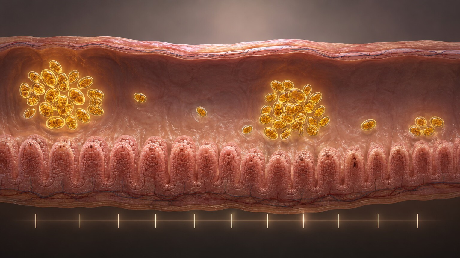
Figure 1. Many intestinal parasites shed eggs or cysts in irregular cycles rather than continuously, meaning a single stool sample can easily land during a "quiet" window with nothing detectable present.
Intermittent Shedding: The Single Biggest Cause
The most common reason a real infection produces a negative test result comes down to one specific biological quirk: many intestinal parasites don't release eggs, cysts, or larvae into your stool at a constant, steady rate. Instead, shedding often happens in cycles, with active release alternating with quieter stretches where little or nothing detectable makes it into any given sample. Giardia, one of the most common intestinal protozoan parasites worldwide, is a textbook example — infected individuals can shed cysts heavily for a few days, then shed almost nothing detectable for the next several days, in a pattern that can repeat unpredictably throughout the course of the infection.
This matters enormously for how a single stool sample gets interpreted, because a lab technician examining that sample under a microscope can only report what's physically present in the specific portion they examine. If your sample happens to be collected during one of the quiet windows in this cycle, the organism can be very much alive and active inside your intestines while the sample itself shows absolutely nothing — not because the test failed, and not because the lab made an error, but simply because the timing of collection landed on the wrong day of an inherently irregular biological cycle.
The underlying biological reason for this cyclical shedding pattern varies somewhat by organism, but a common thread involves the parasite's own reproductive and developmental timing within your intestinal tract. Many protozoan parasites alternate between an actively feeding, dividing form and a more dormant, protective cyst form, and the proportion of each type being released into stool at any given moment shifts as the organism population inside you cycles through its own internal rhythm. Some researchers have also proposed that the shedding pattern may be influenced by factors like your own gut motility, diet, and even immune activity fluctuating day to day, adding yet another layer of biological variability on top of the parasite's own life cycle timing — all of which adds up to a fundamentally unpredictable pattern from the outside, even though it likely follows some underlying logic the parasite itself is, in effect, following.
Why a Single Sample Often Isn't Enough
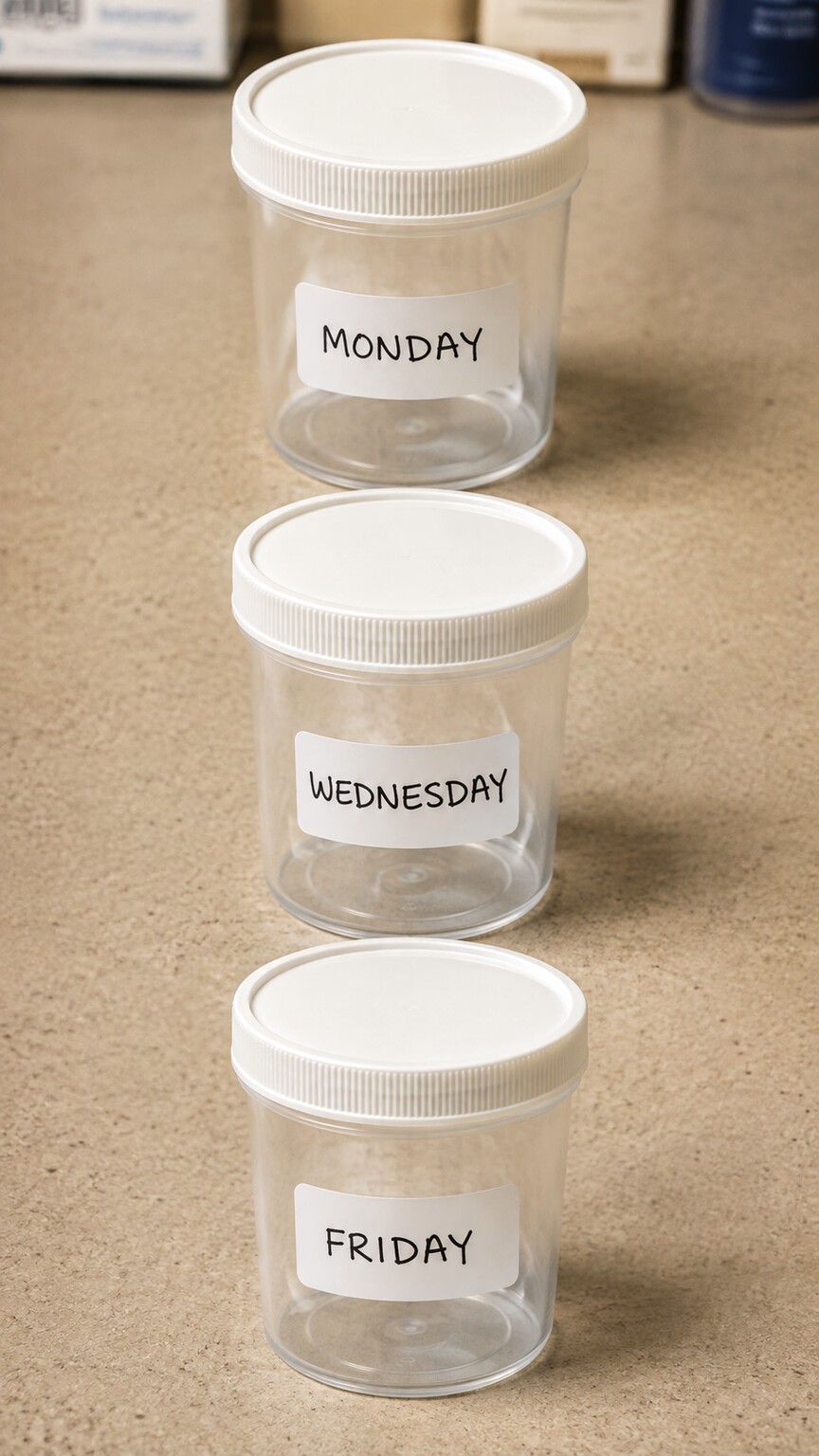
Figure 2. Collecting three separate samples over several different days meaningfully raises the statistical odds of catching a parasite during one of its active shedding windows.
Because of this intermittent shedding pattern, a single stool sample examined once under a microscope has a real, well-documented chance of missing an infection that a series of samples would have caught. This is precisely why the standard protocol for a proper ova-and-parasite exam, when done correctly, calls for three separate stool samples collected on three different days, rather than a single specimen brought in once. Each additional sample, collected on a different day, represents another independent chance of landing during an active shedding window, and the cumulative sensitivity of three separate samples is meaningfully higher than any single one on its own — for some organisms, close to double the detection rate of testing only once.
This is also exactly why a doctor asking for a repeat stool sample after an initial negative result isn't second-guessing the first test or assuming a lab error — they're accounting for a known, quantifiable limitation of single-sample testing that has nothing to do with how carefully that first sample was examined. If your symptoms are ongoing and your first test came back negative, requesting or agreeing to a follow-up sample on a different day is often the single most useful next step, precisely because it directly addresses this specific, well-understood cause of false negatives.
Curious what the rest of your own results are really telling you? Upload them and get a complete, plain-language breakdown in under 15 minutes.
Analyze My ResultsSample Handling and Timing Problems
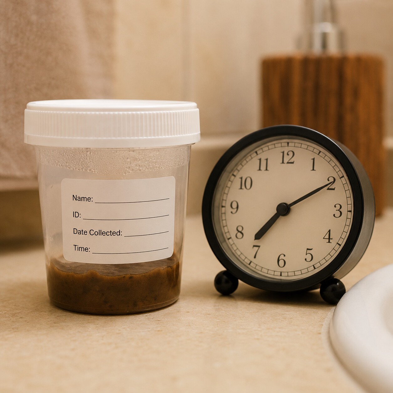
Figure 3. Certain protozoan parasites lose their distinguishing features within a few hours at room temperature, which is why timely delivery to a lab or immediate use of a fixative solution matters more for these organisms than most people realize.
Even when a sample is collected during an active shedding window, what happens to it between collection and examination can still cause a real infection to go undetected. Certain protozoan parasites, particularly the fragile, motile trophozoite form of some species, degrade relatively quickly once outside the body — losing the specific shape, internal structure, or movement pattern a lab technician relies on to identify them under a microscope. A sample left at room temperature for several hours before reaching the lab, especially in warm weather, can allow these delicate forms to break down past the point of reliable identification, even though the organism was genuinely present in the specimen at the moment it was collected.
This is exactly why proper stool ova-and-parasite testing typically requires either prompt delivery to the lab, usually within an hour for the most reliable results, or immediate placement into a preservative fixative solution provided specifically for this purpose. A sample collected correctly but handled carelessly afterward — left in a car, delayed in transit, or stored improperly overnight — can produce a false negative for reasons that have nothing to do with whether the infection is real, which is part of why lab collection kits come with such specific handling instructions printed directly on the container.
Temperature specifically deserves its own mention, since it's one of the most common ways a well-timed sample gets ruined before it ever reaches a microscope. A stool sample sitting in a warm car during a summer errand run, or left on a bathroom counter overnight rather than refrigerated as instructed, accelerates the breakdown of exactly the fragile trophozoite forms that matter most for identifying certain protozoan infections. Conversely, freezing a sample that was meant to stay refrigerated, rather than frozen, can be equally damaging in the opposite direction, rupturing cell structures that a technician needs intact to make a confident identification. This is why collection instructions are typically specific rather than generic — "keep cool" and "keep refrigerated" and "add to this fixative immediately" are meaningfully different instructions, and following the exact one provided for your specific test matters more than most patients realize when they're handed a plastic container and told to bring it back tomorrow.
Light Infections and Low Organism Burden
Not every infection produces the same quantity of detectable eggs or cysts, and this matters more than most people expect. A heavy, well-established infection can shed enormous numbers of eggs consistently, making detection relatively straightforward even from a single sample. An early-stage infection, or one where the parasite burden has always stayed relatively light for reasons related to your own immune response, can shed such a small quantity of eggs that even a technically well-timed, well-handled sample simply doesn't contain enough material for a technician to reliably spot under standard microscopy, especially if only a small portion of the total sample is actually examined, as is standard practice.
This is one of the more genuinely frustrating causes of a false negative, because unlike sample timing or handling, there's no simple fix a patient can control for it beyond, again, repeating the test — a light infection today may shed more heavily in a few weeks as the organism population grows, or a doctor suspecting this scenario based on strong symptoms may move toward a more sensitive test method entirely, discussed next, rather than repeating the same microscopy-based approach that already struggled to find a light burden the first time.
Your own immune system plays a genuinely significant role in how heavy or light an infection stays, which is part of why the same parasite can produce wildly different shedding patterns in two different people. Someone with a robust, well-functioning immune response may keep a parasite population suppressed to a low, hard-to-detect level for a long stretch of time, even while still experiencing real symptoms from the infection's presence. Someone with a weakened immune system — due to an underlying condition, certain medications, or simply individual variation in immune strength — may allow the same organism to reproduce far more freely, producing a heavier burden that's paradoxically easier to detect precisely because the infection is, in an important sense, less well controlled. This is one of several reasons a doctor's overall clinical assessment, not just the raw lab result, matters so much in interpreting what a negative or positive result actually means for any individual patient.
Not All Test Methods Have the Same Sensitivity
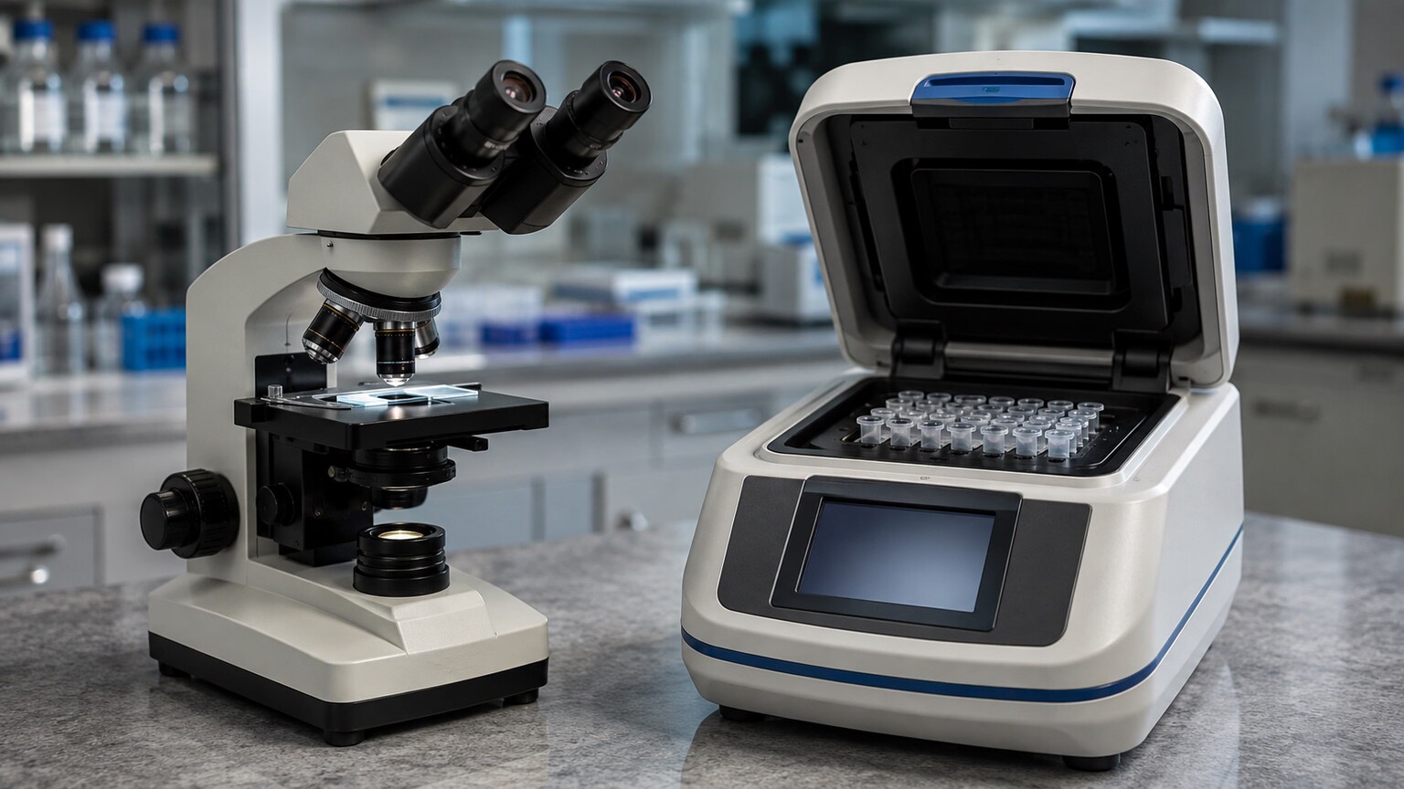
Figure 4. Traditional microscopy relies on a trained technician visually spotting an organism, while newer molecular tests detect fragments of the parasite's genetic material directly, at a sensitivity level microscopy alone often can't match.
Traditional ova-and-parasite microscopy, still the most widely used method because of its low cost and broad ability to detect many different organism types at once, fundamentally depends on a trained human technician visually identifying a specific shape under magnification within the small portion of sample they actually examine. This works well for larger, more distinctive organisms and heavier infections, but it has genuine, well-documented sensitivity limits for smaller organisms, lighter infections, or parasites whose eggs closely resemble debris or other harmless material in the sample.
Newer molecular testing methods, particularly PCR-based stool panels that detect fragments of a parasite's genetic material rather than relying on visual identification, have demonstrably higher sensitivity for several notoriously hard-to-catch organisms, sometimes catching infections that multiple rounds of traditional microscopy missed entirely. These molecular panels aren't universally used as the first-line test everywhere, partly due to cost and availability, which is exactly why a doctor faced with strong clinical suspicion and repeated negative microscopy results will sometimes specifically request one of these molecular panels as a next step, rather than simply repeating the same lower-sensitivity method a third or fourth time and expecting a different outcome.
A third category worth knowing about sits between traditional microscopy and full molecular panels: antigen-detection tests, which use lab-produced antibodies to identify specific proteins on the surface of a particular parasite, such as Giardia or Cryptosporidium, directly within a stool sample. These tests are generally faster and less dependent on a technician's visual expertise than microscopy, and more affordable and widely available than a full PCR panel, making them a genuinely useful middle option — though, because each antigen test is built to detect one specific organism rather than scanning broadly the way microscopy does, they only help if the right organism is actually suspected and specifically tested for in the first place, echoing the same "you only find what you specifically look for" theme that runs through Cryptosporidium detection more broadly.
It's worth being clear-eyed about the tradeoffs involved in choosing between these methods, since none of them is simply "better" in every respect. Standard microscopy remains valuable precisely because it can spot a wide range of different organisms in one pass, including some rarer parasites a targeted antigen or PCR test wouldn't have been built to look for in the first place — a genuine advantage when the specific culprit isn't yet known. Antigen and PCR tests trade that broad coverage for much higher sensitivity toward the specific organisms they're designed to detect. This is exactly why doctors often start with broad microscopy and only move to a targeted molecular or antigen test once the clinical picture — symptoms, travel history, exposure risk — has narrowed the list of likely suspects down to one or two specific organisms worth testing for directly.
Some Parasites Are Simply Harder to Catch Than Others
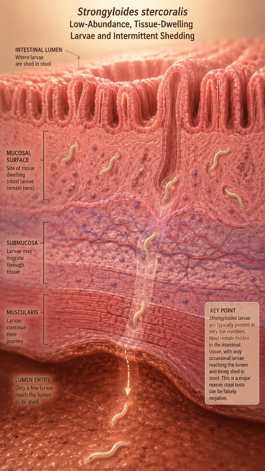
Figure 5. Strongyloides stercoralis is notorious among infectious disease specialists for producing negative stool results even during a genuine, ongoing infection, due to its unusually low and irregular larval shedding pattern.
Beyond general timing and method limitations, certain specific parasites have earned a well-established reputation among infectious disease specialists for being unusually difficult to detect through standard stool testing, regardless of how carefully the sample was collected and handled. Strongyloides stercoralis sits at the top of this list — its larvae are shed into stool in such low, irregular numbers that a single stool exam can miss a genuine, established infection more often than it catches it, which is exactly why serologic (antibody-based) blood testing is often recommended alongside or instead of stool microscopy specifically for this organism, particularly in anyone with a relevant travel or residence history in a region where it's common.
Cryptosporidium, a protozoan parasite that causes significant diarrheal illness, presents a related challenge: its oocysts are small enough, and structurally different enough from other more commonly searched-for parasites, that a standard general ova-and-parasite exam doesn't always specifically look for them unless a specialized stain or test is separately requested. A lab technician scanning a slide for the typical range of intestinal parasites can genuinely miss Cryptosporidium not because it wasn't present, but because the standard screening approach wasn't specifically designed to catch this particular organism without an additional, deliberately requested test.
Entamoeba histolytica adds yet another distinct wrinkle worth knowing about: it looks nearly identical under standard microscopy to a separate, harmless, non-disease-causing species called Entamoeba dispar, to the point that visual identification alone genuinely cannot reliably tell the two apart. Historically, this meant a technician could correctly see amoeba cysts under the microscope and still be unable to determine whether the infection was the disease-causing form worth treating or the harmless look-alike, a genuinely different problem from simply missing an organism outright. Modern antigen or PCR-based testing has largely solved this specific identification problem where it's available, but it's a useful reminder that "detection" and "correct identification" aren't always the same accomplishment, even when something is clearly seen under the microscope.
When the Infection Lives Somewhere the Sample Never Reaches
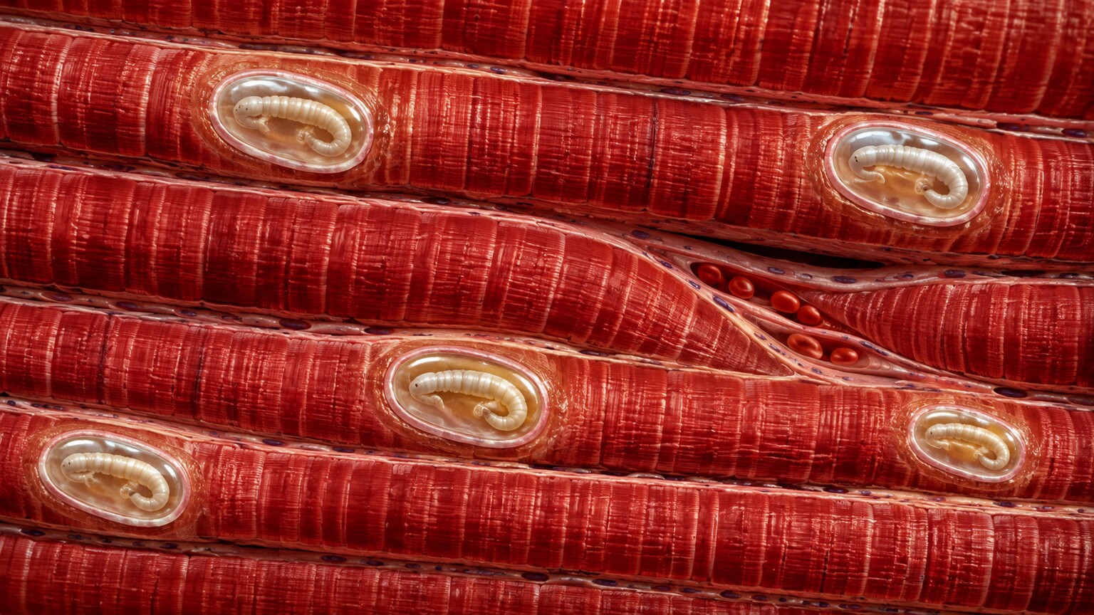
Figure 6. Trichinella larvae encyst directly within muscle fibers rather than the intestinal tract, which is why no amount of stool testing, however carefully repeated, can ever detect this stage of the infection.
Some of the most confusing false negatives happen for a reason that has nothing to do with shedding cycles, sample handling, or test sensitivity at all: the parasite simply doesn't live anywhere a stool sample would ever detect it. Trichinella, acquired through undercooked meat, encysts directly within muscle tissue rather than living in the intestinal tract in its later stages, meaning a stool exam was never going to find it regardless of how many samples were collected or how promptly they were processed — diagnosis instead relies on blood antibody testing or, in specific cases, muscle biopsy. Toxoplasma gondii behaves similarly in its chronic form, forming cysts within tissue such as the brain and muscle rather than shedding detectable material into stool.
This distinction matters enormously for interpreting a negative result correctly: a negative stool parasite test only tells you that no detectable organism material was found in your intestinal tract at that moment — it says nothing at all about parasites that live exclusively in blood, tissue, or other body compartments entirely outside where that specific sample type was ever capable of looking. This is exactly why a doctor taking a detailed history of your specific symptoms, dietary exposures, and travel background before ordering testing matters so much — it determines not just which parasites are plausible, but which sample type and test method actually has a chance of finding them in the first place.
Trichinella's life cycle helps explain exactly why this happens, and it's worth understanding as a concrete example of the broader "wrong sample type" problem. In its earliest stage after being consumed in undercooked meat, the larvae do briefly pass through the intestinal tract, where in theory a very precisely timed stool sample might catch some evidence. But within roughly a week, the larvae migrate out of the intestines entirely, traveling through the bloodstream to reach skeletal muscle, where they physically encyst — burrowing into individual muscle fibers and forming a protective capsule around themselves that can persist for years. Once this migration has occurred, which happens quickly relative to how long it typically takes for symptoms to become severe enough to prompt testing, no stool sample collected afterward, however many times repeated, has any biological chance of finding the organism, because it is no longer anywhere the stool passes through. Diagnosis at this stage depends entirely on blood testing for the antibodies your immune system produces in response to the encysted larvae, or in select cases a small muscle biopsy examined directly for the encysted forms themselves.
A Worked Example: Why the Same Symptoms Took Three Rounds of Testing
Walking through a realistic sequence of events makes all of these individual causes click into place far better than discussing them in the abstract. Picture someone who returns from several weeks of travel with persistent, watery diarrhea, bloating, and fatigue that doesn't resolve after a couple of weeks back home. A first stool sample, collected once and sent to a standard lab, comes back negative for ova and parasites. Symptoms continue, so a second single sample is collected two weeks later — also negative. At this point, it would be easy to conclude there's simply no parasitic cause and move on to investigating something else entirely, but the pattern described throughout this article suggests a more useful next step first.
A doctor familiar with these detection limitations, rather than accepting the second negative as final, orders the full three-sample protocol collected on three separate days within a single week, explicitly explaining to the patient why timing matters this much. The first of the three comes back negative again. The second, collected two days later, finally shows Giardia cysts under microscopy — the same organism that could have been present all along, simply shedding in the irregular cyclical pattern described earlier in this article, and finally caught only because enough independent sampling attempts were made across enough different days to land during an active window.
Notice what didn't change across this entire sequence: the patient's symptoms were consistent and real throughout, the organism was very plausibly present the whole time, and nothing about the first two negative results was a lab error or a sign the patient was imagining their illness. What changed was simply the number of independent chances given to catch a genuinely intermittent signal — a distinction worth holding onto any time an early negative result doesn't match what your body is actually telling you.
This kind of sequence — two single negative tests followed by a positive result only once proper multi-sample protocol was actually followed — happens often enough in real clinical practice that it's a well-recognized pattern rather than a rare fluke, and it illustrates precisely why persistence and the right testing approach matter more than simply accepting an early negative at face value when the underlying symptoms haven't gone anywhere.
Why Cryptosporidium and Giardia Deserve Their Own Specific Mention
Giardia and Cryptosporidium are worth returning to and treating as a matched pair, because together they cause a substantial share of the parasitic gastrointestinal illness seen in developed countries, and both illustrate detection challenges in genuinely different ways. Giardia's challenge, as covered earlier, is primarily about timing — its cyclical shedding pattern means the organism is often present but simply not in the specific sample collected. Cryptosporidium's challenge is different and more structural: its oocysts are unusually small, thick-walled, and don't stain or appear the same way under a routine wet-mount preparation that a standard ova-and-parasite exam uses for most other organisms, which is why labs typically need a specific request, and often a specific stain such as a modified acid-fast stain, to reliably identify it at all.
This means that if a doctor doesn't specifically suspect Cryptosporidium and doesn't order the corresponding specific test, a standard general stool parasite panel can come back completely negative for it even when it's genuinely present in an easily detectable quantity — not because the organism evaded detection through any biological trick, but because nobody actually asked the lab to look for it in the way it needs to be looked for. This is a particularly clear illustration of a broader point worth remembering throughout this entire article: a negative "parasite test" is never truly a blanket statement covering every possible parasite — it's always a negative result for the specific organisms that specific test method was actually designed and requested to detect.
The Role of Recent Antiparasitic or Antibiotic Treatment
One more practical cause of a false negative deserves mention, since it's entirely within a patient's control to avoid: recent use of antiparasitic medication, or even certain antibiotics and antidiarrheal medications taken for an unrelated reason, can suppress or temporarily clear enough of a parasite population to produce a negative test result without necessarily achieving full, lasting eradication of the infection. Bismuth-containing antidiarrheal medications, certain antibiotics, and antimalarial drugs have all been documented to interfere with the ability of a subsequent stool exam to detect specific organisms, sometimes for a week or more after the last dose.
This is exactly why a thorough stool collection instruction sheet typically asks patients to avoid these categories of medication for a specified window, often one to two weeks, before providing a sample — and why it's worth proactively mentioning any recent antibiotic course, antidiarrheal use, or even certain vitamin or mineral supplements like bismuth subsalicylate to whoever orders your testing, even if it doesn't feel directly relevant to your digestive symptoms. A negative result obtained while one of these interfering substances is still active in your system carries meaningfully less weight than one obtained after an adequate washout period, a nuance that's easy to overlook but genuinely changes how confidently that negative result should be interpreted.
What to Do If You Test Negative but Symptoms Continue
A negative parasite test alongside ongoing, unexplained symptoms — persistent digestive upset, unexplained weight loss, chronic fatigue, or a rash that developed after relevant travel or exposure — is a genuinely reasonable situation to pursue further rather than simply accept at face value, especially once you understand how many legitimate ways a real infection can slip past a single test. The most straightforward next step is usually repeating the stool exam, ideally spaced a few days apart as part of the standard three-sample protocol described earlier, rather than assuming the first negative was the final word.
Beyond simple repetition, it's worth specifically mentioning any relevant details you might not think to volunteer unprompted: recent international travel, drinking water from an untreated source, contact with soil in specific regions, occupational exposure to livestock, or eating undercooked meat or freshwater fish. Any of these details can meaningfully change which specific test — stool microscopy, a molecular panel, or blood-based serology — actually has the best chance of finding the organism responsible, since as this article has shown, "testing for parasites" isn't one single uniform test but a whole family of different methods, each with its own specific strengths and blind spots.
It's also worth explicitly asking whether your specific sample was tested with a general screening panel or a targeted test for a particular suspected organism, since the answer changes how much weight a negative result should carry. A general screening panel that found nothing tells you less than a specific test for a particular organism strongly suspected based on your symptoms and history — the first is a broad "nothing obvious was seen," while the second is a much more confident "specifically checked for this and it wasn't there." Understanding which type of negative you actually received is often the single clearest way to judge how reassured you should genuinely feel, or how reasonable it is to keep pursuing an answer.
Frequently Asked Questions
Why would my doctor ask for three separate stool samples instead of just one?
Because many parasites shed eggs or cysts in irregular cycles rather than continuously, a single sample can easily land during a quiet window and miss a real infection. Three samples collected on different days meaningfully raises the odds of catching the parasite during an active shedding period.
Can a stool test miss a parasite that's genuinely there?
Yes. Intermittent shedding, sample handling delays, low organism burden in early or light infections, and the inherent sensitivity limits of standard microscopy can all cause a real infection to produce a negative result.
Why do some parasites need a blood test instead of a stool test?
Some parasites, like Trichinella or chronic Toxoplasma, live in muscle or other tissue rather than the intestinal tract, so a stool sample was never capable of detecting them. Strongyloides is also often tested through blood antibodies since its larval shedding into stool is unusually low and unreliable.
Is a PCR parasite test better than a traditional stool exam?
PCR-based molecular panels generally offer higher sensitivity for several hard-to-detect organisms since they identify genetic material directly rather than relying on visual identification, but they aren't universally used as a first-line test everywhere due to cost and availability, and doctors often reserve them for cases with strong clinical suspicion and repeated negative microscopy results.
How quickly does a stool sample need to reach the lab?
For the most reliable results, ideally within about an hour of collection, or immediately placed into a preservative fixative solution if provided. Delayed or improperly stored samples can allow certain fragile parasite forms to degrade past the point of reliable identification.
Can recent medication use cause a false negative parasite test?
Yes. Certain antibiotics, antidiarrheal medications containing bismuth, and antiparasitic drugs can temporarily suppress detectable organism levels without necessarily achieving full eradication, which is why collection instructions often ask patients to avoid these for one to two weeks before testing.
Does a general parasite panel test for every possible parasite?
No. A general screening panel checks for a defined set of commonly tested organisms using methods suited to detecting them, but some parasites, like Cryptosporidium, require a specifically requested stain or test to be reliably identified, and others live entirely outside the intestinal tract where a stool sample was never capable of finding them.
Conclusion
A negative parasite test is genuinely reassuring in most cases, but it isn't an infallible guarantee, and understanding exactly why takes away a lot of the confusion when symptoms and results don't seem to match. Intermittent shedding cycles, the number of samples collected, how quickly and carefully a sample was handled, how heavy the actual infection is, which test method was used, and where in the body the specific parasite actually lives all shape whether a real infection gets caught or slips through. If your symptoms are telling a different story than a single negative result, that's not a contradiction to dismiss — it's a legitimate reason to talk to your doctor about repeating the test, trying a different method, or providing exposure details that might redirect the investigation toward where the answer is actually hiding.
The broader lesson underneath all of these specific causes is one worth carrying into how you read any lab result, not just parasite testing: a negative result answers the specific question a specific test was actually designed to ask, under the specific conditions that sample was collected — it was never a blanket, universal statement covering every possible explanation for your symptoms. Approaching a confusing or unconvincing negative result with that framing, rather than either dismissing your symptoms or assuming the test simply failed, is what turns a dead end into the next useful step toward an actual answer.
None of this means every unresolved symptom deserves an indefinite chain of repeat parasite testing — at some point, ruling out other causes becomes the more productive path, and that judgment call is exactly what an ongoing conversation with your doctor is for. But knowing the real, specific, well-documented reasons a genuine infection can hide from a single test at least ensures that conversation starts from an accurate understanding of what a negative result actually does and doesn't tell you, rather than from the common but mistaken assumption that one clean report closes the book entirely once and for all.
Still Not Sure What Your Results Mean?
Upload your labs and get a complete, visual, plain-language interpretation of every biomarker — delivered to your inbox in under 15 minutes.
Get My ReportThis article is for educational purposes only and does not constitute medical advice. Always consult your healthcare provider regarding your specific test results and symptoms.