
Understanding the Difference Between Protozoa and Worm Infections
A single stool ova and parasite exam can detect two categories of organisms that have almost nothing in common biologically: protozoa, single-celled microorganisms invisible without a microscope, and helminths — worms — multicellular animals that in some cases grow large enough to be seen with the naked eye. They end up on the same test purely because both happen to pass through, or live within, the digestive tract, not because they're related in any meaningful biological sense. Understanding this fundamental split matters well beyond biology trivia, since it directly shapes how a positive result gets interpreted, which medications actually work, and how urgently treatment is typically pursued.
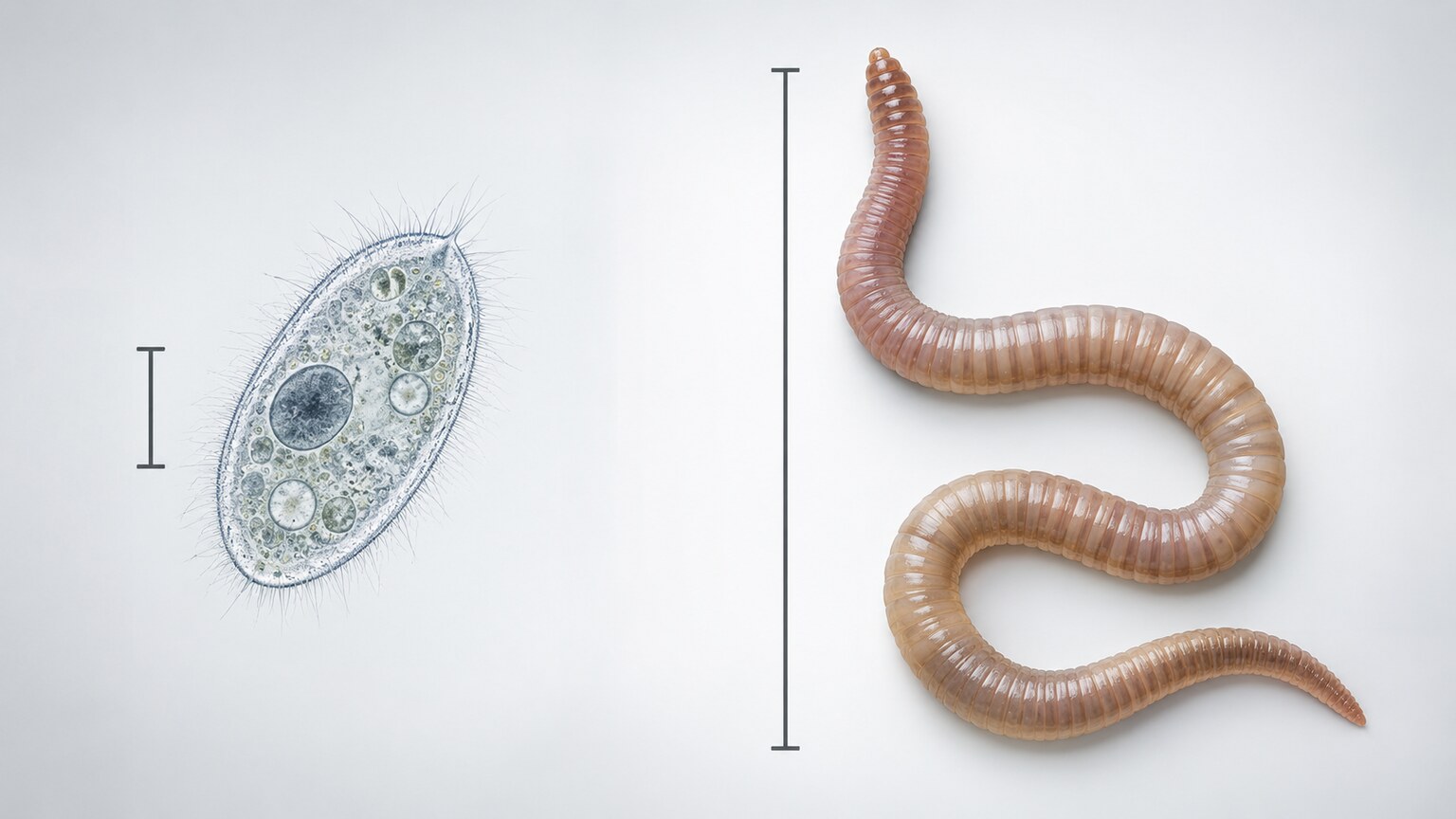
Figure 1. The size difference alone is dramatic: a single protozoan cell measures roughly 10 to 50 micrometers, while an adult worm can reach several centimeters or, in the case of some tapeworms, several meters in length.
Single-Celled Versus Multicellular: The Core Biological Divide
Protozoa are single-celled eukaryotic organisms — meaning each one is a single, complete cell with its own nucleus and internal structures, capable of independently carrying out every function needed to survive, move, feed, and reproduce, all within that one cell. Organisms like Giardia, Entamoeba histolytica, and Cryptosporidium fall into this category, and despite being genuinely complex at the cellular level, an individual protozoan is entirely invisible without magnification, typically measuring somewhere between 10 and 50 micrometers, smaller than the width of a human hair.
Helminths, by contrast, are multicellular animals with specialized organ systems, a genuine nervous system, muscles, and, in most species, dedicated reproductive organs capable of producing enormous numbers of eggs. The three major categories — roundworms (nematodes), tapeworms (cestodes), and flukes (trematodes) — vary considerably in size and shape, but all share this fundamental multicellular, organized-tissue structure that no protozoan possesses. This isn't a minor technical distinction: it means treating a worm infection and treating a protozoal infection are, at a pharmacological level, entirely different problems, since medications effective against a single-celled organism's internal machinery often have no meaningful effect on a multicellular animal's tissues, and vice versa.
This taxonomic split reflects an evolutionary distance that's genuinely difficult to overstate. Protozoa belong to a broad, ancient group of single-celled eukaryotes that diverged from other life forms over a billion years ago, more closely related in a strict biological sense to organisms like amoebas found in pond water than to any animal. Helminths, despite being parasitic and often looking utterly unlike anything most people picture as "animals," are genuine members of the animal kingdom, sharing the same fundamental multicellular animal body plan — nerve tissue, muscle tissue, an oriented head-to-tail body axis in most species — that connects them, at a deep evolutionary level, to every other animal on the planet, humans included, however distant that specific evolutionary relationship actually is. Two organisms sharing a stool sample, in other words, can be less closely related to each other than either one is to entirely different, unrelated branches of life.
Size alone, while dramatic, doesn't fully capture how different these two categories are structurally. A protozoan accomplishes everything it needs — sensing its environment, moving, feeding, defending itself, reproducing — using specialized structures within that single cell: a whip-like flagellum for movement in some species, a flexible outer membrane that can extend into temporary limb-like projections in others, internal vacuoles for digesting food particles. A worm accomplishes these same broad categories of function using dedicated, specialized tissues and organs spread across its entire multicellular body, an organizational strategy fundamentally unavailable to any single-celled organism regardless of how internally complex that one cell might be.
Why Both Categories Show Up on the Same Test

Figure 2. A single microscopic examination technique — direct visual identification of shape, size, and internal structure — happens to work for spotting both protozoal cysts and worm eggs, which is the practical reason both categories are checked on the same test.
The "ova and parasite" exam groups these two biologically unrelated categories together for a purely practical, technical reason: both happen to be detectable using the exact same core laboratory technique — direct microscopic examination of a processed stool sample, looking for organisms or their eggs based on distinctive shape, size, and internal structure. A trained laboratory professional scanning a slide is simultaneously watching for protozoal cysts and trophozoites (the two forms protozoa can take, covered in more detail below) and for helminth eggs, larvae, or occasionally adult worm segments, all within the same visual examination process, using the same equipment and the same basic sample preparation.
This shared testing method is precisely why the exam carries the combined name "ova and parasite" — "ova" referring specifically to the eggs that represent how most worm infections are actually detected, and "parasite" serving as the broader term covering the protozoa detected directly in their living or dormant cellular forms. Despite sharing this test, the biological reality remains that identifying a protozoan and identifying a worm egg are, from that point forward, two completely separate diagnostic and treatment pathways.
It's worth understanding that "parasite" itself is a broader biological category than either protozoa or helminths individually — the term technically describes any organism living on or within another organism (the host) at that host's expense, a definition broad enough to include not just protozoa and worms but also certain external organisms like lice and ticks, which fall entirely outside the scope of a stool exam altogether, since they don't live within the digestive tract at all. Within the specific context of a stool ova and parasite exam, "parasite" is used in its narrower, practical sense — referring specifically to the intestinal protozoa the test is designed to detect directly, as distinct from the "ova" half of the name covering worm eggs.
This terminology can genuinely cause confusion on an actual lab report, since some laboratories list findings under a single combined "parasites identified" heading regardless of which biological category a given organism actually belongs to, while others separate results into distinct protozoa and helminth sections explicitly. Neither reporting convention is inherently better than the other, but understanding that the underlying biological distinction exists regardless of how a specific lab happens to format its report is what allows a patient or provider to correctly interpret what a given positive finding actually means, and which treatment pathway it should lead toward.
Curious what your own stool test results actually mean for your specific situation? Upload your results and get a complete, plain-language breakdown in under 15 minutes.
Analyze My ResultsHow Each Category Actually Reproduces and Spreads
Protozoa reproduce through simple cell division, splitting one cell into two genetically identical daughter cells, a process that can happen remarkably quickly under the right conditions, allowing a small initial infection to expand into a large population within the intestine over a relatively short period. Many protozoa also cycle between two distinct forms during their life cycle: an active, feeding trophozoite form that lives and reproduces within the host, and a hardier, dormant cyst form that gets passed in stool and is specifically built to survive outside the body long enough to infect a new host through contaminated water, food, or surfaces.
Helminths reproduce sexually in most cases, requiring specialized reproductive organs, and many species require passage through the external environment, and sometimes through an entirely different intermediate host species, before their eggs or larvae become capable of infecting a new human host. A single adult female roundworm, for instance, can produce hundreds of thousands of eggs daily, but those eggs typically need weeks of specific environmental conditions — the right soil temperature and humidity — to mature into an infectious form, a genuinely different reproductive strategy than a protozoan's rapid, direct cell division within the host itself.
Tapeworms illustrate an even more elaborate reproductive strategy worth understanding specifically, since they combine sexual reproduction with a body plan built almost entirely around egg production. An adult tapeworm's body consists of a small attachment structure (the scolex) followed by a long chain of repeating segments called proglottids, with each individual segment functioning as something close to a self-contained reproductive unit, capable of producing its own eggs. As segments mature and fill with eggs, they detach from the end of the chain and pass in stool, sometimes visibly, meaning a single tapeworm can continuously generate enormous numbers of new eggs over its entire lifespan — which for some species can extend for years within a single host — through this segmented, modular reproductive structure entirely unlike anything found in protozoa or in most other worm species.
Some protozoa also demonstrate a genuinely fascinating reproductive variation worth mentioning: rather than simple, symmetric cell division, certain species undergo a process where the two resulting daughter cells aren't perfectly identical, allowing for a degree of genetic variation to accumulate across generations even without full sexual reproduction. This matters clinically in select cases, since it can contribute to variation in how a given protozoal species behaves or responds to treatment across different geographic populations, an added layer of biological complexity that a purely "simple cell division" description doesn't fully capture, even though it remains fundamentally distinct from the specialized sexual reproduction helminths rely on.
Diagnostic Differences: What the Lab Is Actually Looking For
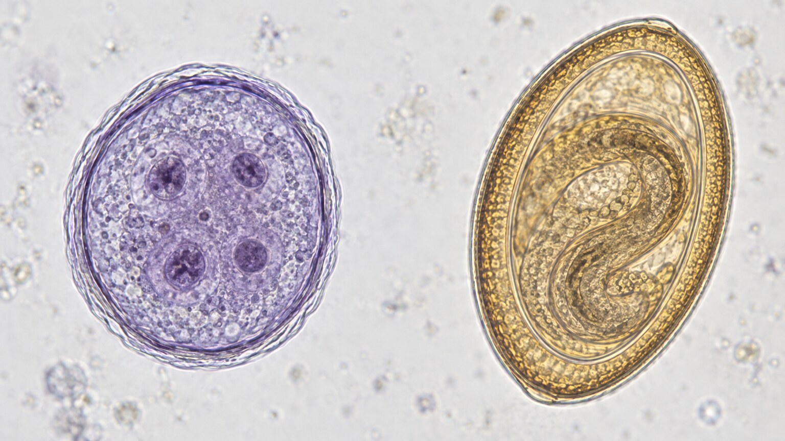
Figure 3. Protozoal cysts and worm eggs look nothing alike under the microscope, each with distinctive, species-specific features a trained technologist uses to identify exactly which organism is present.
Identifying a protozoan on microscopy involves recognizing the specific size, internal nuclear structure, and shape characteristic of either the trophozoite or cyst form of a given species, since different protozoa look meaningfully different from one another even at this microscopic level. Identifying a worm infection, by contrast, generally means recognizing a distinctive egg shape, size, and shell characteristics specific to that worm species, since it's the eggs — not the adult worm itself — that pass consistently and reliably enough in stool to serve as the primary basis for diagnosis in most cases. This is precisely why intermittent shedding, covered in more detail elsewhere on this site, affects both categories similarly: a worm may not be actively laying eggs at the exact moment a sample happens to be collected, just as a protozoan's cyst-shedding cycle can also vary from day to day.
Newer molecular testing methods, including PCR-based stool panels, are increasingly used alongside or instead of traditional microscopy, and these methods work by detecting organism-specific genetic material rather than relying on visual identification at all. This approach offers meaningfully higher sensitivity for many organisms in both categories and doesn't depend on a technologist's direct visual pattern recognition, though traditional microscopy remains valuable specifically because it can detect organisms a targeted molecular panel wasn't specifically designed to look for in the first place.
Antigen testing represents a third diagnostic approach worth understanding, sitting conceptually between traditional microscopy and molecular testing. Rather than visually identifying an organism's physical structure or detecting its genetic material directly, antigen tests use laboratory-manufactured antibodies designed to bind specifically to a protein unique to a particular organism, producing a detectable signal when that protein is present in the sample. This method has become particularly important for Giardia and Cryptosporidium specifically, since both can be genuinely difficult to reliably identify through microscopy alone given their small size and the intermittent shedding pattern both organisms share, and antigen testing for these two protozoa in particular has become a standard, frequently used complement to traditional stool microscopy in many clinical laboratories today.
Specimen preservation and handling differ somewhat between the two categories as well, worth knowing since it affects how a sample needs to be collected and transported. Protozoal trophozoites are notably fragile once outside the body, degrading within a couple of hours at room temperature unless placed into a specific fixative preservative solution designed to maintain their structure long enough for accurate identification. Worm eggs are considerably hardier structures, generally tolerating a wider range of handling and transport conditions without degrading nearly as quickly, since their protective shell is specifically built by nature to survive extended periods in the external environment before hatching — the same biological toughness that makes them infectious in the first place also happens to make them more forgiving specimens to work with in a laboratory setting.
Treatment: Two Genuinely Different Medication Classes

Figure 4. Antiprotozoal drugs and anthelmintic (anti-worm) drugs work through entirely different mechanisms, targeting the specific biological vulnerabilities of single-celled organisms versus multicellular animals respectively.
Antiprotozoal medications, like metronidazole and tinidazole, generally work by disrupting specific metabolic processes essential to a single cell's survival, mechanisms that simply don't exist within a worm's more complex, multicellular biology. Anthelmintic medications, the class used against worms — including albendazole, mebendazole, and praziquantel depending on the specific worm involved — work through entirely different mechanisms, often targeting a worm's ability to absorb nutrients through its own specialized tissue, paralyzing its muscular attachment mechanisms, or disrupting its energy metabolism in ways specific to multicellular parasite physiology.
This is precisely why correctly identifying which category, and ideally which specific organism, is present matters so directly for treatment: a medication effective against a protozoan generally does nothing meaningful against a worm, and treating with the wrong medication class doesn't just fail to help — it delays effective treatment while the actual infection continues. Some broader-spectrum medications do have activity against limited members of both categories, but this is the exception rather than the rule, and accurate identification remains the foundation that determines which specific medication gets prescribed in the first place.
Praziquantel, used specifically against tapeworms and flukes, illustrates just how targeted anthelmintic mechanisms tend to be even within the worm category itself. This particular medication works by disrupting calcium regulation specifically within the worm's own muscle tissue, causing sustained, uncontrolled muscle contraction that paralyzes the parasite's attachment mechanism and allows it to be dislodged from the intestinal wall and passed naturally. This mechanism depends entirely on the worm having the specific type of muscle tissue targeted by the drug — tissue no protozoan possesses at all — which is precisely why praziquantel, despite being genuinely effective against these specific worm categories, has no meaningful activity against protozoal infections whatsoever.
Treatment duration also differs meaningfully between the two categories in many cases. Several protozoal infections respond to a relatively short course of medication, sometimes just a single dose or a few days of treatment, since the goal is simply to disrupt an actively multiplying single-celled population enough that the body's own immune system can clear whatever remains. Certain worm infections, particularly tapeworms attached firmly to the intestinal wall or migrating larval forms of certain species, can require a longer treatment course, sometimes with a planned follow-up dose weeks later specifically timed to catch any larvae that were in a treatment-resistant developmental stage during the initial round, a nuance related to a worm's more complex life cycle that simply doesn't apply to the more straightforward reproductive pattern most protozoa follow.
Eosinophilia: The Blood Clue That Points Toward Worms Specifically
One of the more genuinely useful diagnostic clues distinguishing these two categories doesn't come from the stool sample at all, but from a routine blood test. Eosinophils, a specific type of white blood cell, characteristically rise in response to multicellular parasitic invaders — worms — in a way that simple protozoal infections generally don't trigger to nearly the same degree, since eosinophils are specifically part of the immune system's toolkit for combating larger, tissue-invading organisms rather than single-celled ones. A patient presenting with unexplained symptoms and a notably elevated eosinophil count on a routine complete blood count is a classic pattern that specifically raises suspicion for a helminth infection, sometimes prompting stool testing that wouldn't otherwise have been ordered, or specifically guiding a clinician toward considering worm-specific causes even before stool results come back.
This distinction is genuinely useful in practice, since it means a patient's blood work can offer an early hint about which broad category of parasite is more likely involved, well before the specific organism has been formally identified through stool microscopy, helping guide the overall diagnostic approach and level of urgency from the very first visit.
The underlying biological reason eosinophils respond so specifically to worms, but not meaningfully to protozoa, relates directly back to the fundamental size difference covered earlier in this article, one of the clearest practical consequences of the single-cell-versus-multicellular divide these two categories represent. Eosinophils are specifically equipped to release toxic proteins onto the surface of a target too large to be internalized and destroyed the way a white blood cell would handle a single bacterium or protozoan — essentially attacking large invaders from the outside rather than consuming them whole. A worm, or even a worm larva migrating through tissue, is far too large for this internalization approach, making it a specific, characteristic target for this particular immune strategy, while a single protozoan cell is small enough to be handled through entirely different immune mechanisms that don't meaningfully elevate circulating eosinophil counts to nearly the same degree.
It's worth noting that eosinophilia isn't perfectly specific to worm infections alone — allergic conditions, certain skin conditions, and some other medical conditions entirely unrelated to parasites can also elevate eosinophil counts, meaning an elevated result on its own doesn't automatically confirm a worm infection is present. Rather, it's best understood as one meaningful piece of supporting evidence that, combined with a patient's specific symptoms, travel history, and exposure risk factors, helps a clinician decide how strongly to suspect a helminth infection and how aggressively to pursue confirmatory stool testing, imaging, or other diagnostic steps to reach a definitive answer.
Chronicity and Symptom Patterns: How the Two Categories Typically Differ
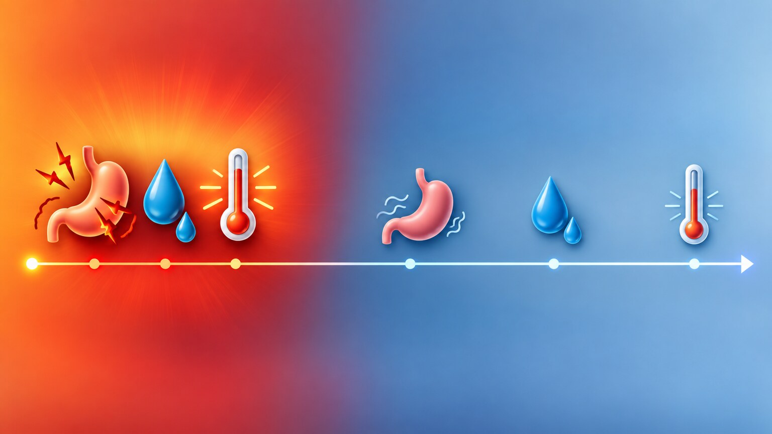
Figure 5. Protozoal infections often produce a more acute, noticeable illness relatively soon after exposure, while many worm infections progress silently for months or years before symptoms become significant enough to prompt evaluation.
Protozoal infections, particularly Giardia and Cryptosporidium, frequently produce a relatively acute onset of gastrointestinal symptoms — diarrhea, cramping, bloating — appearing within one to three weeks of exposure, prompting many patients to seek care specifically because the timeline connects clearly enough to a recent, identifiable exposure like travel or a specific water source. Worm infections, by contrast, often progress far more silently, since a single adult worm or even a modest population typically doesn't produce the same kind of acute, obvious gastrointestinal disruption a rapidly multiplying protozoal infection can cause. Many worm infections are instead identified incidentally, or only after months to years of subtler, easier-to-overlook symptoms like mild fatigue, gradual weight loss, or unexplained iron deficiency anemia (particularly with hookworm, which feeds directly on blood from within the intestinal lining).
This difference in typical presentation is part of why the two categories often prompt genuinely different clinical suspicion in the first place: acute, recent-onset diarrhea after a specific exposure understandably points a clinician's initial thinking toward protozoa, while chronic, vague, slowly progressive symptoms without an obvious acute trigger more often eventually lead toward considering a worm infection, particularly once other more common explanations have already been reasonably ruled out.
Migratory symptoms represent a genuinely distinctive presentation worth understanding specifically, since they occur almost exclusively with certain worm infections and have no real protozoal equivalent at all. Some roundworm species, after hatching from ingested eggs, don't simply settle directly into the intestine — their larvae migrate through the bloodstream, pass through the lungs, and are then coughed up and swallowed again before finally maturing in the intestine, a genuinely elaborate journey that can produce a transient cough, chest discomfort, or even a brief pneumonia-like illness during the lung phase specifically, well before any intestinal symptoms or a positive stool test would ever appear. Recognizing this specific pattern — a mysterious, self-limited respiratory illness followed weeks later by gastrointestinal symptoms — is one of the more distinctive clinical clues pointing toward this particular category of worm infection, a presentation pattern entirely absent from the typical protozoal infection timeline.
Cutaneous larva migrans offers another genuinely distinctive worm-specific presentation, occurring when certain hookworm larvae penetrate human skin but, unlike the species that successfully complete their full migration into the intestine, become stuck wandering just beneath the skin's surface, unable to complete their normal life cycle in a human host. This produces a visibly serpentine, intensely itchy, slowly advancing red track on the skin, tracing the larva's actual physical movement beneath the surface over days to weeks — a striking, visually distinctive presentation with genuinely no protozoal equivalent, since protozoa simply don't have the physical size or independent mobility to produce this kind of visible, tracked movement through tissue.
Transmission Routes: Where Exposure Risk Actually Diverges

Figure 6. Some worm species, like hookworm, have transmission routes with no protozoal equivalent at all — larvae in soil that penetrate intact skin directly, rather than requiring ingestion through the mouth.
Protozoa are, with very few exceptions, transmitted through the fecal-oral route — contaminated water, food, or surfaces that eventually make contact with the mouth, whether directly or indirectly through unwashed hands. Nearly every major protozoal pathogen relevant to human intestinal infection follows this same basic pattern, which is part of why hand hygiene and safe water practices remain the single most consistently effective prevention strategy across this entire category, regardless of which specific protozoan is involved.
Worms follow a genuinely more varied set of transmission strategies, some of which have no protozoal equivalent whatsoever. Hookworm larvae, as mentioned earlier, penetrate intact human skin directly, most commonly through bare feet in contact with contaminated soil, entirely bypassing the fecal-oral route altogether. Tapeworms require an intermediate animal host — livestock like pigs or cattle, or in some cases fish — meaning transmission depends on consuming that intermediate host's undercooked flesh rather than direct contact with human waste at all. Roundworm eggs, by contrast, do follow something closer to the fecal-oral pattern, typically through contaminated soil or produce, aligning more closely with how most protozoa spread. This genuine diversity in transmission strategy across the worm category is one more reflection of just how biologically varied helminths are as a group, compared with the more consistent fecal-oral pattern protozoa overwhelmingly share.
The requirement for an intermediate host in tapeworm transmission specifically deserves a bit more explanation, since it represents a genuinely unique evolutionary strategy compared with essentially everything else covered in this article. A tapeworm's dormant larval stage develops specifically within the muscle tissue of an intermediate animal host after that animal consumes tapeworm eggs from contaminated feed or environment, and the larval form remains dormant, essentially waiting, until a human eats that animal's undercooked flesh, at which point the larva activates, attaches to the human intestinal wall, and matures into a full adult tapeworm capable of producing its own eggs. This two-host requirement means proper cooking temperature, thoroughly destroying any dormant larvae present in meat, functions as an entirely separate and independently effective prevention strategy from the water-treatment and hand-hygiene measures most relevant to protozoa and to soil-transmitted worm species instead.
Geographic and environmental factors also shape transmission risk somewhat differently between the two categories in practice. Protozoal transmission risk correlates most strongly with water sanitation infrastructure and general hygiene conditions, meaning risk can vary considerably even within short geographic distances depending on local water treatment quality. Soil-transmitted worm risk correlates more strongly with climate — warm, humid conditions favor the extended external development period many worm eggs and larvae require to mature into an infectious form — meaning risk tends to concentrate more predictably in specific climate zones globally, an important consideration for anyone assessing personal risk based on travel plans or regional residence.
Frequently Asked Questions
Can the same medication treat both protozoa and worms?
Generally not. Antiprotozoal drugs target mechanisms specific to single-celled organisms, while anthelmintic drugs target mechanisms specific to multicellular worm physiology. A small number of broader-spectrum medications have limited activity against both, but this is the exception.
Why did my blood test show elevated eosinophils but my stool test was negative?
Eosinophilia is a classic clue for a worm infection specifically, but a single stool sample can miss an infection due to intermittent egg shedding. This pattern often prompts repeat stool testing or additional testing methods to confirm or rule out a worm infection.
Are worm infections always more serious than protozoal infections?
Not necessarily. Severity depends on the specific organism, the person's overall health, and how long an infection has gone untreated, rather than which broad category it falls into. Both categories include organisms ranging from mild to genuinely serious, and the specific species involved matters more than the broad category alone.
Why do some protozoa not require treatment while worms almost always do?
Several protozoa are considered harmless commensal organisms that coexist without causing disease in most people. Worms, as larger, actively feeding, tissue-attached organisms, generally cause at least some degree of harm by their nature, which is why confirmed worm infections are typically treated regardless of symptom severity at the time of diagnosis.
Can I have both a protozoal and a worm infection at the same time?
Yes, this is entirely possible, particularly with shared exposure routes like contaminated water or soil. When this occurs, treatment typically requires two separate medications, one from each drug class, addressed either sequentially or simultaneously depending on the specific organisms involved.
How soon after treatment should a follow-up stool test be done?
Timing varies by organism, but protozoal infections are often reasonably confirmed cleared within a few weeks of finishing treatment. Certain worm infections with a more complex life cycle sometimes require a longer follow-up interval to fully confirm resolution.
How Long Treatment Confirmation Actually Takes
Follow-up testing timelines differ somewhat between the two categories as well, worth understanding to set realistic expectations after completing treatment. Protozoal infections are often confirmed cleared relatively quickly, sometimes with a follow-up stool sample collected just a few weeks after finishing treatment, since the shorter reproductive cycle and generally quicker treatment response of single-celled organisms allows for a reasonably prompt confirmation of successful clearance. Worm infections, particularly those involving a longer life cycle or requiring a staged treatment approach to catch larvae in different developmental phases, sometimes require a longer follow-up interval, occasionally with a second confirmatory stool sample collected a month or more after treatment specifically to allow enough time for the full life cycle to have been meaningfully disrupted before declaring the infection successfully resolved.
Cost and insurance coverage for treatment generally reflect standard outpatient prescription costs for both categories, since the medications involved in both antiprotozoal and anthelmintic treatment are widely available, well-established drug classes rather than specialized or unusually expensive therapies, making cost a relatively minor consideration compared with the more clinically important question of correctly identifying which specific organism and category is actually present in the first place.
Family and household screening deserves brief mention as an additional practical consideration relevant to both categories, though for somewhat different reasons. Protozoal infections spread readily through shared households given their fecal-oral transmission route and often prompt consideration of testing close contacts, particularly young children in the same household, who may be sharing the same exposure source or spreading the infection between family members through imperfect hygiene. Worm infections, depending on the specific transmission route involved, sometimes warrant similar household consideration, particularly for species with a direct fecal-oral component, though soil-transmitted or intermediate-host-dependent species are less likely to spread directly between household members in quite the same way as a fecal-oral protozoan infection typically does.
Immune status also shapes how each category behaves, and the difference is meaningful enough to mention here. In someone with a healthy, fully functioning immune system, many protozoal infections eventually clear on their own or stay mild even without treatment, and even worm burdens often stay low-level and manageable for years. In someone who is immunocompromised — due to chronic illness, certain medications, or an underlying condition affecting immune function — the same organisms can behave very differently, with some protozoa capable of causing severe, even life-threatening disease that would barely register as a nuisance in a healthy host, which is one more reason a lab report identifying the exact organism, rather than simply flagging "parasite detected," matters as much as it does for guiding an appropriately tailored treatment decision.
Re-infection risk is another practical distinction worth flagging before moving on. Protozoal infections, because they typically spread through fecal-oral contamination in shared water, food, or close personal contact, can recur relatively quickly if the original exposure source — a contaminated well, an infected household member, an unresolved daycare outbreak — is not identified and addressed alongside treatment. Worm infections tied to a specific soil, water, or intermediate-host exposure, by contrast, generally will not recur unless that same specific exposure pattern repeats, which makes a single environmental or travel-related exposure less likely to produce an ongoing cycle of reinfection than a household-level protozoal source often does.
Conclusion
Protozoa and worms end up on the same stool test purely as a matter of laboratory convenience, both detectable through the same core microscopic examination technique, despite being fundamentally different kinds of organisms — one a single cell, the other a complex, multicellular animal with its own organ systems. This biological gap explains nearly every practical difference that follows: how each is diagnosed, which medications actually work, how quickly symptoms typically appear, and even what a routine blood test can hint at before stool results are available. Understanding which category a specific finding belongs to turns a potentially confusing stool report into something considerably easier to interpret and act on, and helps make sense of why two seemingly similar "parasite" findings on the same report can require entirely different next steps depending on which biological category each one actually belongs to.
Still Not Sure What Your Results Mean?
Upload your labs and get a complete, visual, plain-language interpretation of every biomarker — delivered to your inbox in under 15 minutes.
Get My ReportThis article is for educational purposes only and does not constitute medical advice. Always consult your healthcare provider regarding your specific lab results.