
Understanding the Difference Between Hyaline and Granular Casts
A hyaline cast and a granular cast are actually the exact same kind of thing, made the exact same way, in the exact same tiny tube inside your kidney — the only real difference is what got trapped inside while it was forming. Picture a clear jelly mold: if nothing but clear jelly went in, you get a see-through, smooth shape. If little bits of debris got mixed into the jelly before it set, you get a speckled, grainy shape instead. A hyaline cast is the clear one, and it shows up in healthy people all the time — after a hard workout, after not drinking enough water, even after standing up for a long time. A granular cast is the speckled one, and while a few of them can also just mean you were a bit dehydrated, larger numbers of them are one of the ways a doctor can tell that kidney cells themselves are actually being stressed or injured. Same mold, same tube, very different meaning — and that difference is exactly what this article walks through.
Curious what the rest of your urinalysis is telling you? Upload your results and get a complete, plain-language breakdown in under 15 minutes.
Analyze My ResultsFirst, What a "Cast" Even Is
Before comparing the two types, it helps enormously to know what a cast actually is, because the word itself is confusing — it has nothing to do with a broken bone. Deep inside each of your two kidneys, tucked away where you'd never feel or notice them, are millions of tiny, thin tubes called renal tubules. Their job is to take the fluid your kidneys have filtered from your blood and fine-tune it, pulling back the good stuff your body still needs and letting the rest continue on its way to become urine. The cells lining these tiny tubes constantly release a protein called Tamm-Horsfall protein (scientists also call it uromodulin), which is a normal, everyday part of how the kidney works — not a sign of anything wrong on its own.
Here's the key event: under the right conditions, that protein can gel up and harden, right there inside the tube, taking on the exact cylindrical shape of the tiny tunnel it formed in — like pouring liquid gelatin into a drinking straw and letting it set. Once it's solid, it eventually breaks free and washes downstream with the urine, all the way out of the body, still holding that same tube-shaped form. That solid, tube-shaped structure is a cast. Finding one under a microscope is actually a very useful clue in itself, regardless of what type it is, because a cast is one of the only things in urine that proves, for certain, it came from inside the kidney tissue itself — nothing else downstream (the bladder, the urethra) has the right shape of tunnel to make one.
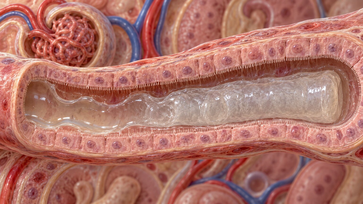
Figure 1. Tamm-Horsfall protein gelling inside a renal tubule, taking on the tube's exact cylindrical shape as it hardens into a cast.
Why a Regular Dipstick Test Can't Find Casts at All
Most people's first experience with a urine test is the quick dipstick kind — a thin plastic strip with small colored squares that gets dipped into a urine sample and changes color within a minute or two. It's fast, cheap, and genuinely useful, but it works by detecting chemicals dissolved in the liquid part of urine, like glucose, protein, or blood pigment. A cast is not a dissolved chemical. It is a solid, physical object, shaped like a tiny cylinder, floating in the liquid the same way a strand of hair floats in a glass of water. No chemical color-change strip, no matter how sensitive, can "see" a solid shape like that.
Finding a cast requires a completely different, more hands-on step: spinning a urine sample in a centrifuge (a machine that spins very fast, like a salad spinner, to force anything solid to settle to the bottom), pouring off the clear liquid on top, and placing a drop of the concentrated material left at the bottom onto a glass slide under an actual microscope. A trained person then has to look through the eyepiece, focus carefully, and identify what they see with their own eyes. This is exactly why a dipstick result can come back completely normal while a microscopic exam of the very same sample reveals casts, cells, or crystals the dipstick was never built to detect — the two tests are answering two different kinds of questions, one chemical and one physical, and a full urinalysis relies on doing both.
The Hyaline Cast: The "Nothing Extra Got Trapped" Version
A hyaline cast is the plain version — just the hardened protein itself, with nothing else caught inside it. Under a microscope, it looks smooth, colorless, and almost see-through, kind of like a tiny sliver of glass or a piece of clear fishing line cut to a short length. Because it's so pale and transparent, lab technicians actually have to dim the microscope's light and adjust the focus carefully just to spot one at all — a hyaline cast is genuinely easy to miss if you're not looking for it the right way.
What makes hyaline casts so reassuring, most of the time, is exactly how common they are in people with perfectly healthy kidneys. A small number of hyaline casts is considered a completely normal finding, not a red flag. They show up more often after hard exercise, after a stretch of not drinking enough water, after standing for a long time, during a fever, or even just first thing in the morning when overnight urine has been sitting more concentrated in the bladder for hours. In every one of these situations, the fluid moving through the kidney's tubules gets a little slower and a little more concentrated, which gives the Tamm-Horsfall protein more opportunity to gel up before it gets flushed along — without anything ever being wrong with the kidney tissue itself.
The Granular Cast: The "Something Got Trapped Inside" Version
A granular cast starts out exactly the same way — the same protein, gelling inside the same tube — but this time, small bits of debris get caught in the protein before it fully hardens. That debris is usually made of broken-down pieces of the kidney tubule's own cells, or trapped white and red blood cells that have started to break apart. Instead of looking smooth and clear, the finished cast looks speckled, grainy, and darker, almost like a piece of clear gelatin that had coffee grounds stirred into it before it set. Depending on how much debris got trapped and how far along the breakdown process is, a granular cast can look finely speckled or coarsely chunky — both are still called granular, just at different points along the same spectrum.
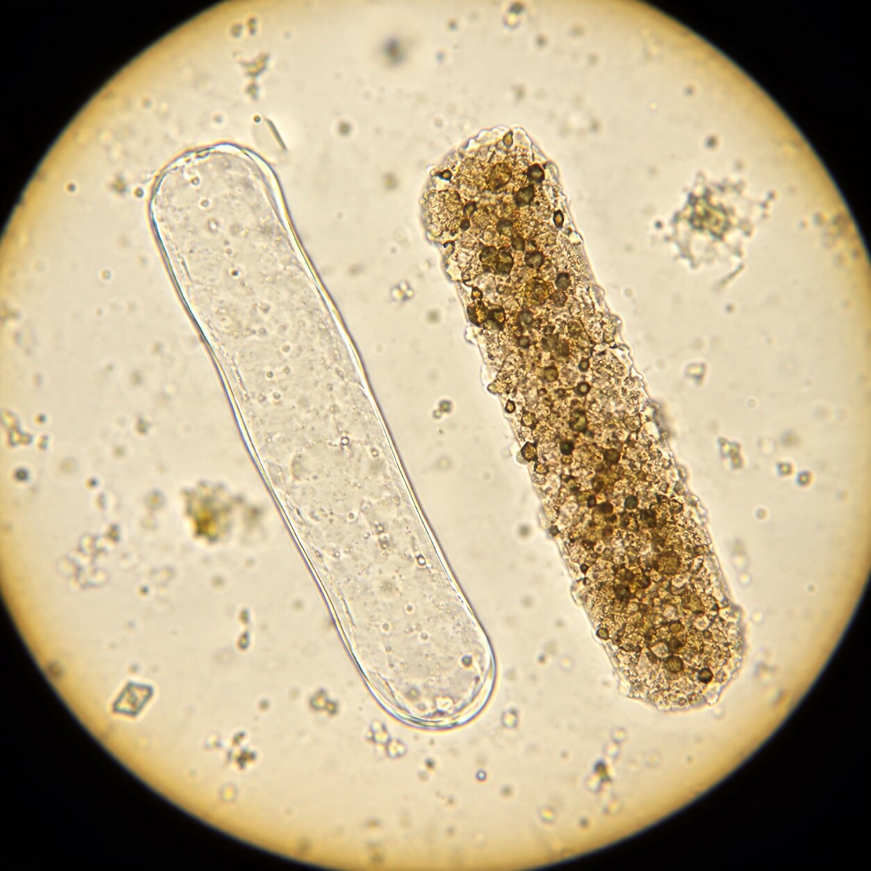
Figure 2. A smooth, translucent hyaline cast (left) beside a speckled, debris-filled granular cast (right), both viewed on the same urine sediment slide.
The debris trapped inside a granular cast is the whole reason it carries more weight clinically than a hyaline cast does. That debris didn't come from nowhere — it means kidney tubule cells, or blood cells that made their way into the tubule, were actually breaking down at the exact spot where that cast formed. A urine sample can't lie about that detail: if a granular cast is present, something inside a tubule really was falling apart when it formed, even if the cast itself has long since washed out of the body by the time it's examined. That single fact is why doctors pay closer attention to granular casts than to hyaline ones, and why finding a large number of them changes the conversation.
It's worth being clear, though, that a few granular casts on their own are not an emergency. Like hyaline casts, small numbers of granular casts can show up after intense exercise, dehydration, or even a viral illness with fever, situations where a bit of ordinary cellular wear-and-tear inside the tubules is expected and temporary. What actually raises concern is a combination: many granular casts, especially the darker, "muddy" kind described below, showing up together with other signs that the kidney itself is under real stress — a drop in urine output, rising creatinine on a blood test, or symptoms like swelling and fatigue.

Figure 3. Intense exercise and dehydration briefly concentrate fluid inside the kidney's tubules, a common, harmless cause of hyaline (and occasionally a few granular) casts in healthy people.
This is exactly why a lab report almost never treats a cast finding as an isolated yes-or-no fact. The report records how many casts were seen per low-power field of the microscope (a specific, standardized way of counting so results can be compared from one test to the next), what type they were, and what else was found alongside them — protein, blood cells, or bacteria. A doctor reads all of that together, the same way a detective reads several small clues together rather than jumping to a conclusion from just one. One or two hyaline casts next to an otherwise completely normal urinalysis, in someone who mentions they just finished a long run, tells a very different story than a report showing many granular casts next to elevated protein and blood in someone who has been feeling unusually tired and swollen.
The runner in the photo above is actually a useful example to hold onto for the rest of this article. If that same person gave a urine sample within an hour of finishing their run, a lab technician might spot a hyaline cast or two under the microscope — and that finding, on its own, would say nothing more than "this person's kidneys were briefly running on concentrated, fast-moving fluid," exactly as expected after hard exercise. No doctor would call them back about it. Keep that low-stakes picture in mind, because the next section moves toward the opposite end of the same scale.
Everyday Causes That Have Nothing to Do With Kidney Disease
Because hyaline and lightly granular casts are so closely tied to how concentrated and slow-moving the fluid inside the tubules is, a whole list of completely ordinary, everyday situations can trigger them without any disease being involved at all. Knowing this list is often the fastest way to make sense of an unexpected finding. Vigorous exercise, especially long-distance running, is one of the best-documented causes — the combination of fluid loss through sweat and a temporary dip in blood flow directed toward the kidneys during intense activity is enough to concentrate the tubule fluid and trigger cast formation in people with completely healthy kidneys. This is well known enough that some laboratories will specifically ask, on request forms for athletes, whether a sample was collected shortly after a workout.
Simple dehydration from not drinking enough water works through the same basic mechanism, just without the exercise component — less water intake means less fluid moving through the tubules, more concentration, and a higher chance that protein has time to gel before it's flushed out. Fever from any cause, even something as ordinary as a cold or the flu, can produce the same effect, since fever increases fluid loss through sweat and breathing while often also reducing how much someone feels like drinking. Prolonged standing, a factor less well known to most people, slows blood return from the legs and can mildly affect kidney blood flow in a similar way over several hours. Even the first urine sample of the morning is more likely to show a hyaline cast or two than a sample given in the afternoon, simply because urine sits more concentrated in the bladder overnight while everyone is asleep and not drinking anything.
None of these everyday causes require any treatment beyond the obvious — drinking enough water, resting after exercise, or letting a mild fever run its course. They're mentioned here specifically because an isolated hyaline cast finding, especially in someone who can identify one of these ordinary explanations, is one of the clearest examples in all of laboratory medicine of a technically abnormal-sounding result that reflects nothing wrong with the body at all.
The "Muddy Brown" Granular Cast — A Specific Warning Sign
Within the granular cast category, there is one especially well-known variation that deserves its own explanation: the muddy brown granular cast. As the name suggests, instead of looking pale and speckled, this cast looks distinctly brownish and dense, almost like a tiny smear of wet mud under the microscope. That darker, muddier color comes from a higher concentration of dying tubule cells packed into the cast, further along in the process of breaking apart than in an ordinary granular cast.
Muddy brown granular casts are strongly associated with a specific kidney problem called acute tubular necrosis, often shortened to ATN — a condition where the cells lining the kidney's tubules are actively dying, usually because of a sudden drop in blood flow to the kidneys (from severe dehydration, major blood loss, or a serious infection affecting the whole body) or from exposure to something toxic to the tubule cells, including certain medications or contrast dye used in some imaging scans. Finding several muddy brown granular casts in someone whose kidney function is also dropping on blood tests is considered one of the more classic, recognizable patterns in all of kidney medicine — a finding doctors are specifically trained to look for and take seriously.
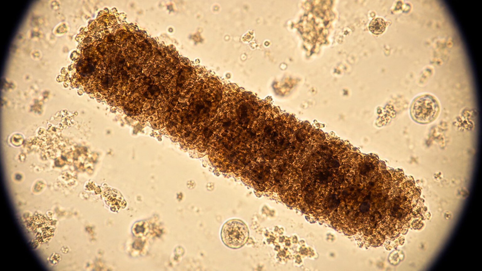
Figure 4. A muddy brown granular cast, densely packed with dying tubule cells — a finding closely associated with acute tubular necrosis.
What Can Actually Trigger Acute Tubular Necrosis
Acute tubular necrosis doesn't come from nowhere, and understanding what actually causes it helps explain why muddy brown granular casts show up in some very specific, recognizable situations rather than randomly. Broadly, there are two main routes to the same end result: the tubule cells either lose their blood supply, or they get directly poisoned by something toxic to them.
The blood-supply route happens whenever the kidneys are suddenly and significantly starved of the blood flow they need, even for a relatively short period. Severe dehydration, major blood loss from an injury or surgery, a body-wide infection serious enough to cause a large drop in blood pressure (called sepsis), and severe heart failure that can't pump enough blood forward can all starve the kidney's tubule cells of oxygen long enough for them to start dying. The toxin route happens when a substance that is specifically harmful to tubule cells reaches the kidneys in high enough concentration — certain antibiotics, some chemotherapy drugs, the iodine-based contrast dye used in many CT scans, and even a large, sudden release of muscle breakdown products after a severe crush injury (a condition called rhabdomyolysis) are all well-documented tubule toxins. In many real hospital cases, more than one of these factors overlaps at once — for example, someone who is already dehydrated and then receives a contrast dye scan is at meaningfully higher risk than either factor would be alone.
This is exactly why doctors ask specific questions when muddy brown granular casts show up: Has this person been seriously ill recently? Did they just have a scan with contrast dye? Are they on any new medications, especially certain antibiotics? Has there been a major injury or surgery? The pattern of casts under the microscope tells doctors that something is happening; the patient's recent history is usually what reveals exactly what that something was.
Why Doctors Think of This as a Spectrum, Not Two Separate Boxes
One of the most useful ways to actually understand hyaline and granular casts is to stop thinking of them as two completely separate categories and instead picture them as two points along a single sliding scale of kidney tubule stress. At one end sits the hyaline cast: pure protein, no debris, essentially a "nothing happening here" signal. Moving along that scale, a little bit of debris starts appearing, creating a finely granular cast — a small amount of cellular wear-and-tear. Moving further still, more debris packs in, creating a coarsely granular cast. At the far end sits the muddy brown granular cast, so packed with dying cellular material that it barely resembles the clear original protein it started as.
This spectrum idea matters because it explains why the same person can have completely different urinalysis results depending on the day, and why finding one type doesn't necessarily mean the others are impossible too. A kidney under mild, temporary stress (dehydration, a hard workout, a passing fever) might show a hyaline cast today and nothing unusual tomorrow. A kidney under a genuinely serious, ongoing injury tends to show more casts, further along that same scale toward granular and muddy brown, and tends to keep showing them across repeat tests rather than clearing up in a day. Thinking in terms of a sliding scale, rather than a strict either/or, is also exactly how a nephrologist (a kidney specialist) actually reads these results in practice.

Figure 5. A lab technician counts casts per low-power field, a standardized method that lets results be compared meaningfully from one test to the next.
This is also why a single urinalysis is often just the starting point rather than the final answer. If a report comes back showing a meaningful number of granular casts, especially the muddy brown kind, a doctor will typically want a repeat sample, a blood test to check creatinine and other kidney function markers, and a closer look at recent events — a bout of illness, a new medication, a scan that used contrast dye, or a period of unusually low fluid intake. Trends across several tests, not one single snapshot, are what actually confirm whether a kidney is recovering, staying stable, or getting worse.
Where Hyaline and Granular Casts Fit Among All the Other Cast Types
Hyaline and granular casts are the two most commonly seen types, but they aren't the only kinds a lab report can mention, and knowing that a wider family exists helps put the two main types in perspective. Red blood cell casts form when red blood cells themselves get trapped inside the tubule before the protein sets, and they point toward bleeding happening specifically inside the kidney's own filtering units — a much more specific and serious finding than a granular cast, usually linked to inflammation of the kidney's tiny filters rather than simple tubule stress. White blood cell casts, similarly, point toward inflammation or infection reaching all the way up into the kidney tissue itself, rather than staying lower down in the bladder. Waxy casts, a further step along the same spectrum as granular casts, look dense, brittle, and sharply outlined, and tend to show up when urine flow through a tubule has been slow for an extended period, often in longer-standing kidney conditions rather than sudden ones. Fatty casts, containing droplets of fat, are specifically associated with a condition where the kidney's filters leak large amounts of protein.
None of these other cast types are the focus of this article, and each has its own separate set of causes and implications worth its own explanation. They're mentioned here only to make one point clearly: hyaline and granular casts sit at the mild, common end of a much larger family of possible findings, and a lab report that mentions only these two types, especially just hyaline ones, is generally describing a far less concerning picture than one that also mentions red blood cell, white blood cell, waxy, or fatty casts.
What Symptoms, If Any, Go Along With These Findings
Hyaline casts on their own essentially never cause any symptoms, because they reflect completely normal kidney function under slightly concentrated conditions — the kind of thing that resolves on its own once someone rehydrates or rests. Nobody feels a hyaline cast forming, and there's nothing to notice from the outside.
Granular casts, especially in larger numbers, are more likely to show up alongside symptoms tied to the underlying cause rather than causing symptoms of their own. Reduced urine output is one of the more telling accompanying signs, since it reflects the same tubule stress that's producing the casts in the first place. Swelling in the legs, ankles, or around the eyes, unusual fatigue, nausea, and a general sense of feeling unwell can also appear when kidney function is genuinely being affected, though these symptoms are broad and can point to many things besides the kidneys specifically. This is precisely why the urine finding and the symptom picture are interpreted together rather than either one alone — a granular cast finding with zero accompanying symptoms points toward a much milder, likely temporary explanation than the same finding in someone who has also noticed swelling and much less urine than usual.

Figure 6. Falling urine output, tracked closely in a hospital setting, is one of the clinical signs doctors weigh alongside a granular cast finding.
In a hospital setting, this combination is watched especially closely in people who are already at higher risk for sudden kidney stress — someone recovering from major surgery, someone who has had a serious infection, or someone who recently received contrast dye for a CT scan. In these situations, urine output is often tracked hour by hour precisely because a sudden drop, paired with granular casts on a urinalysis, can be one of the earliest available clues that the kidneys are struggling, often appearing before a blood creatinine test has had time to rise enough to show the same problem clearly.
The measuring container itself is usually nothing more elaborate than a simple graduated cup like the one shown here, but the number a nurse reads off its side, hour after hour, is one of the plainest, most direct windows anyone has into how the kidneys are coping in real time. A steady, adequate volume is quietly reassuring even before any lab result comes back; a volume that keeps shrinking is often the very first signal that prompts a doctor to ask for a fresh urinalysis and a same-day creatinine, rather than waiting for a routine follow-up.
What Happens After the Result Comes Back
If a urinalysis comes back showing only a few hyaline casts and nothing else unusual, the most common next step is simply nothing at all — no further testing, no treatment, just a normal result reflecting a normal, healthy kidney doing exactly what it's supposed to do under everyday conditions like exercise or mild dehydration. Doctors do not chase down or repeat testing for isolated hyaline casts in someone who otherwise feels fine.
If granular casts show up in more meaningful numbers, especially paired with other findings like protein or blood in the urine, the typical next step is a blood test checking creatinine and BUN (blood urea nitrogen), both of which reflect how well the kidneys are currently filtering waste from the blood. A doctor will also usually ask about recent events that could explain a temporary kidney stress: a stomach bug with vomiting or diarrhea, a new medication, reduced fluid intake, or a recent scan involving contrast dye. If the story fits a clear, temporary cause and the creatinine level looks reassuring, the plan is often just to recheck things after adequate hydration and see whether the picture improves.

Figure 7. Doctors interpret a cast finding alongside a creatinine trend across multiple tests, rather than relying on a single urinalysis snapshot.
When muddy brown granular casts appear together with a clearly rising creatinine level and a drop in urine output, the situation is treated with more urgency, since this combination is the classic pattern for acute tubular necrosis. Management at that point usually focuses on identifying and correcting the underlying cause as quickly as possible — restoring blood flow and hydration, stopping any medication that might be contributing, and closely monitoring kidney function until it stabilizes or recovers, which it often does once the original trigger is resolved, since tubule cells have a genuine capacity to regenerate over time.
One genuinely reassuring fact worth holding onto through all of this: the renal tubule cells responsible for making these casts are among the more regenerative cell types in the human body. Unlike some tissues that scar permanently after injury, tubule cells that survive an episode of acute stress can often divide and replace the cells that were lost, gradually restoring normal tubule function over days to weeks once the original cause is removed. This is a major part of why acute tubular necrosis, despite sounding permanent and alarming by name, is frequently a recoverable condition when it's caught and managed appropriately — the casts that first flagged the problem are, in a real sense, evidence of a repair process already underway, not only evidence of damage.
How Lab Reports Actually Word This Finding
Reading a real urinalysis report can be confusing because casts are often listed in a dense, abbreviated way that was designed for other clinicians to read quickly, not for patients. A typical line might read something like "hyaline casts: 1-2/LPF" or "granular casts: 5-10/LPF," where LPF stands for low-power field, the specific, standardized microscope setting used for counting casts (a different, higher-power setting called HPF, or high-power field, is used for counting individual cells instead, which is why cast counts and cell counts on the same report use different units). A result reading "0-2/LPF" for hyaline casts is generally considered within the range seen in healthy people, while climbing numbers, especially into double digits, or any mention of "many" or "numerous" casts is what tends to prompt a closer look.
It's also worth knowing that these numbers can vary somewhat between labs and even between technicians, since counting is done by a human eye rather than an automated sensor for the final confirmation step, even though many labs now use an automated pre-screening instrument first. This is a normal, accepted amount of variability in the field, and it's part of why a single mildly elevated count is rarely treated as a definitive answer on its own — a repeat sample, especially one collected under better conditions (well hydrated, first thing in the morning, without recent heavy exercise), often gives a clearer picture than fixating on one number from one moment in time.
Frequently Asked Questions
Can hyaline casts turn into granular casts over time?
Not exactly — each individual cast forms once, over a matter of minutes, and doesn't change type afterward. But a person can produce hyaline casts on one day and granular casts on another, since the type reflects what was happening inside the tubule at the moment of formation, which can shift day to day depending on hydration, activity, and overall kidney stress.
Do I need to worry if my urinalysis shows a few granular casts?
A small number of granular casts, especially without other abnormal findings and without symptoms, is often not concerning and can reflect temporary causes like dehydration or recent illness. Larger numbers, muddy brown casts specifically, or granular casts appearing alongside protein, blood, reduced urine output, or a rising creatinine level are what typically prompt closer follow-up.
Why do casts only come from the kidneys and not the bladder?
Casts can only form inside the narrow, tube-shaped renal tubules, where the protein has both the right shape of tunnel to mold into and enough time in contact with tubule cells to gel. The bladder and urethra are wider, open spaces with no equivalent tunnel structure, so a genuine cast cannot form there.
Is a repeat urinalysis usually needed after finding granular casts?
Often, yes, especially if the first result also showed reduced kidney function on a blood test or if symptoms are present. A repeat test after appropriate hydration or after resolving a likely temporary cause helps confirm whether the finding was a one-time event or part of an ongoing pattern that needs further evaluation.
Can drinking more water before a urine test change the result?
Yes, meaningfully. Because hyaline and mildly granular casts form more easily when tubule fluid is concentrated, being well hydrated before a test can genuinely reduce how many are seen, while showing up dehydrated can increase the count without any underlying kidney problem. If a doctor has asked for a repeat sample, drinking normally beforehand (not excessively) usually gives the most representative result.
Are granular casts always related to the kidneys and nothing else in the body?
The cast itself always forms in the kidney's tubules, but the underlying cause isn't always a primary kidney disease — body-wide problems like severe infection, major blood loss, or dehydration from any source can reduce blood flow to the kidneys enough to trigger tubule stress and granular casts, even when the kidneys were completely healthy before that event started.
Bringing It Back to the Two Casts This Article Started With
After walking through everything a cast can mean, it's worth circling back to the two specific types this article set out to explain, because the contrast between them is really the whole point. A hyaline cast tells you that the tubule fluid was briefly concentrated enough for protein to gel — a passive, mechanical event that says nothing bad about the tissue itself, the way condensation forming on a cold glass says nothing bad about the glass. A granular cast tells you that actual cellular material was present and breaking down at the moment of formation — an active, biological event that reflects something real happening to living tissue, even if that something turns out to be mild and temporary. Holding onto that one distinction — passive concentration versus active cellular breakdown — is enough to correctly interpret the overwhelming majority of hyaline and granular cast findings a person is ever likely to encounter on a real lab report, whether that report shows a single reassuring hyaline cast after a weekend hike or a cluster of granular casts worth a follow-up conversation with a doctor.
Conclusion
A hyaline cast and a granular cast begin life the exact same way, hardening from the same everyday kidney protein inside the same microscopic tube — the only thing separating them is whether cellular debris happened to get trapped inside before the protein set. That single difference in composition is what turns a routine, often-harmless finding into a genuine clinical clue about kidney tubule stress, especially when granular casts show up in larger numbers, look distinctly muddy brown, or appear alongside other warning signs like reduced urine output or a rising creatinine level. Understanding casts as points along one continuous spectrum, rather than as two unrelated categories, is the clearest way to make sense of why the same word — "cast" — can describe something as unremarkable as a hard workout or as significant as a kidney actively trying to recover from injury.
Still Not Sure What Your Results Mean?
Upload your labs and get a complete, visual, plain-language interpretation of every biomarker — delivered to your inbox in under 15 minutes.
Get My ReportThis article is for educational purposes only and does not constitute medical advice. Always consult your healthcare provider regarding your specific lab results.