
What Causes Crystals to Appear in a Urine Microscopic Exam?
When a lab technician spins down your urine sample and slides a drop of the sediment under a microscope, crystals are one of the most common things they see. If your report came back listing a crystal type — maybe "calcium oxalate," maybe "amorphous urates" — it's natural to wonder what your body did wrong to produce them. Most of the time, the honest answer is: nothing at all. Crystals form in urine through the same basic chemistry that makes rock candy form in a jar of sugar water left on a counter, and that chemistry is happening in nearly everyone's urine, nearly every day, in amounts too small to ever get flagged. What actually determines whether a crystal shows up on your slide — and whether it means anything — comes down to concentration, temperature, acidity, and a handful of specific substances your kidneys are constantly filtering. This article walks through exactly what's happening at the microscope, the chemistry behind why crystals form at all, the specific types a lab technician might identify, the everyday and medication-related reasons they show up, and the difference between a crystal that's background noise and one that's actually telling you something worth a follow-up conversation with your provider.
Curious what the rest of your urinalysis is telling you? Upload your results and get a complete, plain-language breakdown in under 15 minutes.
Analyze My ResultsWhat Actually Happens During the Microscopic Portion of a Urinalysis
A standard urinalysis has three parts, and crystals only ever show up in the last one. The first part is a visual check — color and clarity, done just by looking at the cup. The second part is the dipstick, a plastic strip with small color-coded pads that react chemically with your urine to flag things like glucose, protein, or blood. The third part, the microscopic exam, is the only one that involves an actual microscope, and it's a physical rather than chemical process. A technician pours a measured amount of your sample — usually around 10 to 12 milliliters — into a conical tube and spins it in a centrifuge for several minutes at a set speed. Centrifugal force pushes anything denser than liquid urine, including cells, bacteria, and any crystals present, down into a small pellet at the bottom of the tube. The clear liquid on top, called the supernatant, gets poured off, and the technician resuspends that small leftover pellet — the sediment — in a drop or two of fluid, places it on a glass slide, and examines it under a standard light microscope, typically scanning at least ten different fields of view at two magnification levels to get a representative count. Anything solid that was floating in your urine at the moment of collection, or that formed afterward while the sample sat waiting to be processed, ends up concentrated in that tiny pellet, which is exactly why the microscopic exam can pick up crystals that were far too small and far too dilute to ever be visible to the naked eye or to trigger a dipstick reaction.
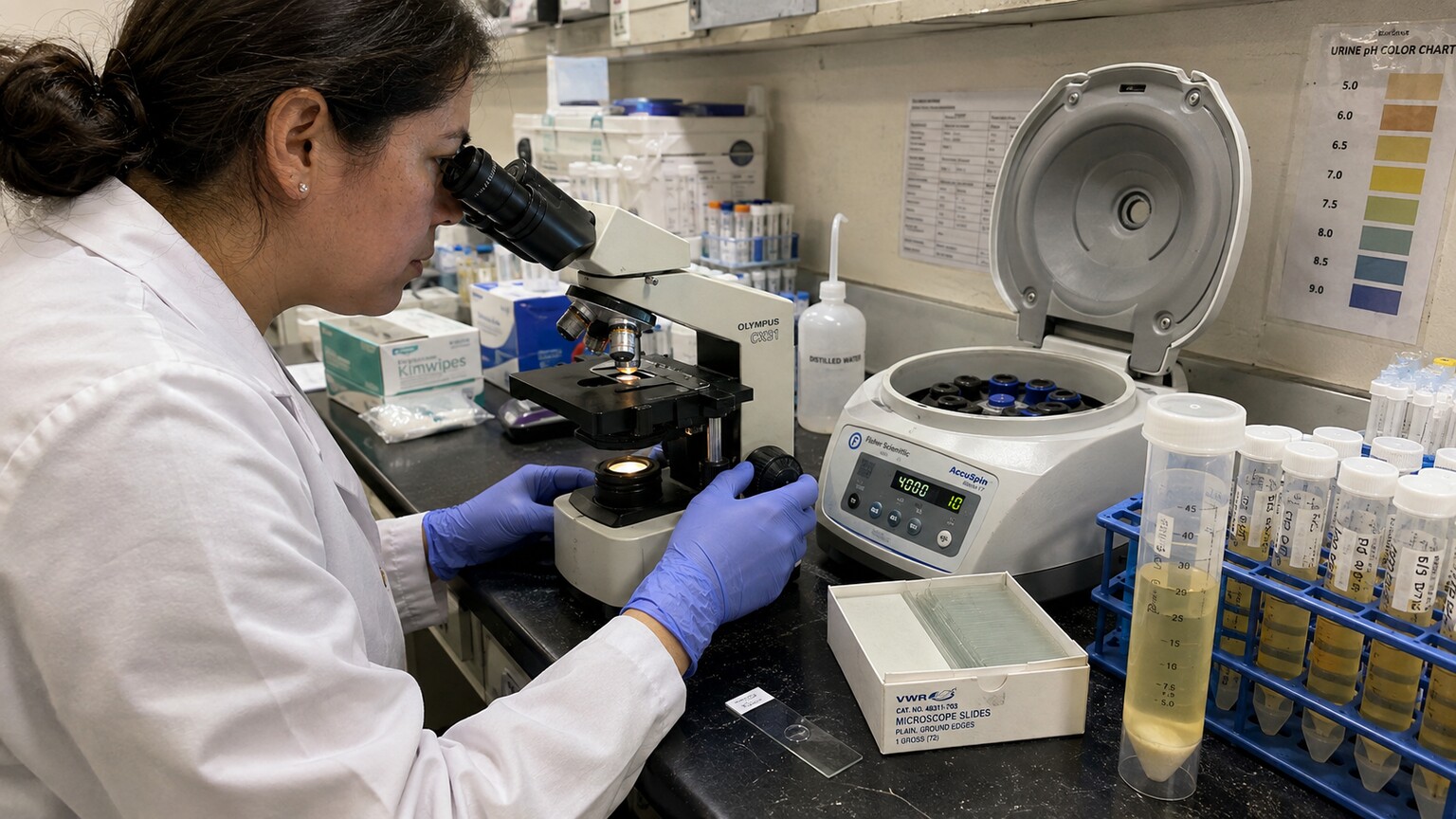
Figure 1. The urine microscopic exam concentrates sediment through centrifugation before it is examined under a light microscope.
How Your Lab Report Actually Grades a Crystal Finding
One detail that trips people up when they read their own results is that a crystal finding almost never comes back as a simple yes-or-no. Instead, most labs report crystals using a semi-quantitative scale — typically "rare," "few," "moderate," or "many" — based on how many crystals the technician counted per high-power field, meaning the view seen through the microscope at its higher magnification setting, usually 400x. This matters more than it might seem, because the word "rare" sitting next to "calcium oxalate" on your report is a fundamentally different statement than "many" sitting next to the same crystal name. A rare finding is often just the ordinary background chemistry described throughout this article — the kind of thing that shows up in a slightly concentrated sample and would likely be absent if the same person gave a second, well-hydrated sample an hour later. A finding graded as moderate or many, especially if it repeats across separate visits, is a meaningfully stronger signal that the underlying urine chemistry is consistently pushing past its saturation point, which is exactly the pattern that eventually builds toward an actual stone rather than staying as isolated microscopic debris. Some labs supplement this scale with a rough per-field number range instead of a word (for example, 0 to 2, 3 to 5, or greater than 10 crystals per high-power field), but the underlying logic is the same either way: quantity is doing a lot of the interpretive work, and a single word like "present" without any grading attached is close to meaningless on its own.
Because this grading depends on how many fields the technician scans and how the sediment was resuspended, results can vary slightly between two labs analyzing urine from the same person on the same day — one more reason a single crystal finding, particularly a low-grade one, is rarely treated as a standalone diagnosis. Reference labs generally cross-check unusual or high-grade findings by having a second technician confirm the count and crystal type before it's finalized on the report, precisely because the shape-based identification described earlier in this article does involve a degree of trained judgment rather than a purely mechanical measurement.
Manual Microscopy vs. Automated Urinalysis Analyzers
Many larger labs now run a preliminary pass on urine sediment through an automated analyzer before a human ever looks through a microscope. These instruments typically use one of two approaches: digital flow imaging, which captures thousands of high-resolution photographs of particles as they stream past a camera and sorts them into categories using pattern-recognition software, or flow cytometry, which shines a laser through the sample and classifies particles based on how they scatter and fluoresce light. Both methods are excellent at flagging obvious categories like red blood cells, white blood cells, and bacteria at high speed and low cost, which is why they've become standard in high-volume hospital and reference labs. Crystals, however, remain one of the categories where automated systems are least reliable on their own. The instruments are good at recognizing that a particle is crystalline in nature, but distinguishing a look-alike amorphous phosphate cluster from an early, poorly formed uric acid crystal — or catching a rare but clinically important cystine or drug-induced crystal — still leans heavily on the shape recognition, color judgment, and pH cross-referencing that a trained human eye performs far more reliably than current software. Because of this, most lab protocols use the automated analyzer as a fast first-pass screen and route any sample flagged for crystals, or any sample with an unusual overall pattern, to a technician for manual microscopic confirmation. That manual confirmation step is precisely the process described earlier in this article — the centrifuged pellet, the glass slide, the trained eye scanning multiple fields — and it's the reason a crystal finding on your report, even at a lab that runs mostly automated equipment, has typically passed through direct human verification rather than being generated by a machine alone.
Why Crystals Form at All: the Chemistry of Saturation
Urine is not simply water with waste floating in it — it's a working solution, carrying dissolved minerals, salts, and metabolic byproducts your kidneys have pulled out of your bloodstream. As long as each of those substances stays fully dissolved, none of it is visible under any microscope. But every solution has a ceiling. Picture stirring salt into a glass of warm water: at first it disappears completely, but keep adding spoonfuls and eventually the water simply can't hold any more, and the excess collects as visible grains at the bottom of the glass. Chemists call the point where a solution can't dissolve any more of a substance its saturation point, and once you push past it, you've entered supersaturation — the exact unstable state in which the extra material has nowhere to go except to come back out of solution as a solid. Urine crystals form the same way. When the concentration of a specific substance, most often calcium, oxalate, uric acid, or phosphate, climbs high enough relative to the amount of water around it, molecules of that substance start finding each other and locking together into an organized, repeating structure — a crystal lattice. This first cluster is called a nucleus, and once it forms, additional molecules attach to it in a predictable geometric pattern, which is exactly why different crystal types have such distinctive, almost textbook shapes under the microscope: the geometry isn't random, it's a direct reflection of how that particular molecule packs together at the atomic level.
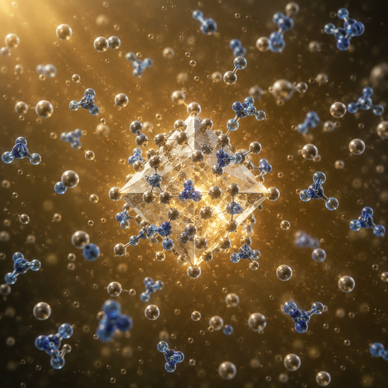
Figure 2. Once a urine solution becomes supersaturated with a mineral like calcium oxalate, molecules begin locking together into an organized crystal lattice.
Three variables control how easily this happens, and all three shift constantly throughout a normal day. The first is concentration itself — how much of the substance is dissolved relative to how much water is diluting it, which is directly tied to hydration. The second is temperature, because warm liquids can hold more dissolved material than cold ones, the same reason hot tea dissolves sugar more readily than iced tea does. The third is pH, the measure of how acidic or alkaline the urine is, because many of these substances are far more soluble in one pH range than another. None of these three variables has to reach an extreme to tip a small amount of urine into supersaturation for a few hours; a slightly concentrated morning sample, a slightly cooler collection cup, or a slightly more acidic stretch after a high-protein dinner is often enough on its own. That's the core reason crystals are such a common, usually unremarkable finding: the chemistry that produces them is operating on a knife's edge in everyone's urine, essentially all the time.
The Main Crystal Types a Microscopic Exam Can Identify
Laboratories generally sort urine crystals into two broad categories: the common types that show up in people with no disease at all, and the rarer types that are considered abnormal no matter how few are seen. Calcium oxalate crystals are by far the most frequently reported of the common group, and under the microscope they typically appear as small, colorless octahedrons that look remarkably like tiny envelopes, or occasionally as elongated dumbbell shapes. They're closely tied to dietary oxalate from foods like spinach, rhubarb, nuts, and chocolate, and they're also the single most common mineral component of actual kidney stones, though finding a few on a microscopic exam is a completely different situation from having a stone. Uric acid crystals form in more concentrated, more acidic urine and can look quite variable — rhombic plates, rosettes, or barrel shapes with a characteristic yellow-brown or reddish tint that comes from urine pigments adhering to their surface. Triple phosphate crystals, also called struvite, have one of the most recognizable shapes in urine microscopy: clear, prism-like structures that genuinely resemble the slanted lid of a coffin, and they form specifically in alkaline urine, frequently in connection with urinary tract infections caused by bacteria that produce an enzyme called urease. Amorphous urates and amorphous phosphates round out the common group — these aren't organized crystals with a defined shape at all, just fine, granular precipitate, pink-tinged in acidic urine or white-tinged in alkaline urine, and on their own they're considered essentially meaningless.
The second category is smaller but clinically far more significant, because these crystal types are considered abnormal in any quantity, regardless of how the sample was collected or handled. Cystine crystals are flat, colorless hexagons that point toward cystinuria, a genetic condition in which the kidneys fail to properly reabsorb a specific amino acid, allowing it to build up in urine to the point of crystallizing — and cystine stones are notoriously prone to recurring throughout a person's life if the underlying condition isn't managed. Tyrosine and leucine crystals, seen far less often, are markers of significant liver dysfunction or certain inherited metabolic disorders, and their presence on a microscopic exam typically prompts immediate further evaluation rather than a routine follow-up. Cholesterol crystals, recognizable by their flat, notched rectangular plates, can appear in urine affected by significant kidney disease involving protein loss. None of these five types are things a healthy person's urine chemistry would casually drift into; unlike calcium oxalate or amorphous urates, seeing even one is enough for a technician to flag it for closer attention.
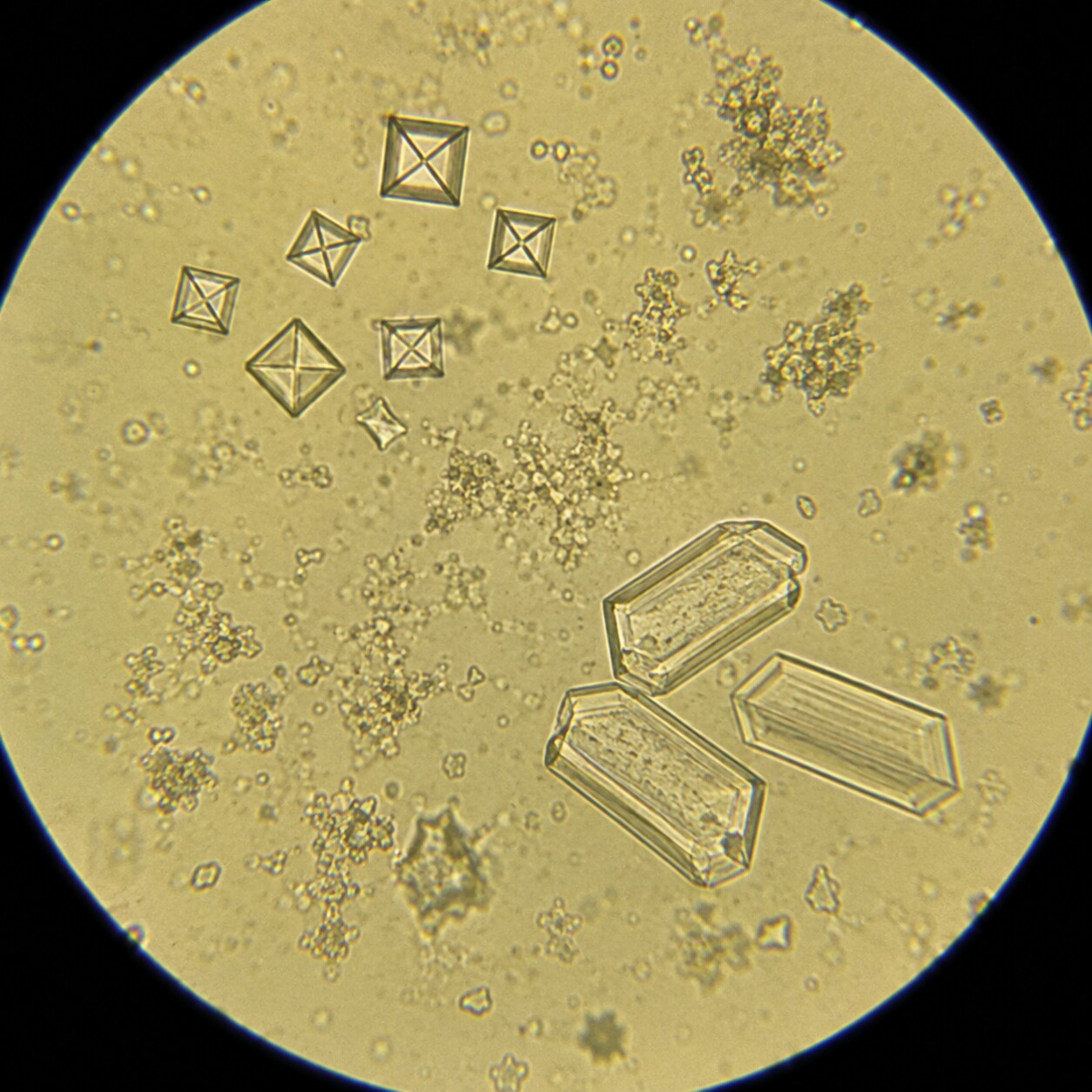
Figure 3. Calcium oxalate crystals form distinctive envelope shapes, while struvite crystals form the coffin-lid shapes technicians use to identify them.
Identifying which of these shapes is present is a genuine, learned skill, and it's part of why the microscopic exam is still done by a trained eye rather than fully automated, even in labs that use image-analysis instruments to pre-screen samples. Shape, color, and the surrounding pH context all factor into the call, and technicians cross-reference the dipstick's pH reading against what they're seeing under the lens, since several crystal types have near-identical cousins that only the pH context can reliably distinguish. A borderline shape seen in acidic urine might be read one way, while that same borderline shape seen in alkaline urine points toward a different substance entirely — which is one more reason a crystal finding is rarely interpreted in isolation from the rest of the urinalysis panel.
Why Urine pH Decides Which Crystals You're Likely to See
If concentration determines whether crystals form at all, pH determines which specific ones show up. Urine pH normally swings somewhere between about 4.5 and 8.0 over the course of a day, shifting with diet, hydration, medications, and even the time since your last meal — a phenomenon called the "alkaline tide" briefly raises urine pH after eating, as your stomach ramps up acid production for digestion and your kidneys compensate. Uric acid crystals and amorphous urates are solubility-sensitive to acidity, so they crystallize almost exclusively in acidic urine, generally below a pH of about 5.8; a diet heavy in animal protein, which tends to acidify urine, makes this environment more likely. Triple phosphate crystals and amorphous phosphates sit at the opposite end, forming in alkaline urine, generally above a pH of about 7.2, which is also the range where urease-producing bacteria thrive during a urinary tract infection, since that enzyme actively raises urine pH by breaking urea down into ammonia. Calcium oxalate is the outlier of the common group, since it can crystallize across a wider pH range and isn't nearly as pH-dependent as the other two — part of why it's the single most frequently seen crystal type regardless of a person's typical urine chemistry. This pH relationship isn't just academic trivia; it's the actual clinical tool a provider uses when a crystal finding needs interpretation, since a pattern of recurring uric acid crystals in someone whose urine consistently runs acidic tells a very different story than an isolated finding in an otherwise neutral sample.
The Body's Own Brakes: Why Urine Doesn't Crystallize More Often
Given how easily urine tips into supersaturation, a fair question is why crystals aren't found on nearly every single urinalysis. Part of the answer is that your body actively works against crystal formation, not just passively avoids it. Urine naturally contains a set of substances known as crystallization inhibitors, which interfere with the process at a molecular level even when concentration, temperature, and pH would otherwise favor it. Citrate is the best studied of these — it binds directly to free calcium in urine, effectively tying it up so it's less available to pair with oxalate or phosphate and form a crystal lattice in the first place, which is exactly why low urinary citrate is one of the most common findings in people who form calcium stones repeatedly, and why citrate supplements are a standard preventive treatment for recurrent stone formers. Magnesium performs a related function, competing with calcium for binding sites on oxalate molecules. Tamm-Horsfall protein, the most abundant protein normally excreted in urine, and a handful of other specialized proteins such as nephrocalcin and osteopontin, physically coat the surface of tiny crystal nuclei as they form, interfering with their ability to grow larger or stick to the walls of the urinary tract and to each other. This is part of why two people can have nearly identical urine concentration and pH and yet only one of them shows crystals on a microscopic exam: the inhibitor side of the equation, not just the mineral side, varies meaningfully from person to person, and it's influenced by genetics, overall hydration habits over months rather than a single day, and certain chronic conditions that alter how much of these protective substances the kidneys produce.
Everyday, Non-Disease Reasons Crystals Show Up
A meaningful share of crystal findings have nothing to do with what was happening inside your body at the moment you provided the sample — they form afterward, in the cup, simply because urine keeps cooling and concentrating even after it leaves you. The most common trigger is dehydration, which is really just concentration by another name: less water diluting the same amount of dissolved mineral pushes urine that much closer to its saturation ceiling, so crystals that would never form in well-hydrated urine appear routinely in a dehydrated sample. Diet plays a comparably large role — a dinner heavy in spinach, beets, or nuts can push oxalate high enough for calcium oxalate crystals to show up the next morning, while a diet heavy in red meat and shellfish raises uric acid production and acidifies urine at the same time, a combination that favors uric acid crystal formation. But timing and temperature matter just as much as anything you ate. The moment urine leaves your body, it begins cooling toward room temperature, and cooler liquid simply can't hold as much dissolved mineral as urine held at body temperature — the same solubility principle behind the tea analogy earlier in this article, just running in reverse. If a sample sits in its cup for even thirty to sixty minutes before it reaches the lab, especially without refrigeration, crystals can form directly in the container that were never present, in any form, while the urine was still inside you. Laboratories know this well enough that guidelines generally call for examining urine within about two hours of collection specifically to minimize this kind of after-the-fact, in-vitro crystal formation — and it's exactly why an isolated, small amount of a common crystal type, found with no other symptoms, is so often treated as background noise rather than a diagnosis.
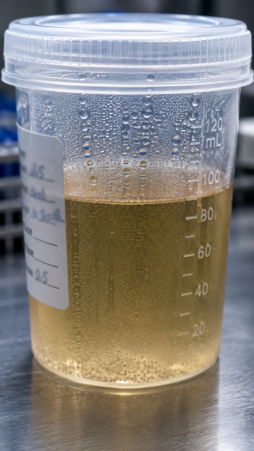
Figure 4. As a urine sample cools after collection, its capacity to hold dissolved minerals drops, which can trigger crystal formation that never occurred inside the body.
This is also why some collection instructions matter more than they might seem to at first glance. A "first morning" sample is deliberately the most concentrated urine your body produces all day, since it represents everything filtered overnight without any water intake diluting it — useful for certain tests precisely because of that concentration, but also the sample most likely to show incidental crystals that a midday, well-hydrated sample from the same person would never produce. Providers who specifically want to rule out in-vitro crystal formation will sometimes request a fresh, promptly processed sample rather than relying on one that sat around, and a repeat test under better-controlled conditions is one of the simplest, lowest-cost ways to distinguish a real chemical tendency from a one-off artifact of how the first sample was handled.
Medications That Can Trigger Crystal Formation
A smaller but clinically important cause of urine crystals is medication itself. Certain drugs are excreted by the kidneys in a form that's poorly soluble in urine, and at high enough doses or with insufficient fluid intake, they can crystallize directly inside the urinary tract in a process called drug-induced crystalluria. Sulfonamide antibiotics were one of the first drug classes recognized to do this, forming sheaves of needle-like crystals, and it's part of why patients prescribed these medications are typically told to drink extra water throughout the course of treatment. Acyclovir, an antiviral used for herpes infections, can form fine, needle-shaped crystals when given in high intravenous doses, particularly if a patient becomes even mildly dehydrated during treatment. Certain HIV medications, including some older protease inhibitors like indinavir, are well known for producing distinctive crystals and, in some patients, contributing directly to kidney stone formation. High-dose intravenous vitamin C can also raise urinary oxalate enough to encourage calcium oxalate crystal formation in some people. None of this means these medications are unsafe as prescribed — for the overwhelming majority of patients, they're taken without any crystal-related complication at all — but it does mean that if you start a new prescription and your next urinalysis flags an unfamiliar crystal type, mentioning your current medication list is one of the most useful pieces of context you can give your provider, since drug-induced crystals have a distinct shape and clinical story compared with the ordinary supersaturation crystals discussed earlier in this article, and recognizing that story early is usually enough to adjust the dose, increase fluid intake, or switch medications before any lasting kidney irritation develops.

Figure 5. Certain medications, including sulfonamide antibiotics, are excreted in a form that can crystallize directly in the urinary tract if fluid intake is insufficient.
When a Crystal Finding on Your Microscopic Exam Actually Warrants Follow-Up
Most crystal findings genuinely don't require any action beyond noting them, but a handful of patterns shift a finding from background noise into something a provider will want to look at more closely. The clearest trigger is crystal type: cystine, tyrosine, leucine, cholesterol, or drug-induced crystals are considered abnormal in any quantity, since a healthy person's normal urine chemistry doesn't produce them, unlike calcium oxalate or amorphous urates. Quantity matters for the common crystal types too — a lab report grading crystals as "many" or "numerous" across repeated tests suggests an active, ongoing tendency toward supersaturation rather than a one-time artifact from a concentrated or delayed sample. Persistence across multiple, separately collected samples is another strong signal, since it rules out the in-vitro, after-collection formation discussed earlier — if the same crystal type keeps showing up on fresh, promptly processed urine, that's a real chemical pattern inside your body, not a fluke of how one sample happened to sit around. And context from the rest of the urinalysis changes the picture entirely: crystals found alongside red blood cells, white blood cells, or bacteria point toward either stone formation actively causing irritation or an infection driving the chemistry, and crystals accompanied by flank pain, blood visible in the urine, or burning with urination warrant prompt evaluation regardless of how common the crystal type is. When any of these flags are present, a provider may order imaging such as a renal ultrasound or CT scan to check for an actual stone, or request a 24-hour urine collection, which measures the total amount of each mineral your kidneys excrete over a full day rather than relying on a single concentrated snapshot. Absent those flags, though, the standard advice is genuinely simple: drink more water, and depending on the specific crystal type, make a small, targeted adjustment to diet — one of the few situations in lab medicine where the most effective response is also the least complicated one.
When a provider does want more certainty before deciding anything, a repeat urinalysis on a fresh, promptly examined sample, collected a few days to a couple of weeks later under normal hydration, is usually the first and simplest step — it directly answers whether the original finding was a one-time artifact of concentration or timing versus a genuine, repeatable pattern. If the second sample comes back clear or shows only a rare, low-grade finding, most providers are comfortable closing the loop there without further testing. If it persists, that's the point where the conversation moves toward imaging or a 24-hour urine collection, since a confirmed, repeatable crystal tendency is worth understanding on its own terms rather than dismissing as noise a second time.
Crystals in Pregnancy and in Children
Two groups deserve a quick, separate note, because crystal findings mean something slightly different in each. During pregnancy, the kidneys filter a larger blood volume than usual and hormonal shifts relax the smooth muscle lining the ureters, the tubes carrying urine from the kidneys to the bladder — a combination that can slow urine flow and make it easier for a concentrated sample to sit longer inside the urinary tract before being expelled. Pregnant patients are also more prone to dehydration from morning sickness in early pregnancy, which independently raises the odds of an incidental crystal finding on a routine prenatal urinalysis. None of this makes crystals dangerous in pregnancy by default, but because kidney stones during pregnancy carry added complexity for both imaging (ultrasound is preferred over CT to avoid radiation exposure to the fetus) and treatment, a provider is often somewhat more attentive to a recurring or heavily graded crystal finding in a pregnant patient than they would be in someone who isn't pregnant.
In children, the same core chemistry applies, but the reference point for what counts as "excessive" hydration or diet differs from adults, and inherited metabolic conditions that produce crystals — including cystinuria and several rarer disorders that produce distinctive crystal shapes — are more likely to be caught for the first time during childhood, since they're present from birth rather than developing gradually with age. A pediatric urinalysis showing an unusual crystal type, particularly cystine, is generally taken seriously enough to prompt further metabolic workup, since catching these conditions early meaningfully changes how manageable they are over a lifetime. As with adults, though, the overwhelming majority of crystal findings in children's urine samples are the same ordinary, benign calcium oxalate or amorphous crystals seen in adults, usually tied to a concentrated sample or a snack heavy in oxalate-rich foods rather than anything requiring concern.
Frequently Asked Questions
Can crystals in a urine microscopic exam form after the sample is collected, even if nothing is wrong with me?
Yes, this happens routinely. Urine cools and can slowly concentrate as it sits in the collection cup, and cooler liquid holds less dissolved mineral than urine at body temperature. If a sample isn't examined within roughly two hours of collection, crystals — most often calcium oxalate or amorphous urates — can form in the cup that were never present inside your body.
Does finding crystals mean I'll develop a kidney stone?
Not on its own. A stone forms when crystals stick together and keep growing over time rather than passing out in normal urine flow. An isolated finding of a common crystal type, without symptoms or repeated findings, is not the same as active stone formation, though a consistent pattern of a particular crystal does reflect the same underlying chemistry that builds stones.
Are some crystal types always considered abnormal, no matter how few are found?
Yes. Cystine, tyrosine, leucine, cholesterol, and drug-induced crystals are treated as abnormal findings regardless of quantity, since a healthy person's normal urine chemistry doesn't produce them. This is different from common types like calcium oxalate or amorphous urates, which are only a concern when they appear in large amounts or persist across repeated, properly handled samples.
What does it mean if my report says "rare" crystals versus "many" crystals?
That word reflects how many crystals the technician counted per high-power field under the microscope. A "rare" grading is usually incidental, ordinary background chemistry with little clinical weight on its own. A "moderate" or "many" grading, especially if it repeats on a fresh, promptly processed sample, points toward a more consistent tendency toward supersaturation and is more likely to prompt a closer look from your provider.
Can I do anything at home before my next test to reduce the chance of a crystal finding?
Staying well hydrated in the hours before collection is the single most effective step, since it directly lowers urine concentration. Providing a fresh sample and getting it to the lab promptly rather than letting it sit also reduces the chance of crystals forming after collection rather than reflecting anything happening inside your body.
Conclusion
A crystal finding on a urine microscopic exam is, more often than not, exactly what it looks like on paper: a small, ordinary snapshot of chemistry that's constantly balancing on the edge of saturation in everyone's urine. Concentration, temperature, and pH decide whether that balance briefly tips into a visible crystal, your body's own inhibitors like citrate and Tamm-Horsfall protein work quietly against that same process, and diet, hydration, sample handling, and certain medications all nudge those variables around from day to day — which is why the same person can show crystals on one test and none on the next without anything meaningful having changed in between. What actually deserves attention is the pattern behind the finding: an abnormal crystal type, a grading of moderate or many that repeats across separate samples, or a crystal appearing alongside blood, infection, or pain — rather than the mere word "crystals" appearing anywhere on the report. If your own results mentioned a crystal type you don't recognize, the most useful next step is simply asking your provider which category it falls into, how it was graded, and whether it fits into a larger picture, rather than assuming the worst from a single line on a lab form.
Still Not Sure What Your Results Mean?
Upload your labs and get a complete, visual, plain-language interpretation of every biomarker — delivered to your inbox in under 15 minutes.
Get My ReportThis article is for educational purposes only and does not constitute medical advice. Always consult your healthcare provider regarding your specific lab results.