
Why Are Epithelial Cells Present in My Urine Sample?
If your urinalysis report came back mentioning "epithelial cells," take a breath — in the vast majority of cases, this finding is not a red flag at all. Epithelial cells are simply the cells that line every surface of your urinary tract, from the kidneys all the way down to the tip of the urethra, and like the skin on the outside of your body, that lining is constantly shedding old cells and replacing them with new ones. A few of those shed cells almost always end up floating in your urine, and finding "a few" or even "a moderate number" on a microscopic exam is considered a completely normal, expected part of a routine urine test. What actually matters — and what your lab report is quietly trying to tell you — isn't just that epithelial cells are present, but which of three very different types they are, because that distinction is the difference between "your urine sample brushed against skin on its way out" and "your kidneys are trying to get your attention."
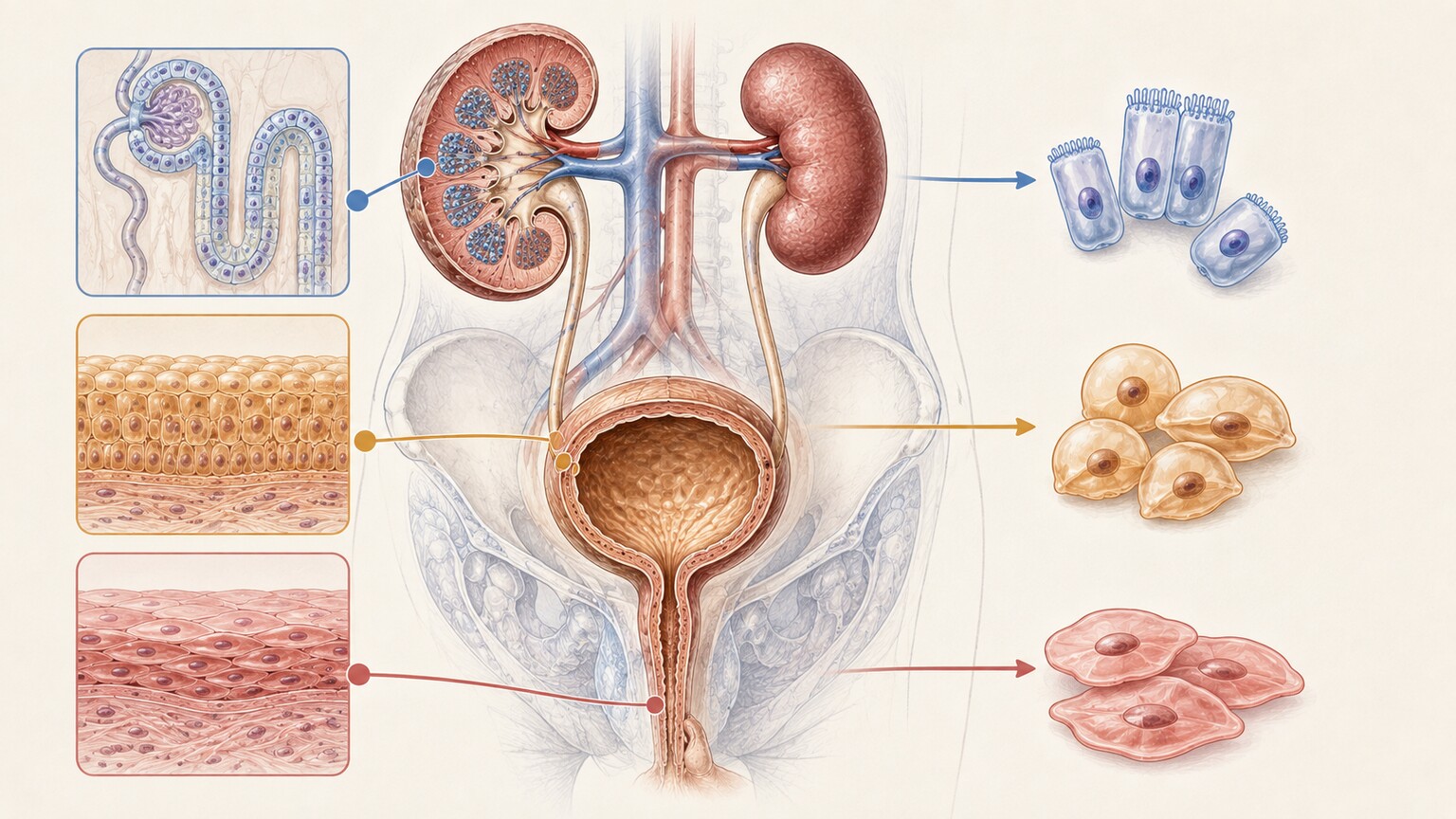
Figure 1. Epithelial cells in urine can come from three distinct locations along the urinary tract, and each source carries a different clinical meaning.
What Epithelial Cells Actually Are, and Why Your Urine Is Supposed to Have a Few
Every internal surface of your body that comes into contact with the outside world — your skin, the inside of your mouth, the lining of your gut, and yes, the entire length of your urinary tract — is covered by a protective sheet of cells called epithelium. Think of epithelium as the wallpaper lining a hallway: it takes the daily wear and tear of everything passing through, and rather than letting that wear damage the structure underneath, the wallpaper itself is designed to be replaced. Your skin does this constantly, shedding roughly a million cells a day without you ever noticing, and the lining of your urinary tract works the exact same way. Old cells on the surface naturally loosen, detach, and get swept away by the fluid moving past them — in the case of your urinary tract, that fluid is urine. This is not a sign of injury or disease; it is simply routine cellular turnover, the same process that keeps every lining tissue in your body fresh and functional. A urine sample, especially one collected in the normal way, is expected to carry a small number of these shed cells along with it, which is exactly why every standard urinalysis report includes a section for "epithelial cells" as one of its routine microscopic findings, right alongside red blood cells, white blood cells, and crystals.
What changes the meaning of this finding isn't the mere presence of epithelial cells — it's the type of cell involved and, in some cases, how many of them show up in a single high-power view under the microscope, commonly abbreviated on lab reports as "HPF." A laboratory scientist examining your urine sediment isn't just counting cells; they're identifying them, because the three cell types that can appear come from three very different depths of the urinary system, and only one of those depths is genuinely tied to the kidneys themselves. Understanding which type your report is referring to turns a vague, slightly alarming-sounding term into something you can actually interpret.
The Three Types of Epithelial Cells — and Why the Difference Matters More Than the Number
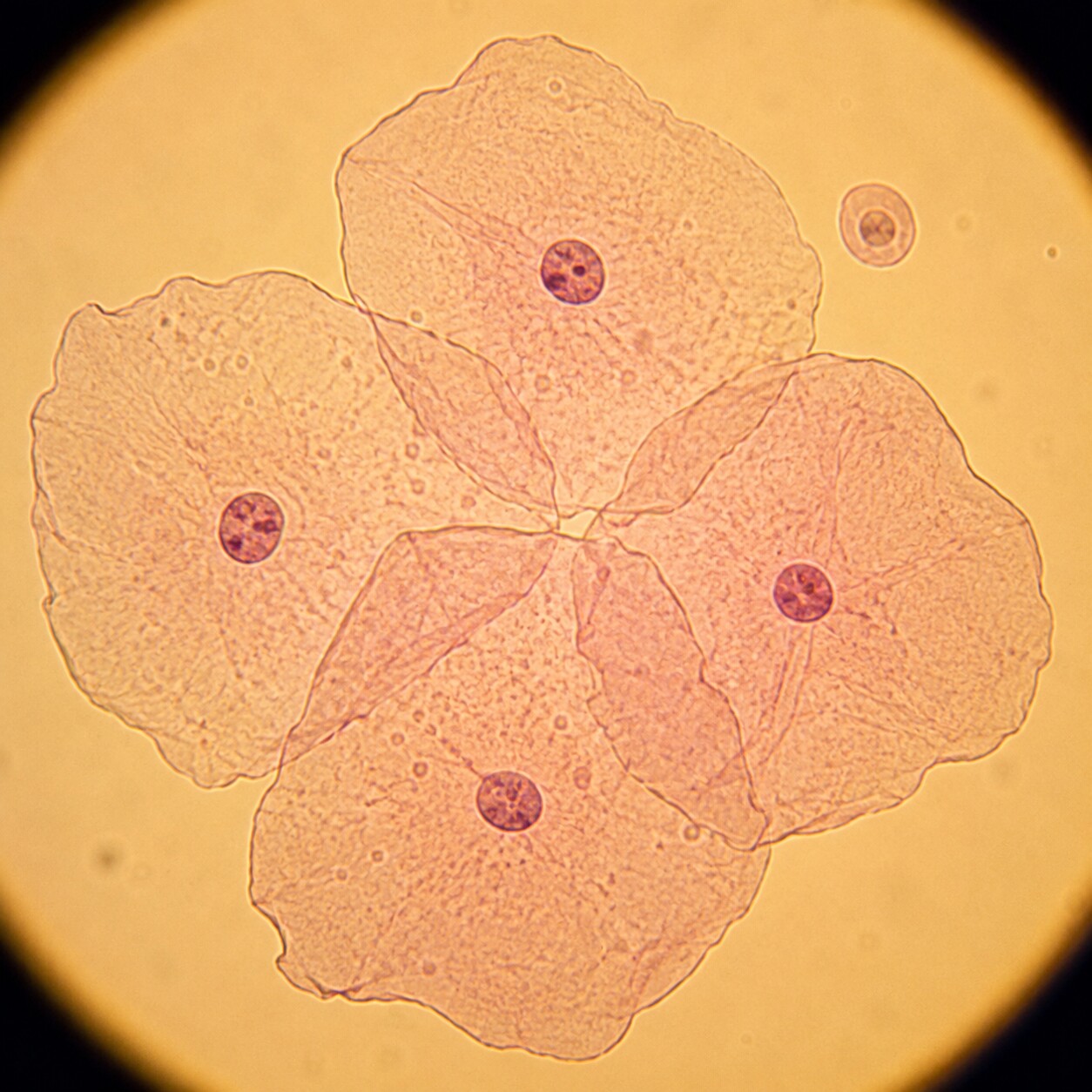
Figure 2. Squamous epithelial cells are the largest cell type seen in urine, easily identified by their broad, flat, irregular shape and small central nucleus.
Laboratories sort epithelial cells in urine into three categories based entirely on where in the urinary tract they came from, and each one looks distinct enough under a microscope that a trained technologist can usually tell them apart on sight. The first and by far the most commonly seen type is the squamous epithelial cell. These are large, flat, irregularly shaped cells with a small, round nucleus sitting roughly in the center — under magnification, they genuinely resemble flattened paving stones or fried eggs scattered across the field of view. Squamous cells line the outermost portion of the urinary tract: the distal urethra (the last stretch of the tube urine travels through before leaving the body), as well as the vaginal walls and vulva in women and the foreskin in uncircumcised men. Because these areas sit right at the exit point, cells from them are the ones most likely to fall into a urine sample simply through ordinary contact during collection, which is why squamous cells are, overwhelmingly, the type your lab report is referring to when it lists "a few epithelial cells" as a routine finding.
The second type is the transitional epithelial cell, sometimes labeled on a report as a "urothelial cell." These cells are smaller than squamous cells and more rounded, oval, or occasionally pear-shaped, and they line a much deeper stretch of the urinary system: the renal pelvis (where urine first collects as it leaves each kidney), both ureters (the tubes carrying urine down to the bladder), the bladder itself, and the proximal, upper portion of the urethra. This lining is called "transitional" for a good reason — it's built from several layers of cells specifically designed to stretch and compress as the bladder fills and empties, similar to the way an accordion's pleats let it expand and contract without tearing. A small number of transitional cells turning up in urine is still considered normal, since this tissue also undergoes routine turnover, but because these cells sit closer to the kidneys and are more often linked to catheter use, bladder irritation, or urinary tract infection, a lab and your provider will generally pay a bit more attention when their numbers rise.
The third and least common type — but the one that carries the most clinical weight — is the renal tubular epithelial cell, often abbreviated RTE or RTEC. These are the smallest of the three cell types, roughly the size of a white blood cell, with a round-to-polygonal shape and a nucleus that takes up a comparatively large portion of the cell. Renal tubular cells line the tiny tubules deep inside the kidney itself — the microscopic structures responsible for the actual, hands-on work of filtering your blood, reclaiming water, salts, and nutrients your body needs, and concentrating waste into what eventually becomes urine. Because these cells come directly from functioning kidney tissue rather than from a passive lining tube, finding more than an occasional renal tubular cell in a urine sample is a meaningfully different finding than seeing plenty of squamous cells, and it's the one type of "epithelial cells in urine" result that most reliably prompts a closer look at how the kidneys themselves are doing.
Squamous Cells — Almost Always a Collection Story, Not a Disease Story

Figure 3. Properly performed clean-catch collection — cleaning first, then catching the sample midstream — meaningfully lowers squamous cell counts and reduces contamination.
If your report shows squamous epithelial cells, the honest, evidence-backed explanation is almost always mechanical rather than medical: your urine picked up a few cells from the skin or mucosal tissue it passed by on its way into the collection cup. This is such a well-recognized part of urine testing that most labs report squamous cells using a rough scale — "rare," "few," "moderate," or "many" — rather than an exact reference range, because the number is expected to vary quite a bit from person to person and even from one collection to the next in the same person. It's especially common, and especially unremarkable, in women, simply because the urethral opening sits closer to the vaginal walls, giving vaginal squamous cells an easy path into a midstream sample. Men are not exempt — uncircumcised men in particular can show similar contamination from foreskin tissue.
This is exactly why proper collection technique exists as a standard part of urine testing instructions. A "clean-catch midstream" sample means cleaning the area around the urethral opening with the provided wipe, beginning to urinate into the toilet for a second or two to flush out cells sitting right at the opening, and only then moving the collection cup into the stream to catch the middle portion. Skipping any part of that sequence — not cleaning first, or catching the very first part of the stream — reliably increases squamous cell counts on the resulting test. Menstruation is another well-documented and completely benign cause of elevated squamous cells (often paired with a few red blood cells as well), which is part of why providers generally recommend avoiding routine urine testing during a menstrual period when it can be scheduled around, or at minimum, mentioning it on the collection form when it can't. None of this reflects anything happening inside your urinary tract — it reflects what happened in the two or three seconds before the sample reached the cup.
Where squamous cells do become practically relevant is when a very heavy number of them shows up on a sample that was also sent for a urine culture — the test used to identify a bacterial urinary tract infection. A culture is essentially asking, "what's actually living and growing inside the urinary tract?" and a sample dense with skin and vaginal cells is also likely to be carrying skin and vaginal bacteria that have nothing to do with the bladder. Because of that, a lab may flag a culture from a squamous-heavy sample as "likely contaminated" and a provider may simply request a fresh sample rather than treating the result as reflecting a true infection.
Curious what the rest of your urinalysis is telling you? Upload your results and get a complete, plain-language breakdown in under 15 minutes.
Analyze My ResultsTransitional Cells — What Rising Numbers From the Bladder and Ureters Can Signal
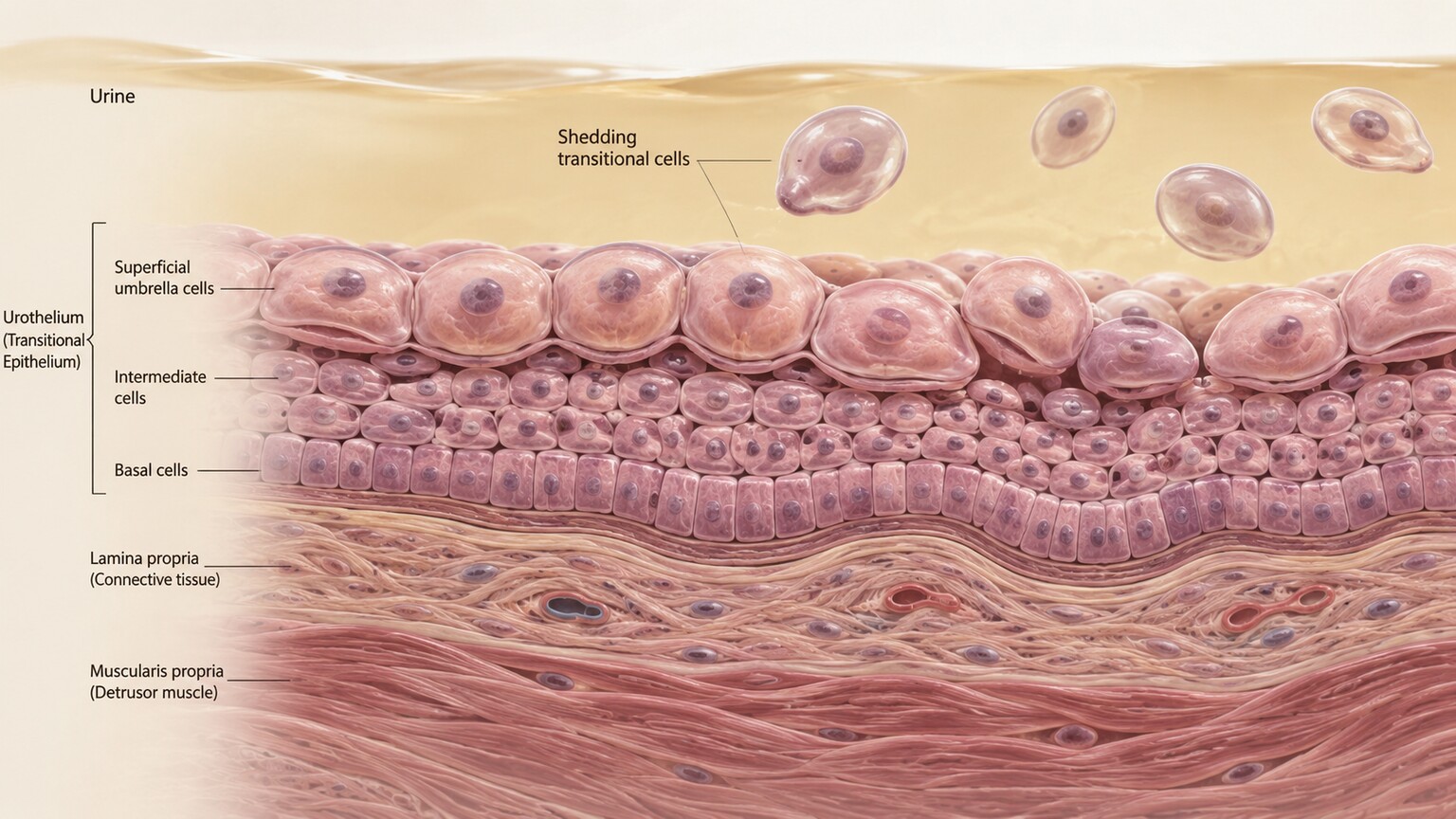
Figure 4. The bladder's multi-layered urothelial lining stretches and compresses with filling and emptying, and mild irritation or infection can accelerate how many transitional cells it sheds.
A small number of transitional cells is a routine finding, the same as squamous cells, and doesn't by itself point to anything wrong. What tends to move this from "background noise" to "worth a second look" is a meaningful increase in how many appear per high-power field, especially when it shows up alongside other findings like white blood cells, bacteria, or blood. One of the most common, benign causes of a temporary jump in transitional cells is recent catheterization or cystoscopy — any time an instrument or tube has physically passed through the urethra and bladder, it mechanically dislodges extra cells from that lining, similar to how running a squeegee over a window pulls off more than just water. This effect typically settles within a day or two as the lining recovers.
Beyond instrumentation, a genuine urinary tract infection is one of the more frequent reasons transitional cell counts climb, because the infection itself irritates and accelerates turnover of the bladder lining as the immune system responds to it — which is also why a UTI so often shows elevated white blood cells and bacteria in the very same sample. Bladder stones, chronic bladder irritation, and inflammatory bladder conditions can produce a similar pattern. In older adults, particularly those with a history of smoking or prior urothelial disease, a provider may occasionally want transitional cells examined more closely if their shape looks unusual under the microscope — not their number, but their appearance — since visibly abnormal, atypical urothelial cells can occasionally prompt a referral for urine cytology, a more specialized test built specifically to evaluate cell appearance rather than simply count cell types. It's worth being clear that this is a distinct, deliberately ordered follow-up test, not something a routine urinalysis is designed to diagnose on its own — a standard urine microscopic exam finding "a few transitional cells" is not, by itself, a marker of anything to be worried about.
Renal Tubular Epithelial Cells — the Type That Deserves a Closer Look
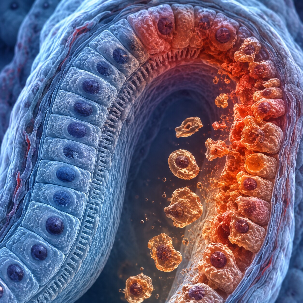
Figure 5. Renal tubular epithelial cells line the microscopic tubules where the kidney does its actual filtering work — when this lining is injured, cells detach directly into the urine.
Renal tubular epithelial cells are where this topic shifts from "almost certainly nothing" to "genuinely worth understanding," because unlike squamous or transitional cells, these come straight from active, working kidney tissue rather than from a passive lining tube further downstream. A helpful way to picture the difference: squamous and transitional cells are more like drywall dust from a hallway — a normal, expected byproduct of foot traffic passing through. Renal tubular cells are more like finding actual fragments of the load-bearing wall itself. A little drywall dust means nothing. Finding pieces of structural wall means someone needs to go check what's happening to the building.
The kidney's tubules are the workhorses of filtration — after blood is initially filtered in a structure called the glomerulus, the resulting fluid passes through a long, winding tubule lined with these renal tubular cells, which reabsorb the water, glucose, amino acids, and electrolytes your body still needs while letting waste products continue on toward the bladder. Because these cells are doing hard, continuous metabolic work, they're also more vulnerable to injury than the tougher lining tissue elsewhere in the urinary tract, and when they're damaged, they detach and appear in the urine. The most common driver of this is a condition called acute tubular necrosis, or ATN — a form of acute kidney injury in which the tubule cells are hurt badly enough that they die and slough off, often from a period of significantly reduced blood flow to the kidneys (such as during severe dehydration, major blood loss, or a period of very low blood pressure) or from exposure to a substance toxic to the kidneys.
That second category, nephrotoxic exposure, covers more everyday situations than people often expect. Certain antibiotics (particularly a class called aminoglycosides), the intravenous contrast dye used for some CT scans, chemotherapy drugs, and even regular heavy use of over-the-counter NSAID pain relievers like ibuprofen can all stress or injure tubular cells enough to cause them to shed into urine. Viral infections that directly affect the kidney, certain autoimmune conditions, and — in someone who has received a kidney transplant — early signs of organ rejection are additional, well-documented causes. This is precisely why finding more than an occasional renal tubular cell on a urinalysis tends to prompt a provider to look further: checking blood tests like creatinine and estimated glomerular filtration rate (eGFR) to see how well the kidneys are currently filtering, reviewing recent medications and any new drug exposures, and looking at the rest of the same urine sample for other supporting clues.
One of the most important of those supporting clues is whether renal tubular cells are showing up loose and scattered, or clumped together inside a structure called a renal tubular epithelial cell cast. Casts form when cells and debris get trapped and molded inside the tubule itself, within a mesh of protein normally produced by healthy tubule cells, then get flushed out as urine flow carries the whole cylindrical clump downstream — essentially a tiny, cell-filled impression of the inside of the tubule. Finding actual renal tubular cell casts, rather than just free-floating renal tubular cells, is considered stronger, more specific evidence of genuine, active kidney tubule injury, because a cast can only form inside the tubule itself, which rules out the possibility that the cells came from contamination somewhere else along the way.
There's one more variation of the renal tubular cell worth knowing about, mostly because it sounds unusual if it ever appears on a report: the oval fat body. This is simply a renal tubular epithelial cell that has absorbed droplets of fat, something that happens when the kidney's filtering barrier is leaking excessive amounts of protein into the urine, a condition known as nephrotoxic-range proteinuria or, when severe and paired with other specific findings, nephrotic syndrome. Under a standard microscope, oval fat bodies look like tubular cells dotted with small, refractile globules; under specialized polarized light, the fat droplets inside them can form a distinctive cross-shaped pattern that laboratory professionals call a "Maltese cross," a visual signature specific enough that its presence is considered a meaningful clue in its own right. Oval fat bodies are a relatively uncommon finding, and seeing them noted on a report is another cue that a provider will want to look at kidney function and urine protein levels together rather than in isolation.
How a Lab Actually Tells These Cell Types Apart Under the Microscope

Figure 6. Distinguishing squamous, transitional, and renal tubular cells requires a trained eye evaluating size, shape, and nucleus-to-cytoplasm ratio under high magnification — it isn't a fully automated step.
After a urine sample arrives at the lab, a portion of it is spun in a centrifuge, which separates the heavier solid material — cells, crystals, casts, and any bacteria present — from the liquid, concentrating everything solid into a small pellet at the bottom of the tube. That sediment is then examined under a microscope at high magnification, and a trained laboratory professional (or, increasingly, a digital imaging system with human review) evaluates what's actually present field by field. Some labs use a special stain, most commonly the Sternheimer-Malbin stain, which colors different structures slightly differently and makes it considerably easier to tell a squamous cell from a transitional cell from a white blood cell at a glance, since without staining, several of these structures can look deceptively similar in a plain, unstained view.
Identification comes down to a consistent set of features: overall size (squamous cells are noticeably the largest, renal tubular cells the smallest), shape (flat and irregular versus round or oval versus polygonal), and the ratio between the nucleus and the surrounding cytoplasm, since renal tubular cells have a comparatively large nucleus relative to their small overall size, which is one of the clearest tells separating them from the other two types even when a cell looks otherwise unremarkable. Cell counts are then reported per high-power field, typically described using a semi-quantitative scale — rare, few, moderate, or many — or as an estimated numeric range, depending on the laboratory's specific reporting convention. This is genuinely one of the more skill-dependent parts of a urinalysis; unlike a chemical strip test that changes color based on a straightforward reaction, sediment identification depends on trained pattern recognition, which is part of why results can occasionally vary slightly between two different technologists reviewing the very same slide, and why a repeat or confirmatory look is sometimes requested for a borderline or unusual finding.
Many larger laboratories now use automated urine microscopy analyzers as a first pass, instruments that push the sample past a camera or a flow-based imaging sensor and use pattern-recognition software to sort and pre-count the particles it sees, flagging cells, casts, and crystals by category in a fraction of the time manual review would take. These systems are genuinely good at high-volume screening and at catching samples that look entirely unremarkable, letting the lab route those through quickly. They are, however, intentionally paired with human oversight rather than left to operate alone: any sample the software flags as unusual, borderline, or containing a cell type it can't confidently classify — which happens fairly often with the less common, more clinically significant renal tubular cells — gets pulled aside for a laboratory professional to examine directly under the microscope before a final result is released. In other words, the technology speeds up the easy calls so that trained human attention can stay focused on the results that actually need it.
Common, Everyday Reasons Epithelial Cell Counts Come Back Elevated
Before assuming an elevated epithelial cell count means something is medically wrong, it's worth walking through the ordinary, non-disease explanations first, since they account for the overwhelming majority of results that get flagged. Collection technique is the single biggest factor — a sample that wasn't a true clean-catch, or where the very first part of the stream was collected instead of the midstream portion, will almost always show more squamous cells than a carefully collected one. Menstruation, as mentioned earlier, is a well-established and completely benign cause of elevated squamous cells, frequently paired with a few red blood cells in the same sample. A delay between collecting the sample and getting it to the lab can also distort results in more subtle ways: urine left at room temperature for more than an hour or two allows cells to begin breaking down and can allow any bacteria present to multiply, which is why proper samples are refrigerated or processed promptly, and why a lab may ask for a fresh sample if a delay is suspected.
Simple dehydration is another everyday factor, though its effect is somewhat different — concentrated urine packs everything present, including epithelial cells, into a smaller volume of fluid, which can make counts look proportionally higher on a very concentrated sample compared with a well-hydrated one, even though the actual rate of cell shedding hasn't changed. This is one of several reasons a provider looks at the full urinalysis picture — including the urine's concentration, measured as specific gravity — rather than reacting to a single number in isolation. In women specifically, vaginal discharge unrelated to menstruation, recent sexual activity, or the use of certain vaginal products can also introduce extra squamous cells into a sample collected without careful technique. None of these everyday causes require treatment; they simply call for a cleaner recollection when accuracy matters, such as before a urine culture.
Epithelial Cells in Children, Pregnancy, and People With Catheters
A few groups of people reliably see different baseline epithelial cell patterns than the general adult population, and knowing this ahead of time can prevent unnecessary worry. In infants and young children who aren't yet toilet-trained, urine is frequently collected using an adhesive collection bag applied to the skin rather than a true midstream catch, and this method is well known to pick up a substantial number of squamous cells from the surrounding skin — often more than would ever be seen in an adult clean-catch sample. Because of this, when a bagged specimen in a young child raises concern for a urinary tract infection, many pediatric guidelines call for confirming the result with a more controlled collection method, such as a catheterized specimen, before starting treatment, specifically because bag samples are so prone to this kind of benign contamination.
Pregnancy is another situation where squamous cell counts tend to run a bit higher than they would outside of pregnancy, largely due to increased vaginal discharge and hormonally driven changes to the vaginal and vulvar tissue that occur throughout gestation. Because urinary tract infections are more common during pregnancy and carry higher stakes for both parent and baby if left untreated, providers pay close attention to urinalysis results during prenatal visits — but they also factor in this normal, pregnancy-related tendency toward more squamous contamination rather than treating every elevated count as evidence of infection on its own.
People living with an indwelling urinary catheter represent a third distinct case. Because a catheter sits continuously against the bladder and urethral lining, it causes ongoing, low-level mechanical irritation that most people without a catheter never experience, and this reliably produces a persistently higher baseline of transitional cells in urine samples collected through or around the catheter. Clinicians who regularly care for catheterized patients are aware of this pattern and adjust their interpretation accordingly, generally looking for a clear change from that person's own usual baseline — a new spike in white blood cells, visible blood, or a change in urine odor and clarity — rather than reacting to transitional cells alone, which are an expected and largely unavoidable part of long-term catheter use.
When Elevated Epithelial Cells Might Point to Something That Needs Follow-Up
The pattern that actually concerns a healthcare provider looks quite different from ordinary contamination, and it's rarely about epithelial cells in isolation. What tends to draw genuine attention is renal tubular cells appearing at more than an occasional, rare level — especially when they show up alongside other supporting findings on the same urinalysis, such as protein in the urine, red or white blood cells, granular casts, or renal tubular cell casts specifically. This combination paints a picture of active injury happening inside the kidney's filtering units rather than a single, isolated lab quirk. It's also meaningfully more relevant in someone with a known risk factor for kidney injury: a recent hospitalization involving low blood pressure or significant blood loss, a new medication known to stress the kidneys, recent IV contrast dye for imaging, chronic heavy NSAID use, or a history of kidney transplant.
For transitional cells, the equivalent concerning pattern is a persistent, meaningful rise that doesn't resolve with a repeat sample, particularly when paired with visible blood, bothersome urinary symptoms like burning or urgency, or unusual cell appearance flagged by the lab. And for squamous cells, there is essentially no numeric level, on its own, that constitutes a medical concern — even a report reading "many squamous epithelial cells" is, in isolation, a comment on sample quality rather than a diagnosis. Providers use context, not a single flagged word on a lab report, to decide what a finding actually means, and epithelial cells are a textbook example of why that context matters so much.
What to Do If Your Report Shows Elevated Epithelial Cells
If your urinalysis flags epithelial cells and you're unsure what to make of it, the most useful first step is figuring out which type is listed, since that single detail does most of the interpretive work. A report noting squamous cells, especially described as "few" to "moderate," generally warrants no action at all beyond, at most, a more carefully collected repeat sample if a urine culture is also pending. A report noting transitional cells in modest numbers is typically just as unremarkable, though your provider may want to know if you've recently had a catheter placed, a cystoscopy performed, or any symptoms of a urinary tract infection. A report specifically calling out renal tubular epithelial cells, or any epithelial cell casts, is the one variation genuinely worth a conversation with your provider, ideally alongside a look at your kidney function blood work and a review of any medications, supplements, or recent procedures that could plausibly be affecting your kidneys.
In every case, a single urinalysis is a snapshot, not a verdict — providers routinely repeat testing, especially with a more carefully collected sample, before drawing any real conclusions from a borderline or unexpected result. Bringing your full lab report, not just the epithelial cell line, to that conversation gives your provider the complete picture they need to tell the difference between an ordinary collection artifact and something that actually deserves further evaluation.
Frequently Asked Questions
Is it normal to see epithelial cells in every urine test?
Yes. Because the entire urinary tract is lined with epithelium that constantly renews itself, a small number of shed epithelial cells — almost always squamous cells from the lower urethra — is an expected, routine finding on nearly every urinalysis. Labs report a reference range specifically because a complete absence isn't necessarily typical either; it simply reflects normal cellular turnover.
Can drinking more water lower my epithelial cell count?
It may help somewhat with concentration-related appearance, since well-hydrated, more dilute urine spreads the same number of shed cells across a larger volume, which can make counts look lower on a repeat test. It won't change the underlying rate at which your urinary tract lining sheds cells, and it has no meaningful effect on renal tubular cell findings, which relate to kidney tissue health rather than hydration.
Should I be worried if my report just says "epithelial cells present" with no further detail?
Generally, no — most routine urinalysis reports use this exact wording for ordinary squamous cells and don't warrant concern on their own. If you'd like clarity on which type was seen and whether the number was within the expected range, that's a reasonable and easy question to bring to whoever ordered your test, since the specific type is usually recorded in the lab's internal data even when the printed summary is brief.
What's the difference between epithelial cells and epithelial cell casts on a report?
Epithelial cells listed on their own simply means loose, individual cells were seen in the sample, most often squamous cells with a benign, mechanical explanation. Epithelial cell casts are a distinctly different finding — cells that became trapped and molded together inside a kidney tubule before being flushed out — and specifically point toward genuine tubular injury rather than surface contamination. The word "cast" is the key detail that changes the significance considerably.
Can a urinary tract infection cause elevated epithelial cells?
Yes, most often affecting transitional cells rather than squamous cells, since a UTI irritates the bladder lining and speeds up how quickly it sheds cells. This typically appears alongside other classic UTI findings on the same urinalysis, including white blood cells, bacteria, and sometimes visible blood, rather than as an isolated finding.
Does a catheter always cause elevated epithelial cells on a urinalysis?
Ongoing catheter use commonly raises transitional cell counts because of continuous, low-level mechanical irritation to the bladder and urethral lining, and this is generally considered a normal baseline for someone with a long-term catheter rather than a concerning finding by itself. Providers watch for a change from that person's usual pattern — such as new white blood cells, visible blood, or symptoms — rather than treating a stable, mildly elevated transitional cell count as a problem on its own.
Conclusion
Seeing the words "epithelial cells" on a urinalysis report can sound more alarming than it usually is, mostly because the term itself doesn't distinguish between three biologically very different situations. In the overwhelming majority of cases, what's being reported is a handful of squamous cells that brushed off during collection — a routine, expected finding that says more about sample technique than about your health. Transitional cells sit a step further up the significance ladder, occasionally worth a closer look when their numbers rise alongside other urinary symptoms. Renal tubular cells are the type that genuinely matters, since they come directly from working kidney tissue and, in meaningful numbers or as casts, can be an early signal of kidney injury worth investigating. Knowing which of the three your report is actually describing turns a confusing, generic-sounding line item into something you can understand — and know exactly when, if ever, it's worth a follow-up conversation with your provider.
Still Not Sure What Your Results Mean?
Upload your labs and get a complete, visual, plain-language interpretation of every biomarker — delivered to your inbox in under 15 minutes.
Get My ReportThis article is for educational purposes only and does not constitute medical advice. Always consult your healthcare provider regarding your specific lab results.