
Why Did My Doctor Order a Microscopic Urinalysis?
A standard urinalysis usually happens in two stages, and most people never realize the second one is optional. The first stage — a dipstick test — is fast, automated, and performed on essentially every urine sample that reaches a lab, screening broadly for things like glucose, protein, blood, and signs of infection using a simple chemically treated strip. The second stage, the microscopic exam, is different: it requires a trained professional to physically spin down a portion of your sample and examine it under a microscope, and it is not run on every single sample by default. Whether your urine actually reaches this second, more detailed stage depends on a specific set of triggers — symptoms you reported, an abnormal dipstick result, a screening context that calls for it, or simply your doctor's clinical judgment about your individual situation. Understanding exactly what those triggers are turns "why did my doctor order this" from a vague question into a specific, answerable one.

Figure 1. A microscopic exam is a distinct second stage of testing, triggered by specific findings from the initial dipstick screen or by your reported symptoms — not run automatically on every sample.
The Two Stages of a Urinalysis, and Why the Second One Isn't Automatic
When your urine sample arrives at the laboratory, the very first thing that happens to it is almost always the dipstick portion — a thin plastic strip coated with small, separate chemically reactive pads is dipped into the sample, and each pad changes color depending on what it detects. This single strip can screen for roughly ten different things simultaneously: glucose, ketones, blood, protein, leukocyte esterase (a marker suggesting white blood cells), nitrites (a marker suggesting certain bacteria), urine pH, specific gravity (a measure of concentration), bilirubin, and urobilinogen. The entire process takes under a minute, can be read by an automated strip reader for consistency, and requires no specialized microscopy equipment or extensive hands-on technologist time.
The microscopic exam is a fundamentally different kind of test. Rather than reading a color change, it requires spinning a measured volume of urine in a centrifuge to concentrate the solid material — cells, bacteria, crystals, and casts — into a small pellet, then examining that pellet under a microscope at high magnification to directly identify what's actually present. This takes meaningfully longer, requires a trained laboratory professional's direct attention (or, at larger labs, a specialized automated imaging analyzer with human oversight), and adds real cost and turnaround time to the overall test. Because of that added cost and effort, most laboratories and clinical guidelines use what's called a "reflex" testing model: the microscopic exam is only automatically performed, or "reflexed," when the dipstick results or your clinical presentation meet specific defined criteria — otherwise, the dipstick result alone is considered sufficient.
This reflex model isn't arbitrary; it's built on decades of accumulated data about which dipstick patterns actually correlate with a meaningful microscopic finding and which almost never do. Laboratory medicine, as a field, is constantly balancing diagnostic thoroughness against cost, turnaround time, and the practical reality that a laboratory processing hundreds or thousands of samples a day cannot devote full manual microscopy to every single one without creating serious bottlenecks elsewhere in the system. The specific criteria that trigger a reflex — which dipstick values, at which thresholds — are typically set by each laboratory's own validated protocol, informed by broader clinical guidelines from professional pathology and laboratory medicine organizations, and are periodically reviewed and adjusted as the evidence base evolves.
It's also worth understanding that the order in which these two stages happen is not interchangeable. A microscopic exam performed without any dipstick context loses some of its interpretive value, since the dipstick provides the chemical backdrop that helps explain what the microscope is likely to show — for instance, a positive nitrite result on the strip makes the presence of bacteria on the microscopic exam far more clinically meaningful than the same bacterial finding on a strip that shows no other supporting chemical abnormality at all. This is one of several reasons the two stages are almost always performed together, in sequence, on the same original sample rather than as two entirely separate, disconnected tests ordered independently of one another.
Symptom-Driven Reasons: What You Told Your Doctor Matters

Figure 2. Reported symptoms like burning, urgency, flank pain, or visible blood are among the most common reasons a microscopic exam is ordered from the very start, rather than waiting on a dipstick trigger.
One of the most direct paths to a microscopic exam has nothing to do with your dipstick result at all — it's the symptoms you described at your appointment. If you reported burning with urination, urgency, unusually frequent trips to the bathroom, cloudy or foul-smelling urine, pain in your side or lower back (flank pain, often associated with a kidney-level infection), or visibly blood-tinged urine, your provider will frequently order the microscopic exam upfront alongside the dipstick, rather than waiting to see if the strip result alone triggers it. This is because a strongly suspected urinary tract infection, or a suspicion of kidney involvement specifically, benefits from the more detailed picture a microscopic exam provides: not just whether white blood cells or blood are present, but roughly how many, what type of bacteria might be visible, and whether cellular casts suggest the kidney itself is involved rather than just the bladder or urethra.
Symptoms suggesting a kidney stone are another common upfront trigger. Sudden, severe flank or groin pain that comes in waves, sometimes paired with nausea, is a classic presentation, and a microscopic exam is useful here for two reasons: it can confirm blood in the urine even when a dipstick reads only mildly positive or trace, and it can identify specific crystal types that sometimes hint at which kind of stone is likely forming, which can shape follow-up recommendations around diet, hydration, or preventive medication.
Recurrent symptoms carry particular weight in this decision as well. A patient who has already had two or three confirmed urinary tract infections within a single year is a very different clinical picture than someone presenting with a first-ever episode of mild discomfort, and providers frequently order microscopy more readily, and sometimes alongside additional tests like a urine culture, for patients with this kind of recurrence pattern. The reasoning is straightforward: recurrent infections raise the possibility of an underlying anatomical issue, a resistant organism, or a factor making reinfection more likely, and the more detailed microscopic picture is one part of building toward that broader answer rather than simply treating each episode as an isolated, unrelated event.
Certain less obviously "urinary" symptoms can also prompt a microscopic exam as part of a broader diagnostic workup. Unexplained fatigue paired with swelling in the legs or around the eyes, for example, can raise a provider's suspicion for a kidney-related process significant enough to warrant a detailed microscopic look at cellular casts and protein-related findings, even though the presenting complaint wasn't a classic urinary symptom at all. Similarly, unexplained fever without an obvious source is a common enough presentation that a urinalysis, including the microscopic component, is often part of the standard workup used to rule out a urinary source before looking elsewhere.
Not sure what a specific finding on your own urinalysis report means? Upload your results and get a complete, plain-language breakdown in under 15 minutes.
Analyze My ResultsDipstick-Triggered Reasons: When the Screening Strip Itself Calls for a Closer Look
Even without any reported symptoms, certain dipstick results reliably trigger a reflex microscopic exam under most standard laboratory protocols, because the strip result alone can't fully explain what's actually happening. A positive result for blood (hematuria) on the strip is one of the strongest triggers, since the strip's chemical reaction can't distinguish between actual red blood cells, free hemoglobin released from ruptured red blood cells, or myoglobin released from damaged muscle tissue — three very different underlying situations that a microscopic exam can usually tell apart simply by looking for intact red blood cells directly.
A positive leukocyte esterase result, a positive nitrite result, or both together, is another near-universal trigger, since these are the strip's two indirect markers for a possible urinary tract infection, and a microscopic exam confirming actual white blood cells and visible bacteria under direct examination adds meaningful diagnostic confidence beyond the chemical screen alone. Protein detected on the strip, particularly at a moderate or higher level, commonly triggers a microscopic look for cellular casts, since certain cast types (like red blood cell casts or heavily granular casts) can point toward a kidney-level process worth investigating rather than a lower urinary tract issue. An unusually high or low specific gravity, or a pH result far outside the typical range, can likewise prompt a microscopic look specifically for crystals, since certain crystal types only tend to form at specific pH extremes.
It's worth understanding why leukocyte esterase and nitrites are treated as indirect markers rather than direct proof of infection, since this distinction is exactly why the microscopic follow-up carries real diagnostic value rather than simply repeating what the dipstick already showed. Leukocyte esterase is an enzyme released by white blood cells, so a positive result tells you white blood cells are likely present somewhere in the sample, but it says nothing about how many, what else might be accompanying them, or whether they came from a genuine urinary tract source versus contamination introduced during collection. Nitrites, similarly, are produced when certain bacteria — specifically ones capable of converting naturally occurring urinary nitrates into nitrites — have had enough time sitting in the bladder to produce a detectable level, meaning a negative nitrite result doesn't rule out infection from bacteria that don't perform this particular conversion, and a positive result doesn't specify which organism is actually present. A microscopic exam directly counting white blood cells and directly visualizing bacteria closes both of these gaps at once.
A dipstick reading positive for glucose can also prompt microscopy in some laboratory protocols, not because glucose itself is a urinary tract finding requiring microscopic follow-up, but because glucose in urine creates a more favorable environment for certain organisms to grow, modestly raising the statistical likelihood of a coexisting infection worth confirming directly. Similarly, a strongly positive bilirubin or urobilinogen result, while primarily relevant to liver and gallbladder function rather than the urinary tract itself, occasionally prompts a more complete microscopic look simply as part of a broader diagnostic workup when liver-related causes are already being investigated through the same sample.
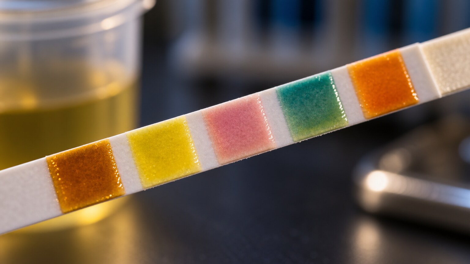
Figure 3. Specific dipstick reactions — blood, leukocyte esterase, nitrites, protein, or an extreme pH — are the most common automatic ("reflex") triggers for a follow-up microscopic exam.
Screening and Monitoring Contexts That Call for It by Default
Beyond individual symptoms or a specific abnormal dipstick reaction, certain broader clinical contexts routinely include a microscopic exam as a standard, built-in part of the ordered panel, regardless of how the dipstick turns out. Prenatal care is one of the clearest examples: because urinary tract infections during pregnancy carry higher stakes for both parent and baby, and because certain pregnancy-related conditions like preeclampsia are partly evaluated through urine findings, many prenatal urinalysis protocols include microscopic examination at multiple points throughout pregnancy as routine practice rather than something reserved for an abnormal screen.
Pre-surgical clearance is another common context, particularly for procedures involving the urinary tract itself or requiring general anesthesia, where a baseline microscopic exam helps rule out an undetected infection or kidney-level issue before a patient goes under anesthesia. Ongoing monitoring for people with chronic kidney disease, a kidney transplant, lupus, or certain other autoimmune conditions affecting the kidneys is another routine-inclusion scenario, since a microscopic exam can catch early cellular casts or cell types signaling a flare or rejection episode well before those changes would show up as a meaningful shift in blood-based kidney function tests. Diabetic patients undergoing periodic kidney health screening may also have microscopic examination included as standard practice, given the elevated baseline risk of kidney involvement that comes with long-standing diabetes.
Occupational and sports-related screening represents a somewhat different, less clinical category where microscopy sometimes appears as standard practice for reasons unrelated to any suspected illness. Some occupational health programs, particularly those involving exposure to industrial chemicals or heavy metals with known kidney toxicity, include periodic urinalysis with microscopic examination specifically as part of monitoring for early, subclinical signs of occupational kidney injury, long before any symptom would otherwise prompt testing. This is a preventive, exposure-driven inclusion rather than a response to any particular finding, and it illustrates how broadly the decision to include microscopy can be shaped by context rather than by an individual patient's presenting complaint.
Post-infection follow-up testing is another routine-inclusion scenario worth mentioning. After treatment for a confirmed urinary tract infection, particularly a more complicated case involving the kidney itself (pyelonephritis) or one that occurred during pregnancy, providers frequently order a follow-up urinalysis with microscopy to confirm the infection has genuinely cleared rather than simply assuming based on symptom improvement alone. This is especially relevant because symptoms can resolve noticeably faster than the underlying inflammatory changes visible on microscopy, and confirming a truly clean follow-up sample provides more complete reassurance than symptom relief by itself.
The Judgment Call: When Your Doctor Orders It "Just to Be Thorough"

Figure 4. A patient's individual risk profile — age, medication list, or a prior history of kidney or urinary conditions — often factors into a provider's decision to order microscopy proactively.
Sometimes a microscopic exam gets ordered even though no specific symptom or dipstick trigger technically demanded it, and this usually comes down to individual clinical judgment based on a patient's broader risk profile. A patient with a personal or family history of kidney stones, recurrent urinary tract infections, or a known structural urinary tract abnormality may have microscopy included routinely by a provider who has learned, from that patient's own history, that dipstick screening alone tends to miss things worth catching early in their specific case. Similarly, a patient recently started on a new medication known to occasionally affect the kidneys — certain antibiotics, some blood pressure medications, or long-term NSAID use, for example — may have a provider proactively add microscopy simply as an extra layer of monitoring during that adjustment period.
Age and general risk factors play a role too. Older adults, who are statistically more likely to have both urinary tract infections that present with atypical or absent classic symptoms and a higher baseline prevalence of kidney function changes, are more likely to have a provider request the more detailed microscopic picture even on a fairly unremarkable-looking dipstick result. None of this means something is necessarily wrong — it reflects a provider erring on the side of gathering more complete information given what they already know about a particular patient, which is a routine and appropriate part of individualized clinical care rather than a signal of concern in itself.
Age and Life-Stage Factors That Shift the Threshold

Figure 6. Very young children and older adults are both more likely to have microscopy ordered proactively, since classic urinary symptoms are frequently absent or hard to recognize in both groups.
Infants and very young children represent a distinct case where the threshold for ordering a microscopic exam is often lower than it would be for an adult presenting with the exact same dipstick result. Young children, particularly those not yet able to describe their own symptoms, frequently present with nonspecific signs like unexplained fever, fussiness, or poor feeding rather than the classic burning or urgency an adult would describe, which means a urinary tract infection can be easy to miss without a more thorough look. Pediatric guidelines in many settings specifically call for confirming a suspected infection with a more detailed, carefully collected sample and full microscopic examination, rather than relying on dipstick findings alone, given how much is potentially at stake with an undiagnosed kidney-involving infection in a very young child.
At the other end of the age spectrum, older adults frequently present with what's sometimes called "atypical" symptoms of a urinary tract infection — confusion, a general decline in function, or reduced appetite, rather than the burning or urgency a younger adult would report. Because these nonspecific presentations can just as easily be explained by an entirely different, unrelated cause, providers evaluating an older patient with this kind of vague presentation will often include a microscopic exam specifically to help rule a urinary source in or out with more confidence than a dipstick screen alone could provide. Older adults also have a higher background rate of asymptomatic bacteria in the urine that doesn't require treatment, which paradoxically means the more detailed microscopic picture, interpreted carefully alongside symptoms, is often more useful in this age group for avoiding unnecessary antibiotic treatment rather than only for catching missed infections.
What a Microscopic Exam Actually Looks For Once It's Ordered
Once a sample reaches the microscopy stage, a laboratory professional is looking for several broad categories of findings, each of which tells a different part of the story. Cells are the first category: red blood cells (suggesting bleeding somewhere along the urinary tract), white blood cells (suggesting inflammation or infection), and epithelial cells (mostly reflecting normal tissue turnover, though the specific type matters). Bacteria, yeast, and occasionally parasites make up a second category, offering a direct look at what organisms, if any, are actually present rather than relying on an indirect chemical marker.
Crystals form a third category, and while many crystal types are entirely normal byproducts of diet and hydration, certain types and patterns can suggest a tendency toward stone formation or, less commonly, a specific metabolic condition. Casts — cylindrical structures formed inside the kidney's tubules themselves — make up perhaps the most clinically significant category, since different cast types (hyaline, granular, red blood cell, white blood cell, or renal tubular epithelial cell casts) each point toward a distinct process happening specifically within kidney tissue, information a chemical dipstick has no way of providing on its own. Together, these categories give a provider a genuinely detailed, cell-by-cell picture of what's happening throughout the urinary tract, which is exactly why this second stage of testing exists as a distinct, deliberately ordered step rather than being folded automatically into every single sample.
Casts specifically deserve a bit more explanation, since they're one of the more distinctive findings a microscopic exam can identify and something a dipstick has no equivalent way of detecting at all. A cast forms when a specific protein, produced continuously by healthy kidney tubule cells, gets concentrated within the tubule itself and takes on the cylindrical shape of the tiny tube it formed inside, essentially acting as a mold. Whatever else happens to be present in that same segment of tubule at the time — nothing at all, in the case of a plain hyaline cast, or trapped white blood cells, red blood cells, or degenerating cellular debris in other cast types — becomes embedded within that protein matrix and gets flushed downstream intact as a single, recognizable structure. Because a cast can only form inside the kidney's own tubules, finding one is considered strong, specific evidence that whatever it contains genuinely originated from kidney tissue, rather than from further downstream in the bladder or urethra where casts cannot form at all. This is precisely the kind of anatomically specific information that makes the microscopic exam so much more informative than the dipstick alone for anyone with a suspected kidney-level process.
Hyaline casts, made of pure protein with nothing else trapped inside, are the most common type and are frequently seen even in healthy people, particularly after exercise, dehydration, or fever — situations that transiently increase how much of this normal protein gets concentrated in the tubules without reflecting any actual injury. Granular casts, containing degenerated cellular debris, are a step up in significance and can appear with more sustained kidney stress, though they too can appear in otherwise healthy individuals under certain conditions. Red blood cell casts and white blood cell casts are considered more specifically meaningful findings, generally prompting a closer look at glomerular inflammation or a kidney-involving infection, respectively, precisely because these particular cast types are rarely seen as an incidental, healthy-person finding the way hyaline casts sometimes are.

Figure 5. The sample is centrifuged to concentrate solid material into a sediment pellet before it can be examined directly under the microscope — an added step that explains the additional turnaround time.
Why This Extra Step Isn't Run on Every Single Sample by Default
It's worth understanding why laboratories don't simply run a microscopic exam on every urine sample as standard practice, since it might seem like the more thorough option would always make sense. The practical reality is one of resource allocation and diagnostic yield: the overwhelming majority of urine samples submitted for entirely routine reasons — a general wellness check-up with no symptoms, a pre-employment screen, or a straightforward dipstick that comes back completely unremarkable — are extremely unlikely to reveal anything on microscopy that wasn't already apparent, or absent, on the dipstick alone. Running detailed microscopy on every single one of these samples would meaningfully slow down laboratory turnaround times across the board and add unnecessary cost, without a proportional improvement in how many clinically meaningful findings actually get caught.
The reflex-testing model exists specifically to concentrate that more detailed, more resource-intensive attention on the samples statistically most likely to benefit from it — those with a reported symptom, an abnormal dipstick trigger, or a screening context known to carry a higher underlying risk. This isn't a cost-cutting shortcut at the expense of patient care; it's the opposite, an evidence-based approach to routing the more detailed test toward the situations where the additional detail is actually likely to change what a provider does next, while sparing unaffected patients an unnecessary delay and expense on a test unlikely to add anything.
This same logic explains why you might notice a microscopic exam appear on one visit's urinalysis but not another's, even for the same person, without anything being wrong on either occasion. A well person's straightforward annual physical might reasonably skip it if the dipstick is entirely clean and no symptoms were reported, while a visit six months later prompted by new urinary discomfort would reasonably include it from the start. The presence or absence of a microscopic exam on any given report is simply a reflection of what specific triggers were present at that particular visit — not a sign that your care is being handled inconsistently.
It's also worth noting that reflex-testing criteria aren't perfectly identical from one laboratory to the next, which explains why the exact same dipstick result might trigger a follow-up microscopic exam at one facility but not at another. A hospital laboratory serving a population with a higher baseline rate of acute, complex presentations might reasonably set a lower threshold for reflexing to microscopy than an outpatient laboratory serving a largely healthy population for routine screening purposes. Neither approach is objectively wrong; each is calibrated to the population and clinical context that particular laboratory most commonly serves, based on validated internal protocols reviewed periodically by laboratory medical directors.
Cost considerations for the patient are generally minimal in practice, since a microscopic exam is typically bundled into the standard urinalysis billing code when it's performed as part of the same order, rather than billed as a wholly separate, additional test. Insurance coverage for a complete urinalysis, including the microscopic component when clinically indicated, is standard across nearly all health plans, given how routine and foundational this testing is across virtually every area of medicine. Patients curious about the specific cost breakdown for their own visit are generally best served checking directly with their laboratory or insurance provider, since exact billing structures can vary somewhat by region and by which specific laboratory processes the sample.
How to Read the Section of Your Report Once It's Included
If your urinalysis report does include a microscopic section, it's typically organized as a straightforward list: red blood cells, white blood cells, epithelial cells, bacteria, crystals, and casts, each reported either as a semi-quantitative estimate (rare, few, moderate, or many) or as a numeric range per high-power field, abbreviated "HPF" on most reports. A completely clean microscopic section, with every category reading within its normal reference range, is a reassuring result regardless of what prompted the test to be ordered in the first place. An abnormal finding in any single category doesn't automatically mean something serious is happening — as covered elsewhere in detail, plenty of microscopic findings (a few squamous epithelial cells, a handful of hyaline casts, or trace crystals of a common, benign type) are considered normal variations rather than red flags.
What tends to matter most is the overall pattern across categories rather than any single line read in isolation. Elevated white blood cells paired with bacteria and a positive nitrite result paints a consistent picture pointing toward infection. Red blood cells paired with certain crystal types support a kidney stone process. Renal tubular epithelial cells paired with granular or cellular casts and elevated protein suggest a kidney-tissue-level process worth further evaluation. A provider reading your full report is essentially looking for whether these different pieces of information tell a coherent, consistent story or whether an isolated single finding stands alone without other supporting evidence, which meaningfully changes how much weight that finding is given.
It's also useful to know that a single microscopic exam, taken on its own, is rarely the final word on any diagnosis. Providers commonly request a repeat sample, ideally collected with careful attention to proper technique, whenever an initial result is borderline, mildly abnormal in a single category only, or inconsistent with the rest of a patient's presentation. This isn't an indication that the first result was performed incorrectly; it simply reflects how much natural variation can occur between individual collections, and how much value comes from confirming a pattern holds up on a second look before treating it as clinically meaningful. A single mildly elevated finding that normalizes on a properly collected repeat sample is generally treated very differently than the same finding that persists consistently across multiple samples.
Frequently Asked Questions
Does every urinalysis include a microscopic exam?
No. Most standard urinalyses start with a dipstick screening test only, and the microscopic exam is added either because of reported symptoms, a specific abnormal dipstick result, a screening context (like prenatal care) that includes it by default, or a provider's individual clinical judgment about a particular patient's risk profile.
Is a microscopic urinalysis a sign my doctor suspects something serious?
Not necessarily. It's frequently included as routine practice in specific contexts like pregnancy, pre-surgical clearance, or chronic condition monitoring, and is also commonly ordered upfront simply because a patient reported urinary symptoms worth investigating more thoroughly — not because something serious is already suspected.
Can I request a microscopic exam even if my dipstick was normal?
Generally, yes, if you have a specific reason for wanting it — ongoing symptoms not reflected in the dipstick result, a personal history that makes you want a more detailed baseline, or general reassurance. Your provider can weigh whether that request is clinically appropriate in your specific situation.
How long does the microscopic portion add to my results turnaround time?
It varies by laboratory, but the microscopic exam typically adds anywhere from a few hours to a full day beyond the near-instant dipstick result, since it requires centrifuging the sample and either automated imaging with human review or fully manual examination by a trained laboratory professional.
What's the difference between a "reflex" microscopic exam and one ordered directly?
A reflex exam is automatically triggered by the laboratory itself when specific dipstick results meet predefined criteria, without your provider needing to separately request it. A directly ordered exam is one your provider specifically requests upfront, typically based on your reported symptoms or a screening protocol, regardless of what the dipstick eventually shows.
Will my report say which reason triggered the microscopic exam?
Not usually. Most lab reports simply present the microscopic findings themselves without documenting why that stage was performed, since the ordering rationale typically lives in your provider's own clinical notes rather than on the printed lab report. If you're curious about the specific reason in your case, asking your provider directly is the most reliable way to find out.
Finally, it's worth remembering that a microscopic exam, however detailed, is still just one piece of a much larger clinical picture. Providers weigh it alongside your reported symptoms, physical exam findings, other blood work, and, in some cases, imaging, rather than treating a single urinalysis result as a standalone diagnosis in itself. Understanding why the test was ordered in the first place, and what its individual findings typically mean in context, helps you engage more meaningfully with that larger conversation rather than fixating on a single flagged line without the surrounding clinical picture that gives it real meaning.
Conclusion
A microscopic urinalysis isn't an automatic add-on to routine urine testing — it's a distinct, more detailed second stage that gets triggered by a specific set of circumstances: symptoms you reported, an abnormal dipstick finding, a screening context built to include it by default, or your provider's individualized judgment based on your broader health picture. Understanding which of these applied at your particular visit turns a seemingly mysterious extra line item on your lab report into something genuinely explainable, and helps put the result itself — whether entirely normal or flagged in some category — into its proper context rather than reading it in isolation.
Still Not Sure What Your Results Mean?
Upload your labs and get a complete, visual, plain-language interpretation of every biomarker — delivered to your inbox in under 15 minutes.
Get My ReportThis article is for educational purposes only and does not constitute medical advice. Always consult your healthcare provider regarding your specific lab results.