
What Does a Borderline High Calcium Result Mean?
A calcium result sitting just a few tenths of a point above the reference range occupies a genuinely different clinical category than a dramatically elevated one. Mild, borderline hypercalcemia rarely announces itself with symptoms, rarely requires urgent action, and is frequently caught entirely by accident on routine bloodwork ordered for something else altogether. That doesn't mean it should be ignored — a persistently borderline calcium is actually the single most common way primary hyperparathyroidism, the leading cause of elevated calcium in otherwise healthy outpatients, first comes to light. Understanding exactly how a borderline result gets confirmed, monitored, and eventually worked up turns a vague, mildly worrying number into something with a clear, sensible next step.

Figure 1. Hypercalcemia exists on a spectrum rather than as a single category — a borderline result sitting just above the normal range is handled very differently than a dramatically elevated one, both in urgency and in what it typically means.
Where "Borderline" Actually Sits on the Number Line
Standard reference ranges for total blood calcium typically fall between about 8.5 and 10.5 mg/dL (roughly 2.1 to 2.6 mmol/L in SI units), though the exact cutoffs vary slightly by laboratory. "Borderline high" generally describes a result somewhere between just above that upper limit and up to roughly 11.0 to 11.5 mg/dL — meaningfully elevated in a statistical sense, but well short of the levels (typically above 12 to 14 mg/dL) associated with acute symptoms or genuine medical urgency. This distinction matters enormously in practice, since the appropriate response to a calcium of 10.7 mg/dL looks almost nothing like the appropriate response to a calcium of 14 mg/dL, even though both technically qualify as "elevated" on a lab report.
It's worth understanding that this borderline zone isn't simply "less serious" hypercalcemia waiting to become more serious — in many cases, it represents an entirely different underlying process than severe hypercalcemia does. The most common cause of severe, acute hypercalcemia is cancer-related, while the most common cause of chronic, mild, borderline elevation in an otherwise healthy outpatient is primary hyperparathyroidism, a condition that can remain stable at this mild level for years or even decades without ever progressing to a dangerous range at all.
This distinction between mild-chronic and severe-acute hypercalcemia reflects two genuinely different underlying biological processes, not just two points on the same continuum measured at different times. Cancer-related hypercalcemia typically develops relatively quickly, often over weeks, and stems from either direct tumor invasion of bone or a tumor producing a hormone-like substance that mimics PTH's effect on calcium release from bone, a mechanism capable of pushing calcium to genuinely dangerous levels quickly because it operates largely outside the body's normal regulatory feedback loop. Primary hyperparathyroidism, by contrast, still operates through the body's normal parathyroid gland tissue, just with impaired feedback regulation, which is part of why it tends to plateau at a mildly elevated level rather than escalating without limit — the underlying gland tissue, while overactive, still retains some of its normal physiological constraints.
Understanding this mechanistic difference helps explain why a borderline result found in a healthy-appearing outpatient during routine screening carries a very different pretest likelihood profile than the same borderline number found in a hospitalized patient with a known cancer diagnosis or significant recent weight loss — the same laboratory value, interpreted through two different clinical contexts, points toward genuinely different next steps, which is precisely why the surrounding clinical picture matters as much as the isolated number itself.
Why a Single Borderline Result Almost Always Gets Repeated First

Figure 2. A single mildly elevated calcium result is almost always confirmed with a repeat draw, ideally fasting and without a recent tourniquet, before any further workup begins.
Before any meaningful workup begins, a mildly elevated calcium is almost always simply repeated. This isn't excessive caution — it directly addresses several well-documented, entirely mundane reasons a single reading can come back mildly high in someone with genuinely normal calcium metabolism. Prolonged tourniquet application during the blood draw itself can concentrate proteins in the local blood sample enough to nudge total calcium slightly upward, since a meaningful portion of blood calcium is bound to protein and protein concentration directly affects the total measured value. Mild dehydration at the time of the draw produces a similar concentrating effect on essentially everything measured in the sample, calcium included, without reflecting any change in the body's actual calcium regulation.
Because of this, standard practice calls for the repeat sample to be drawn under more carefully controlled conditions — fasting, well-hydrated, with minimal tourniquet time — specifically to rule out these mundane explanations before drawing any further conclusions. A repeat result that returns to clearly normal essentially closes the question for the time being, while a repeat result that confirms the elevation shifts the conversation toward genuine, ongoing evaluation.
Timing of the repeat test also matters in a way that's easy to overlook. A single confirmatory repeat drawn just days after the original result mainly rules out acute, one-off artifacts like dehydration or tourniquet effect, but it doesn't yet establish whether the elevation is genuinely chronic and persistent, since even a real, transient cause — a brief period of illness, a short course of a calcium-affecting medication, or a few days of unusually low fluid intake — could still explain a result confirmed only a few days apart. Because of this, many providers specifically wait several weeks to a few months before drawing a second confirmatory sample when the clinical situation isn't urgent, since a result that remains elevated across this longer interval provides considerably stronger evidence of a genuinely chronic, ongoing process worth formally investigating, compared with two closely spaced readings that could still both reflect the same brief, transient cause.
It's also worth understanding what "persistent" actually means in this context, since the term gets used somewhat loosely. Clinical guidelines generally define persistent hypercalcemia as an elevated result confirmed on at least two separate occasions, typically spaced weeks to months apart, ideally with albumin measured alongside each one. A patient with just one elevated reading, even a clearly confirmed one drawn under ideal conditions, technically hasn't yet met the formal definition of persistent hypercalcemia — which is a meaningful distinction, since a genuinely isolated, non-recurring elevation (however that might have occurred) simply doesn't carry the same diagnostic weight as a pattern documented to be genuinely ongoing over time.
Already have a calcium number from a recent test, but not sure if it's actually good or bad for you? Just plug it in and see instantly.
🧮 Try the Free CalculatorWhy Albumin Has to Be Checked Alongside a Borderline Result
Roughly 40 percent of the calcium circulating in blood is bound to albumin, the most abundant protein in blood plasma, meaning a standard total calcium measurement is directly influenced by how much albumin happens to be present at the time of the draw — independent of how much biologically active, unbound "ionized" calcium is actually available to the body's cells. A person with unusually high albumin, from dehydration or certain other conditions, can show a mildly elevated total calcium purely as a reflection of that elevated albumin, without any true increase in the physiologically relevant ionized fraction at all.
This is exactly why a borderline total calcium result is almost always interpreted alongside a same-sample albumin measurement, and often reported alongside a mathematically "corrected" calcium value that adjusts for albumin's specific effect. A borderline total calcium that fully normalizes once corrected for albumin is generally treated very differently — and with considerably less urgency — than one that remains elevated even after this correction is applied, since the corrected value more accurately reflects what's actually happening with the body's true calcium regulation.
The standard correction formula adds 0.8 mg/dL to the measured total calcium for every 1.0 g/dL that albumin falls below its own normal reference value of approximately 4.0 g/dL, and subtracts proportionally when albumin runs above that baseline. This formula, while widely used and genuinely helpful as a quick screening adjustment, is itself an approximation rather than a perfectly precise measurement, and it can become meaningfully less reliable in patients with significantly abnormal albumin levels, severe liver or kidney disease, or certain other conditions that affect protein binding in ways the simple formula doesn't fully account for.
For this reason, when a borderline corrected calcium result remains genuinely ambiguous, or when the clinical picture doesn't quite add up despite a seemingly reassuring correction, providers increasingly turn to directly measuring ionized calcium instead of relying on the calculated correction. Ionized calcium testing measures the biologically active, unbound fraction directly, bypassing the albumin-dependent estimation entirely and providing what's generally considered the more physiologically accurate number in ambiguous or complicated cases. This test does require more careful sample handling, since ionized calcium is sensitive to how a blood sample is collected and processed (exposure to air can falsely lower the reading through a shift in blood pH), which is part of why it isn't used as the default first-line test for every patient despite its theoretical accuracy advantage over the standard total-plus-correction approach.
The Most Common Reason Behind a Persistently Borderline Result

Figure 3. Primary hyperparathyroidism, caused by one or more of the four small parathyroid glands producing too much hormone, is the most common explanation for a persistently mild, borderline-elevated calcium in an otherwise healthy adult.
Once a truly persistent, confirmed borderline elevation is established, primary hyperparathyroidism becomes the leading suspect in an otherwise well outpatient, particularly in postmenopausal women, the population most commonly affected. The four small parathyroid glands, sitting behind the thyroid gland, normally release parathyroid hormone in a tightly regulated way specifically to keep blood calcium within its narrow, precisely maintained normal range at all times. When one or, less commonly, more than one of these glands develops a small, usually benign growth that produces hormone independent of the body's normal feedback signals, the result is a modest, chronic overproduction that nudges calcium just above the normal range — frequently without producing any noticeable symptoms at all, especially in its earlier, milder stages.
This is precisely why primary hyperparathyroidism today is overwhelmingly diagnosed not because a patient reported symptoms, but because a mildly elevated calcium happened to show up on bloodwork ordered for a completely unrelated reason — a pre-surgical panel, an annual physical, or bloodwork drawn while investigating something else entirely. Decades ago, before routine blood chemistry panels became standard practice, this same condition was typically only caught once it had progressed to producing kidney stones, significant bone loss, or other more advanced complications — a genuinely different clinical picture than the mild, asymptomatic elevation caught early through routine testing today.
This shift in how the condition typically gets discovered has genuinely changed what "having primary hyperparathyroidism" means for the average patient today compared with a generation or two ago. The classic teaching phrase used for decades to describe severe, advanced hyperparathyroidism — "stones, bones, groans, and psychiatric overtones," referring to kidney stones, bone pain, abdominal complaints, and mood or cognitive changes — describes a presentation that's become considerably less common as a first sign of the condition, precisely because routine blood testing now catches the overwhelming majority of cases at the mild, asymptomatic stage, long before this classic, advanced symptom cluster would ever have a chance to develop in the first place.
The specific glandular abnormality behind primary hyperparathyroidism also matters for how it's ultimately managed, even though it rarely changes the initial monitoring-versus-workup decision. In roughly 80 to 85 percent of cases, a single parathyroid gland develops a benign growth called an adenoma, functioning independently of the body's normal calcium feedback signal. In most of the remaining cases, multiple glands are affected simultaneously, a pattern called hyperplasia, which behaves somewhat differently from a single adenoma and can carry different implications for surgical planning if surgery does eventually become the recommended path. Parathyroid cancer causing this presentation is genuinely rare, accounting for well under one percent of cases, and is generally suspected only when calcium levels run considerably higher than the mild, borderline range this article is specifically focused on, or when a parathyroid gland is found to be unusually large on imaging.
How Parathyroid Hormone Testing Confirms the Pattern

Figure 4. Measuring parathyroid hormone alongside a confirmed borderline calcium result is the single test that most efficiently separates primary hyperparathyroidism from every other possible cause of mild elevation.
Once a genuine, persistent borderline elevation is confirmed, the single most useful next test is a parathyroid hormone (PTH) measurement drawn from the very same blood sample. In a healthy person, rising calcium should suppress PTH release, since the body's normal feedback loop actively works to bring calcium back down. Finding a PTH level that's normal or, more tellingly, frankly elevated alongside a confirmed high calcium is genuinely abnormal — the parathyroid glands should be shutting down hormone production in response to elevated calcium, not maintaining or increasing it, and this "inappropriately normal or high" PTH pattern is the specific diagnostic signature of primary hyperparathyroidism.
This single paired measurement is remarkably efficient at narrowing the diagnostic field, since nearly every other cause of hypercalcemia — cancer, excess vitamin D, certain medications, and less common causes — appropriately suppresses PTH in response to the elevated calcium, producing a low PTH result instead. A borderline calcium paired with a suppressed PTH therefore points the workup in an entirely different direction than a borderline calcium paired with a normal or elevated PTH, making this one paired result arguably the most important branch point in the entire evaluation.
When the suppressed-PTH pathway is followed instead, the workup shifts toward the specific non-parathyroid causes covered elsewhere in this article and toward additional testing aimed at ruling out malignancy when clinically indicated — imaging studies, additional blood markers, and a careful review of symptoms, medication history, and risk factors, guided by the overall clinical picture rather than a fixed, one-size-fits-all checklist applied identically to every patient regardless of context. It's worth noting that a small subset of patients present with a genuinely intermediate PTH result, technically within the normal reference range but sitting close to its lower edge — a pattern that doesn't cleanly fit either the classic "suppressed" or "inappropriately normal" category, and that sometimes requires additional testing or simply careful clinical judgment and follow-up to interpret confidently.
A related but genuinely distinct condition worth mentioning specifically because it's sometimes confused with primary hyperparathyroidism is familial hypocalciuric hypercalcemia, a rare, inherited condition in which the body's calcium-sensing mechanism itself is set to a slightly different threshold, producing a mild, lifelong, stable calcium elevation alongside an inappropriately normal PTH — a lab pattern that can look genuinely similar to mild primary hyperparathyroidism at first glance. Distinguishing between the two matters considerably, since familial hypocalciuric hypercalcemia doesn't respond to or require parathyroid surgery at all, and mistakenly proceeding with surgery for this condition provides no benefit while still carrying real surgical risk. A 24-hour urine calcium collection, comparing urinary calcium excretion against the blood calcium level, is the specific test typically used to tell the two conditions apart, since this particular hereditary condition characteristically produces unusually low urinary calcium excretion relative to how elevated the blood calcium is — a pattern not typically seen in primary hyperparathyroidism.
Other Explanations Behind a Mild, Borderline Elevation
Beyond primary hyperparathyroidism, several other causes can produce a genuinely confirmed, persistently mild elevation rather than a one-off artifact. Thiazide diuretics, a commonly prescribed class of blood pressure medication, reduce how much calcium the kidneys excrete in urine, which can nudge blood calcium modestly upward in some patients — a well-recognized medication effect rather than a sign of any underlying gland or bone problem, and one that a provider will specifically ask about when reviewing a borderline result. Excess vitamin D from high-dose supplementation, taken consistently over weeks to months, can also produce a mild, sustained elevation by increasing how efficiently the intestines absorb dietary calcium.
Prolonged immobilization — extended bed rest after major surgery, a serious injury, or a long hospital stay — can also mildly elevate calcium over time, since bone constantly remodels even without weight-bearing activity, and immobilized bone tends to release slightly more calcium into the bloodstream than it reabsorbs. Certain granulomatous conditions, including sarcoidosis and some chronic infections, can likewise produce a mild, chronic elevation through their own distinct mechanism involving abnormal vitamin D activation within inflammatory tissue. Each of these alternative explanations tends to come with its own supporting clues in a patient's broader history and other lab findings, which is exactly why a thorough review of medications, recent activity level, and supplement use accompanies the interpretation of any confirmed borderline result.
Lithium, a medication commonly used to manage bipolar disorder, deserves specific mention among the medication-related causes, since it affects calcium regulation through a genuinely distinct mechanism from thiazide diuretics: lithium appears to directly alter how sensitively the parathyroid glands respond to circulating calcium levels, effectively resetting the threshold at which they reduce hormone output, producing a pattern that can closely resemble primary hyperparathyroidism on standard testing. This effect is well recognized among psychiatrists and endocrinologists managing patients on long-term lithium therapy, and a mild, persistent calcium elevation in this specific population is generally interpreted with this medication effect specifically in mind, rather than immediately assuming an unrelated, coincidental parathyroid problem, though genuine underlying primary hyperparathyroidism can also occur in this same population and sometimes requires careful additional evaluation to distinguish from the medication's own direct effect.
Sarcoidosis and other granulomatous conditions work through a genuinely fascinating and distinct mechanism worth understanding a bit further: the inflammatory immune cells that form granulomas — small clusters of immune tissue characteristic of these conditions — are capable of activating vitamin D on their own, independent of the kidney's normal regulatory control over this process. This unregulated vitamin D activation increases intestinal calcium absorption in a way the body's normal feedback mechanisms can't properly rein in, producing a mild, chronic elevation that, notably, tends to worsen with increased sun exposure or vitamin D supplementation — the opposite of what's typically recommended for most other causes of low or borderline-normal vitamin D status, making an accurate underlying diagnosis genuinely important for guiding appropriate lifestyle and supplementation advice.
Why Borderline Elevation Often Means Monitoring, Not Immediate Intervention

Figure 5. Confirmed mild primary hyperparathyroidism without significant symptoms, bone loss, or kidney involvement is frequently managed through structured annual monitoring rather than immediate surgical referral.
Even once primary hyperparathyroidism is confirmed as the underlying cause, mild, asymptomatic disease is very often managed through structured monitoring rather than immediate intervention. Established guidelines specifically define criteria for when surgical removal of the overactive gland (parathyroidectomy) is recommended versus when careful observation is a reasonable, medically appropriate alternative — criteria that include how far above normal the calcium actually sits, results of a bone density scan looking for early bone loss, kidney function and evidence of kidney stones, patient age, and whether the patient can realistically be expected to follow through on consistent long-term monitoring.
For patients who meet criteria for observation rather than surgery, this typically means annual calcium and kidney function testing, bone density scanning roughly every one to two years, and general attention to adequate hydration and avoiding factors that could further elevate calcium, such as very high-dose calcium or vitamin D supplementation without specific guidance. Many patients monitored this way remain stable for years without ever requiring surgery, while others show gradual progression that eventually does meet surgical criteria — which is precisely the reason ongoing, structured monitoring exists in the first place, rather than a single decision made once and never revisited.
Age plays a genuinely significant role in how this monitor-versus-treat decision gets weighed in practice, even independent of any specific bone or kidney finding. Current guidelines generally lower the threshold for recommending surgery in younger patients — those under roughly 50 years old — even with otherwise mild, asymptomatic disease, simply because a younger patient faces many more decades of potential exposure to chronically elevated calcium and parathyroid hormone than an older patient would, meaningfully increasing the cumulative lifetime risk of eventually developing bone loss, kidney stones, or other complications if the condition is simply monitored indefinitely rather than definitively treated while the patient is otherwise young and healthy enough to tolerate surgery with minimal risk.
Parathyroidectomy itself, when it is recommended, is generally a well-tolerated, minimally invasive outpatient or short-stay procedure in appropriately selected patients, particularly since modern preoperative imaging techniques (specialized ultrasound and a nuclear medicine scan called sestamibi imaging) can often localize the specific overactive gland beforehand, allowing a more focused, minimally invasive surgical approach rather than a more extensive exploration of all four glands. Cure rates for this kind of focused, image-guided surgery performed by an experienced surgeon are generally quite high, which is part of why surgery, once genuinely indicated, is typically framed to patients as a definitive, generally low-risk solution rather than something to be feared or avoided reflexively.
When a Borderline Result Actually Warrants Faster Action
Certain features shift a borderline result from "monitor calmly" toward "investigate more urgently," even without the calcium itself reaching a dangerously high absolute level, and recognizing these features early is precisely what allows a provider to appropriately triage a mildly abnormal number rather than treating every borderline result with an identical, one-size-fits-all response regardless of the surrounding clinical context. A borderline calcium accompanied by genuine, otherwise unexplained symptoms — new kidney stones, unexplained bone pain, notable cognitive changes, or significant unexplained fatigue — warrants a more expedited workup than an identical number found purely incidentally in someone feeling completely well. Similarly, a borderline result found alongside other concerning findings on the same panel, or in a patient with risk factors for malignancy such as unexplained weight loss, is handled with more urgency, since even a mild elevation in that specific context carries a different pretest likelihood of a more serious underlying cause.
A borderline calcium that shows a clear, consistent upward trend across sequential testing over months, even while remaining technically within the "borderline" category at each individual measurement, is also treated as more actionable than one that has remained flat and stable across the same interval, since the trajectory itself is meaningful information a single snapshot value can never provide on its own, regardless of how reassuring any individual reading might look in isolation.
Family history carries its own specific weight in this decision as well, particularly a history of multiple endocrine neoplasia (MEN) syndromes, a group of rare inherited conditions in which primary hyperparathyroidism can occur alongside tumors affecting other hormone-producing glands, most commonly the pancreas and pituitary in MEN1, or the thyroid and adrenal glands in MEN2, syndromes that shift how thoroughly a borderline result gets investigated. A borderline calcium result in someone with a known family history of one of these syndromes, or with a personal history suggesting involvement of multiple endocrine glands, prompts a meaningfully more thorough evaluation than the same result in someone without any such family or personal history, since the underlying genetic cause, when present, carries broader implications extending well beyond the calcium and parathyroid findings alone, including genetic counseling considerations for other family members.
Pregnancy represents another context where a borderline calcium result is handled with particular care and urgency, since both untreated maternal hypercalcemia and any surgical intervention carry distinct considerations during pregnancy that don't apply outside of it. Elevated calcium during pregnancy has been associated with increased risk of certain pregnancy complications, and management decisions in this specific population are generally made collaboratively between endocrinology and maternal-fetal medicine specialists, weighing the relative risks of continued monitoring against the risks of surgical intervention at a particular point in the pregnancy, a genuinely specialized decision that falls outside the standard monitoring framework used for the general adult population.
Breastfeeding introduces its own, separate set of considerations worth briefly mentioning, since calcium demands shift again during lactation, and a borderline result identified during this period is generally interpreted with this additional physiological context specifically in mind, alongside the same broader evaluation framework already described.
Bone Density and Kidney Screening: The Two Checks That Guide the Decision
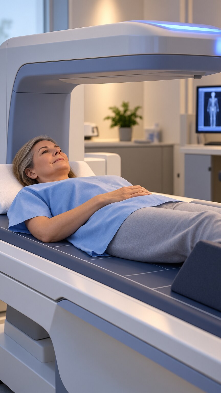
Figure 6. A bone density scan and kidney function assessment are the two standard checks used to determine whether confirmed mild hyperparathyroidism has begun affecting the skeleton or kidneys, directly shaping the monitor-versus-treat decision.
Once primary hyperparathyroidism is confirmed as the cause of a borderline calcium elevation, two specific evaluations do most of the work in determining whether observation or surgical referral is the more appropriate path. A bone density scan (DEXA) checks specifically for early bone loss at the wrist, hip, and spine, since chronically elevated parathyroid hormone gradually draws calcium out of bone to help maintain blood calcium levels, and this effect tends to show up first and most prominently at the wrist — a distinctive pattern that helps distinguish parathyroid-related bone loss from more common causes like postmenopausal osteoporosis, which typically affects the spine and hip more than the wrist.
Kidney function testing and, in some cases, imaging to check for kidney stones (sometimes silent and never previously noticed) forms the second pillar of this evaluation, since chronically elevated calcium increases the risk of stone formation and can, over a long enough period, gradually affect kidney function itself. Finding either meaningful bone loss or kidney involvement on this screening generally shifts a patient from the "monitor" category toward the "consider surgical referral" category, even if the calcium level itself remains only mildly, borderline elevated — since it's evidence the condition is already producing measurable downstream effects rather than sitting quietly at a stable, harmless baseline.
A 24-hour urine calcium collection is often performed alongside these two screenings, serving a dual purpose: it helps distinguish primary hyperparathyroidism from the rare, benign familial condition discussed elsewhere in this article, and it directly quantifies how much excess calcium is actually passing through the kidneys, which is itself a meaningful data point in assessing ongoing kidney stone risk independent of whether a stone has already formed and been detected.
Frequently Asked Questions
Should I be worried about a calcium result that's only slightly above normal?
Not immediately. A single mildly elevated result is commonly repeated first to rule out a testing artifact like dehydration or tourniquet effect. A genuinely confirmed, persistent borderline elevation warrants evaluation, but rarely represents a medical emergency requiring urgent same-day action.
Why does my provider want a PTH test if my calcium is only mildly high?
PTH is the single most efficient test for narrowing down the cause. A normal or elevated PTH alongside high calcium specifically points toward primary hyperparathyroidism, while most other causes of hypercalcemia appropriately suppress PTH instead, making this one paired result a genuinely major diagnostic branch point overall.
Does borderline high calcium always mean I have primary hyperparathyroidism?
No. It's the most common cause in an otherwise healthy outpatient, but medications like thiazide diuretics and lithium, excess vitamin D supplementation, granulomatous conditions like sarcoidosis, and prolonged immobilization can also produce a confirmed, persistent mild elevation.
If I'm diagnosed with mild primary hyperparathyroidism, will I need surgery right away?
Not necessarily. Established guidelines define specific criteria for surgery versus monitoring, based on how elevated calcium is, bone density findings, kidney involvement, and age, with younger patients generally facing a lower surgical threshold given their longer remaining exposure time. Many patients with mild, asymptomatic disease are monitored rather than operated on.
Can dehydration alone cause a borderline high calcium reading?
Yes. Dehydration concentrates protein-bound calcium in the blood sample, which can push total calcium slightly above range without reflecting any true change in calcium regulation, which is one reason a repeat, well-hydrated sample is standard practice.
How long does it typically take to get to a final answer after a borderline result?
It varies, but a full evaluation — including a confirmatory repeat, albumin correction, and a paired PTH measurement — can often be completed within a few weeks to a couple of months, though some providers deliberately space testing further apart in order to confirm a genuinely persistent pattern first.
Turnaround time and cost for the initial evaluation are generally reasonable, since calcium, albumin, and PTH are all routine, widely available laboratory tests typically returning results within a day or two, and covered by essentially all standard health insurance plans when ordered with an appropriate documented clinical indication. Bone density scanning and any imaging needed to localize a specific overactive gland, should surgery eventually be pursued, add somewhat more time to the overall evaluation but remain standard, well-established procedures at most medical centers, generally accessible without significant delay for most patients navigating this specific workup.
Conclusion
A borderline high calcium result occupies a genuinely distinct category from a dramatically elevated one — usually not an emergency, often symptom-free, and frequently explained by primary hyperparathyroidism, a condition that can be safely monitored for years in mild, well-characterized cases. Understanding the standard path a confirmed borderline result actually follows — repeat testing, albumin correction, a paired PTH measurement, and structured monitoring or workup depending on what's found — replaces uncertainty about a mildly abnormal number with a clear, medically sound plan, one built on decades of accumulated clinical evidence about exactly which findings genuinely warrant action and which are best simply watched carefully over time.
Still Not Sure What Your Results Mean?
Upload your labs and get a complete, visual, plain-language interpretation of every biomarker — delivered to your inbox in under 15 minutes.
Get My ReportThis article is for educational purposes only and does not constitute medical advice. Always consult your healthcare provider regarding your specific lab results.