
What Does Low Calcium Mean for Bone and Muscle Health?
Seeing a low calcium result on your lab report — a condition doctors call hypocalcemia — can be alarming, especially once you learn that calcium is the same mineral that builds your skeleton. But here's the twist that surprises most people: a low number on a routine blood test rarely means your bones are somehow running out of calcium. Your body treats blood calcium and bone calcium as two almost entirely separate accounts, protected by one of the most tightly regulated systems in all of human physiology. When blood calcium drops, the more immediate and more dangerous effects usually show up first in your nerves and muscles, not your skeleton — muscle cramps, tingling, and in more serious cases, involuntary spasms. Bone effects, when they happen, tend to build slowly over months or years rather than overnight. This article walks through what a low calcium result actually means, how your body's calcium-control system works, why the number on your report needs context to interpret correctly, what causes it to drop, how it shows up differently across different life stages, and how it's typically worked up and treated.
What "Low Calcium" Actually Means on a Lab Report
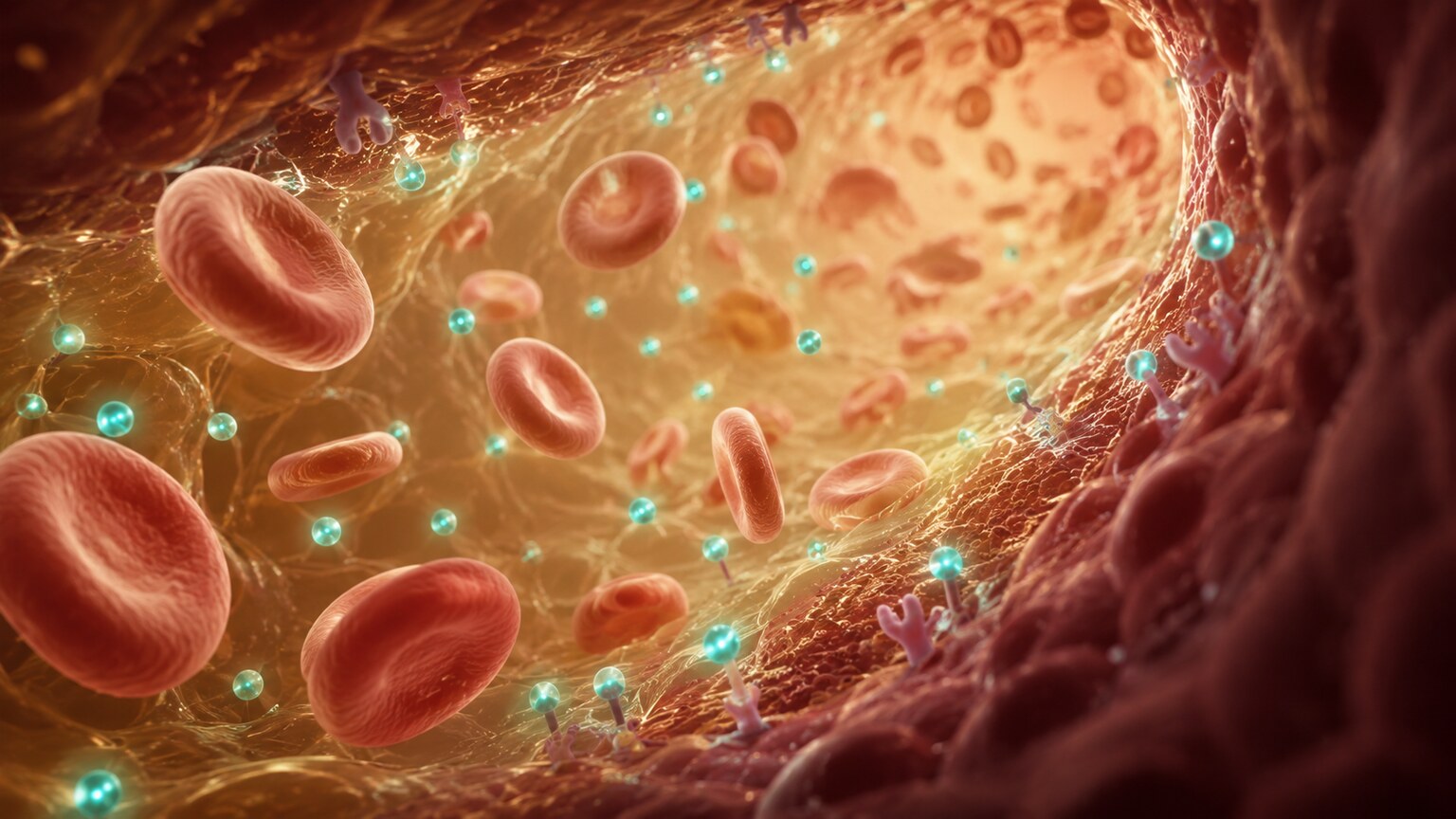
Figure 1. Blood calcium normally circulates within a narrow, tightly regulated range; hypocalcemia describes a level that has dropped below that range.
Calcium is one of the most abundant minerals in your body, but 99% of it is locked away in your bones and teeth, functioning as structural material — think of it as the rebar and concrete of your skeletal framework. The remaining 1% circulates in your blood and soft tissues, where it plays a completely different role: it acts as a messenger, helping nerves fire correctly, muscles contract and relax on command, blood clot when you're injured, and your heart maintain a steady rhythm. When a lab report flags "low calcium," it's almost always referring to that small circulating fraction, not your total body stores. A normal total blood calcium level in adults typically falls between about 8.5 and 10.5 milligrams per deciliter (mg/dL), though the exact reference range can vary slightly between laboratories depending on their equipment and methods. A result below that range is what clinicians call hypocalcemia.
What makes this number tricky to interpret on its own is that roughly 40% of the calcium in your blood travels bound to a protein called albumin, much like a passenger riding in a car rather than walking freely. Only the unbound, "free" portion — called ionized calcium — is actually biologically active, meaning it's the part your nerves and muscles can actually use. Standard total calcium tests measure both the bound and unbound portions together, which means a low total calcium reading can sometimes reflect low albumin rather than a true shortage of usable calcium. This single detail is exactly why an isolated calcium number, without any additional context, can be genuinely misleading — a theme this article returns to throughout, because it's the single most important thing to understand before drawing any conclusions from a low result.
Why Calcium Matters for More Than Bones and Muscles
It's worth pausing on just how many different systems in your body actually depend on that small, tightly guarded 1% of circulating calcium, because it explains why hypocalcemia can produce such a varied and sometimes confusing range of symptoms. Beyond nerve and muscle signaling, calcium is a required ingredient in the blood clotting cascade — several of the clotting factors your body uses to stop bleeding after an injury physically require calcium ions to activate in the correct sequence, which is part of why blood collection tubes used for certain lab tests contain a calcium-binding chemical, to keep the sample from clotting inside the tube before it reaches the laboratory. Calcium is also essential for normal heart muscle contraction, working alongside potassium and magnesium to keep your heart's electrical rhythm steady; significant hypocalcemia can lengthen a portion of the heart's electrical cycle visible on an EKG called the QT interval, which in severe cases raises the risk of a dangerous heart rhythm disturbance.
At the cellular level, calcium acts as what biologists call a "second messenger" — a tiny internal signal that cells use to translate an external trigger (a hormone arriving, a nerve impulse firing) into an actual internal action, whether that's a muscle fiber contracting, a gland releasing its hormone, or an egg cell activating after fertilization. This messenger role is so fundamental that it's shared across nearly every cell type in your body, not just muscle and nerve cells, which is why a significant calcium imbalance can ripple outward into symptoms that, at first glance, seem to have nothing to do with "bones" at all — brittle nails, dry skin, brain fog, or mood changes have all been described in association with more severe or prolonged hypocalcemia, alongside the more classically recognized muscle and nerve symptoms. Understanding this broader role is part of why clinicians take a low calcium result seriously even in someone who feels mostly fine, since the mineral's job description extends so far beyond what its name might suggest.
How Your Body Keeps Blood Calcium So Tightly Controlled
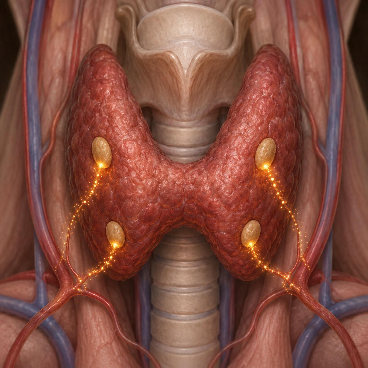
Figure 2. The four parathyroid glands, sitting behind the thyroid, sense falling blood calcium and respond by releasing parathyroid hormone.
To understand why blood calcium stays so remarkably stable — even when your dietary intake varies wildly from day to day — it helps to know about the small but powerful glands responsible for guarding it. Four tiny structures called the parathyroid glands, each roughly the size of a grain of rice, sit tucked behind your thyroid gland in your neck. Their entire job is to monitor blood calcium levels moment to moment and respond the instant it starts to drift, using a hormone called parathyroid hormone, or PTH. When blood calcium falls even slightly, the parathyroid glands sense it within minutes and ramp up PTH release, which then acts on three different target organs simultaneously to pull calcium back up.
First, PTH signals your bones to release some of their stored calcium into the bloodstream, a process that happens through cells called osteoclasts. Second, it signals your kidneys to reabsorb more calcium that would otherwise be excreted in urine, essentially reducing calcium loss rather than actively gaining more. Third, and perhaps most importantly for long-term balance, PTH stimulates your kidneys to activate vitamin D into its most potent form, calcitriol, which then travels to your intestines and boosts how efficiently you absorb calcium from the food you eat. This three-pronged response — bone release, kidney conservation, and enhanced intestinal absorption — usually restores blood calcium to normal within hours, which is exactly why blood calcium levels stay so tightly clustered around a narrow set point in healthy people, regardless of whether they had a dairy-rich breakfast or skipped meals entirely that day.
Why Low Blood Calcium Doesn't Always Mean Your Bones Are Losing Calcium
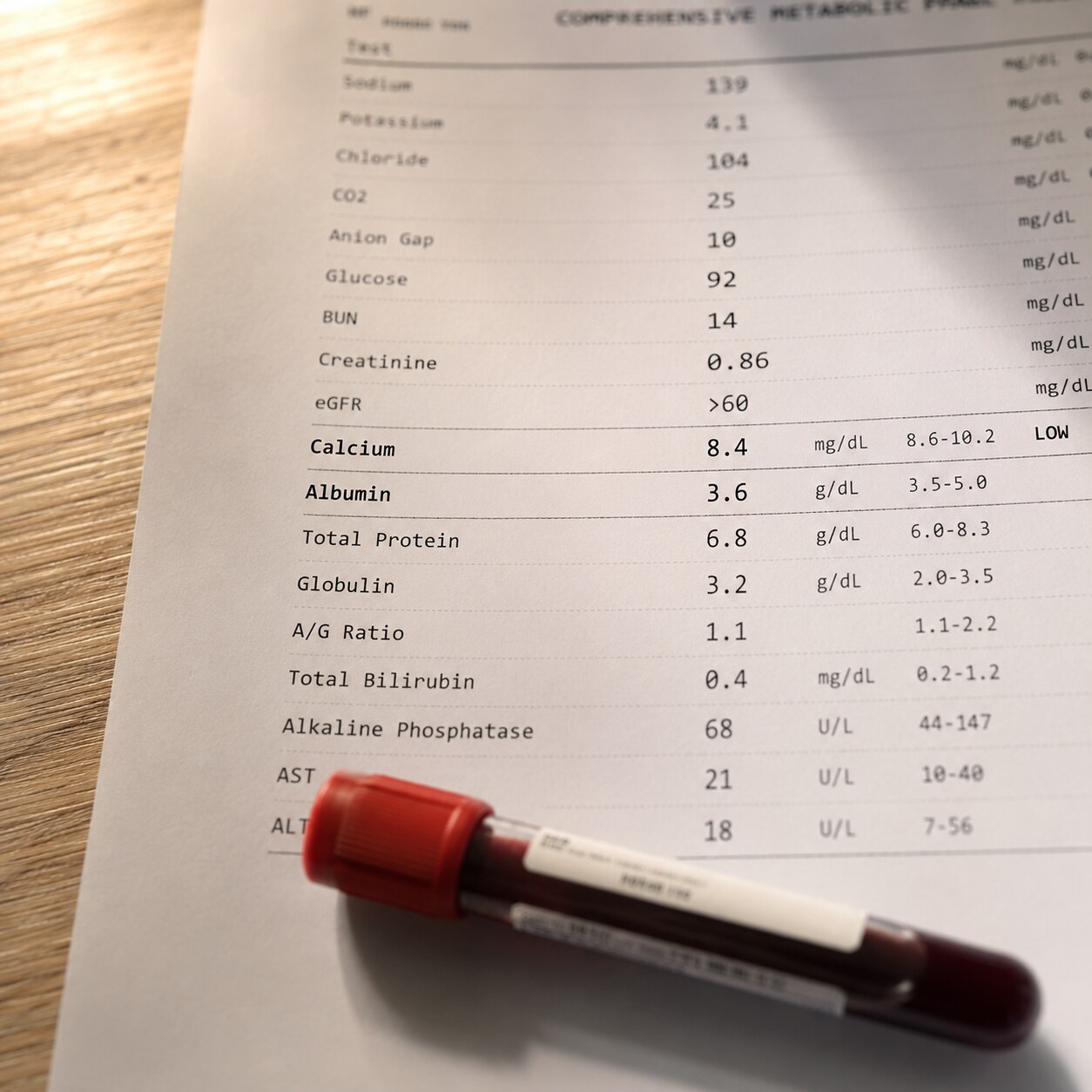
Figure 3. Because roughly 40% of blood calcium travels bound to albumin, a low total calcium reading is often reinterpreted using a corrected calcium calculation.
Given how tightly the parathyroid system defends blood calcium, a genuinely low reading is a meaningful finding — but it's essential to first rule out the far more common, far less alarming explanation: low albumin. Because roughly 40% of your total blood calcium hitches a ride on albumin, anything that lowers your albumin level — malnutrition, liver disease, chronic illness, or simply being in the hospital with intravenous fluids diluting your blood — will mathematically lower your total calcium reading too, even though the free, biologically active ionized calcium your body actually uses remains completely normal. This is such a well-recognized issue that laboratories and clinicians routinely apply a "corrected calcium" formula, adjusting the reported total calcium value based on the patient's actual albumin level, to estimate what the calcium would read if albumin were normal.
When a low calcium result doesn't correct back to normal using this formula, or when there's a need for extra precision — such as in critically ill patients — a direct ionized calcium test can be ordered instead, measuring only the free, active fraction directly rather than inferring it. This distinction matters enormously in practice: a hospitalized patient with low albumin from illness might have a total calcium that looks alarmingly low on paper while their actual, physiologically relevant calcium is fine, requiring no treatment at all. Conversely, someone with a normal-looking total calcium but unusually high albumin could theoretically be masking a real ionized calcium deficiency. This is precisely why an isolated calcium number, pulled out of context from albumin, PTH, and vitamin D values, can lead a patient to either unnecessary worry or false reassurance — and why the full panel matters so much more than any single line on a report. It's a good illustration of a broader principle in lab medicine: individual numbers rarely tell the whole story on their own, and the relationships between values are often more informative than any single result viewed in isolation.
Curious what your own calcium, albumin, and PTH results actually mean together? Upload your results and get a complete, plain-language breakdown in under 15 minutes.
Analyze My ResultsHow Low Calcium Affects Muscle and Nerve Function
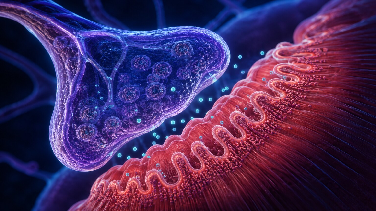
Figure 4. Calcium ions play a direct, physical role at the neuromuscular junction, where nerve signals are converted into muscle contraction.
Calcium's role at the junction between nerves and muscles explains why symptoms show up here first, and often fastest, when blood calcium drops. At a microscopic level, calcium ions help stabilize the membranes of nerve cells, effectively acting as a gatekeeper that controls how easily those membranes let electrical signals fire. When calcium is plentiful, nerve membranes stay appropriately "calm," firing signals only when they're genuinely supposed to. When calcium drops, that gatekeeping function weakens, and nerve membranes become hyperexcitable — firing more easily and more often than they should, sometimes even spontaneously, without any real trigger.
This hyperexcitability is what produces the classic symptoms of hypocalcemia: tingling or numbness, often starting around the mouth or in the fingertips; muscle cramps; and, in more pronounced cases, a condition called tetany, where muscles involuntarily and painfully contract, sometimes pulling the hand into a characteristic clawed position with the wrist flexed and fingers drawn together. Two specific physical exam findings, named after the physicians who first described them, are still used today to check for this heightened neuromuscular excitability at the bedside: Chvostek's sign, where gently tapping the facial nerve near the ear causes an involuntary twitch of the facial muscles, and Trousseau's sign, where inflating a blood pressure cuff on the arm for a few minutes triggers that same clawed hand spasm. In more severe or rapidly developing hypocalcemia, this excitability can extend beyond skeletal muscle to affect the heart's electrical rhythm and, in rare extreme cases, cause seizures — which is why significantly low calcium, especially when it develops quickly, is treated as a genuine medical priority rather than something to simply monitor and wait out.
Recognizing Symptoms: From Mild to Severe
Symptoms of low calcium tend to follow a rough progression tied to both how low the level has fallen and how quickly it got there, and recognizing where on that spectrum a given set of symptoms falls can help someone understand why a clinician is responding with a particular level of urgency. At the mildest end, many people with modestly low calcium notice nothing at all, or only vague, easy-to-dismiss symptoms like fatigue, mild irritability, or brittle nails — subtle enough that they're often only connected to calcium in hindsight, after a lab test explains them. As levels drop further or the deficiency persists, more specific neuromuscular symptoms tend to emerge: numbness or tingling around the mouth, in the fingertips, or in the toes; muscle cramps, particularly in the hands, feet, or calves; and a sensation some people describe as their muscles feeling unusually "twitchy" or prone to spasm with minimal provocation.
At the more severe end of the spectrum, symptoms become impossible to ignore: painful, sustained muscle spasms (tetany), difficulty swallowing if throat muscles are affected, wheezing or a tight feeling in the chest if the muscles around the airway are involved, and, in the most serious cases, seizures or dangerous heart rhythm disturbances. It's worth repeating a point made earlier because it matters so much clinically: the speed of the drop matters as much as the depth. Someone whose calcium fell gradually over months, perhaps from slowly worsening kidney disease, may tolerate a level that would cause dramatic, immediate symptoms in someone whose calcium dropped suddenly, such as within the first day or two after parathyroid surgery. This is why an emergency room evaluating new, severe neuromuscular symptoms will move quickly to check calcium levels directly, rather than waiting for a routine follow-up test days later.
How Low Calcium Affects Bone Health Over Time
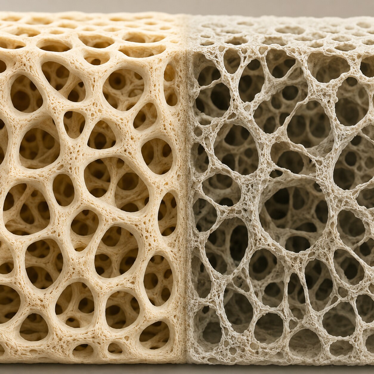
Figure 5. Chronic calcium deficiency gradually thins bone architecture as the skeleton is repeatedly drawn on to defend blood calcium levels.
While the acute, nerve-and-muscle effects of low calcium get most of the immediate attention, the skeletal consequences of a persistent calcium shortfall are just as real, they simply unfold on a much longer timeline. Remember that PTH's first response to falling blood calcium is to pull calcium out of bone — a short-term fix that protects your nerves and heart at the direct expense of your skeleton. If the underlying cause of low calcium isn't corrected and this borrowing continues month after month, year after year, your bones can measurably lose density and structural integrity, a process that contributes to osteoporosis (reduced bone mass and strength) or, in cases tied specifically to inadequate mineralization, osteomalacia (softened bone from poorly mineralized new bone tissue, called rickets when it occurs in children whose bones are still growing).
This slow-motion skeletal cost is one of the main reasons chronic, low-grade calcium or vitamin D deficiency is taken seriously by clinicians even when a patient has no obvious symptoms day to day — the damage accumulates quietly, showing up years later as an unexpected fracture from a minor fall, or as a low score on a bone density scan (DEXA) obtained for an entirely different reason, long after the original deficiency first appeared on a lab report that may have seemed unremarkable at the time. This is also why correcting the underlying cause of chronic hypocalcemia — not just treating the number in isolation with supplements — matters so much for long-term skeletal health, since ongoing PTH-driven bone resorption will continue for as long as the root problem remains unaddressed.
How Diet and Absorption Fit Into the Picture
It's worth separating two questions that often get blurred together: how much calcium someone eats, and how much of that calcium their body actually absorbs and uses — because low blood calcium can stem from either one, and they call for different fixes. Dietary sources of calcium are broader than dairy alone, though dairy remains one of the most concentrated and easily absorbed sources; leafy greens like kale and bok choy, fortified plant milks, canned fish eaten with the soft bones (like sardines or canned salmon), tofu made with calcium sulfate, and almonds all contribute meaningfully to daily intake. The recommended daily amount for most adults falls in the range of 1,000 to 1,200 milligrams, with the higher end typically recommended for women over 50 and men over 70, reflecting the accelerated bone loss that tends to occur after these ages.
But intake is only half of the equation — absorption is where vitamin D becomes essential, since without it, the intestinal cells responsible for actively transporting calcium across the gut wall and into the bloodstream simply can't do their job efficiently, no matter how much calcium arrives in a meal. This is also where certain dietary and lifestyle factors quietly work against absorption even in someone eating plenty of calcium-rich food: excessive intake of foods high in oxalates (like spinach and rhubarb) or phytates (found in some whole grains and legumes) can bind calcium in the gut and reduce how much gets absorbed, though this effect is generally modest for people eating a varied diet. Certain digestive conditions that damage or bypass portions of the small intestine, where most calcium absorption takes place, can have a much larger effect, which is part of why malabsorption disorders appear on the list of causes worth ruling out when dietary intake alone doesn't explain a low result.
Common Causes of Low Calcium

Figure 6. Chronic kidney disease impairs the kidney's ability to activate vitamin D, one of several distinct pathways that can lead to low calcium.
Vitamin D deficiency is, by a wide margin, the most common cause of low calcium worldwide, since vitamin D is essential for absorbing calcium from your diet in the first place — without enough of it, even a calcium-rich diet can leave you effectively deficient, because the calcium you eat simply passes through your digestive system unabsorbed. Hypoparathyroidism, a condition where the parathyroid glands produce too little PTH, is another significant cause; it can be inherited, but it's more commonly acquired after neck surgery, particularly thyroid or parathyroid surgery, where one or more of the small glands can be accidentally damaged or removed in the course of the procedure. Chronic kidney disease is a major contributor as well, since the kidneys are responsible for converting vitamin D into its active form — when kidney function declines, that activation step becomes impaired, reducing calcium absorption regardless of how much vitamin D someone is actually taking in.
Magnesium deficiency deserves particular attention because it's a cause that's easy to overlook: magnesium is required for the parathyroid glands to release PTH properly, so a significant magnesium deficiency can effectively shut down the body's normal calcium-defense response even when the parathyroid glands themselves are structurally healthy — which also means that in these cases, calcium supplementation alone won't fix the problem until the magnesium deficiency is corrected first. This particular interaction often catches people by surprise, since magnesium doesn't intuitively seem connected to calcium or bone health at all, and it's a good example of why a low calcium finding is best treated as an invitation to look at the broader mineral and hormone picture rather than a problem to solve with calcium alone. Malabsorption conditions affecting the digestive tract, such as celiac disease or a history of certain gastric or intestinal surgeries, can reduce calcium and vitamin D absorption directly. Certain medications, including some used to treat osteoporosis (ironically), seizures, and low bone density, along with severe acute pancreatitis and large-volume blood transfusions (due to the citrate used as an anticoagulant in stored blood binding calcium), round out the more common causes seen in clinical practice.
Bisphosphonates, a class of medications commonly prescribed to slow bone loss in osteoporosis, illustrate this medication-related mechanism well: they work by suppressing the very bone-resorption process that PTH relies on to rapidly release calcium during a shortfall, so in someone who is also vitamin D deficient, starting a bisphosphonate can occasionally unmask or worsen hypocalcemia that was previously being compensated for by bone release — which is exactly why clinicians typically check and correct vitamin D status before starting this type of medication. Proton pump inhibitors, widely used for acid reflux, have also been associated with reduced calcium absorption with long-term use, since stomach acid plays a role in making dietary calcium more soluble and absorbable. None of these medication-related causes should discourage anyone from taking a prescribed treatment without first discussing it with their provider — they're included here simply to illustrate how varied and sometimes indirect the pathways to low calcium can be.
Low Calcium in Pregnancy, Older Adults, and Newborns
Certain life stages carry their own particular considerations when it comes to low calcium, worth understanding separately because the causes and stakes can differ meaningfully from the general adult picture described so far. During pregnancy, a growing fetus draws on the mother's calcium supply to build its own developing skeleton, and the mother's body normally compensates by roughly doubling how efficiently it absorbs calcium from food, particularly during the second and third trimesters. When this compensation is inadequate — often due to insufficient vitamin D or dietary calcium — pregnant women can develop hypocalcemia, which is part of why prenatal vitamins routinely include both calcium and vitamin D, and why calcium status is a standard consideration in prenatal care.
In older adults, low calcium takes on added significance because it compounds with the natural, age-related decline in bone density that affects nearly everyone over time. Reduced sun exposure (a key source of vitamin D synthesis), decreased dietary intake, reduced kidney function, and a higher likelihood of taking medications that interfere with calcium metabolism all become more common with age, layering additional risk on top of an already more fragile skeleton — which is part of why unexplained falls or fractures in older adults often prompt a calcium and vitamin D check as a routine part of the workup. Newborns represent a distinct category entirely: neonatal hypocalcemia can occur in the first few days of life, particularly in premature infants, infants of mothers with diabetes, or infants who experienced complications during delivery, since a newborn's own parathyroid system is still maturing and adjusting to functioning independently outside the womb after months of relying on a steady, placental calcium supply. In each of these groups, the underlying biology of calcium regulation is the same one described throughout this article — what changes is which part of the system is most likely to be under strain, and understanding that context helps explain why the same lab number can carry a different degree of urgency depending on who's holding the report.
What Happens After Low Calcium Is Found on a Lab Report
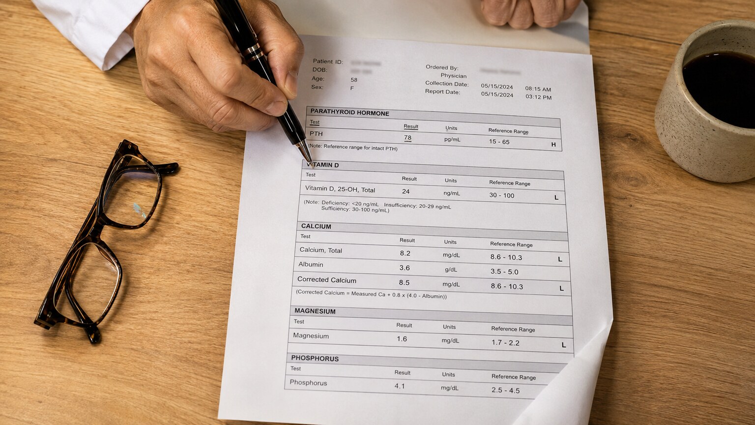
Figure 7. Confirming the cause of low calcium typically involves reviewing PTH, vitamin D, magnesium, and corrected calcium together as one panel.
Once a low calcium result is confirmed and not simply an artifact of low albumin, the practical next step is almost always a small, targeted panel rather than guesswork: albumin (to calculate corrected calcium, if not already done), PTH, vitamin D (specifically 25-hydroxyvitamin D, the standard storage form used to assess overall status), magnesium, and kidney function tests. The pattern these values form together points fairly directly toward a cause. Low calcium with a high PTH suggests the parathyroid glands are working correctly and simply don't have enough calcium or vitamin D to work with — pointing toward a vitamin D or dietary cause. Low calcium with an inappropriately low or normal PTH — a PTH level that should be elevated in response but isn't — points toward hypoparathyroidism or a magnesium problem interfering with PTH release. Low calcium alongside abnormal kidney function shifts the focus toward chronic kidney disease as the underlying driver, while a low magnesium result found on the same panel often explains why PTH isn't rising the way it normally would, redirecting the workup toward correcting magnesium first before reassessing the rest of the picture.
Treatment follows directly from whichever cause is identified: oral or, in urgent cases, intravenous calcium supplementation to correct the immediate deficit; vitamin D repletion when that's the driver; magnesium correction when magnesium deficiency is blocking the normal PTH response; and, for hypoparathyroidism, longer-term calcium and active vitamin D (calcitriol) supplementation to compensate for the missing hormone. Mild, asymptomatic hypocalcemia found incidentally on routine labs is generally manageable and often resolves once the underlying cause is addressed, while more severe or rapidly symptomatic cases — particularly with signs like tetany, seizures, or heart rhythm changes — are treated more urgently, sometimes in a hospital setting, given how quickly and seriously calcium affects nerve and muscle excitability. In an urgent setting, intravenous calcium is typically given slowly and with heart-rhythm monitoring in place, since correcting calcium too quickly carries its own risks; once the acute symptoms are controlled, the focus shifts to identifying and treating the underlying cause so the deficiency doesn't simply recur once the intravenous treatment ends.
Tracking Calcium and Related Markers Over Time
Because calcium is so tightly regulated, a single result — especially one drawn during an acute illness, a hospital stay, or right after a surgical procedure — doesn't always reflect someone's baseline, ongoing status. This is one of the reasons clinicians managing a known calcium disorder, whether that's hypoparathyroidism, chronic kidney disease, or a malabsorption condition, typically recheck levels periodically rather than relying on one snapshot, watching not just the calcium number itself but how it moves alongside PTH, vitamin D, and magnesium over successive visits. A pattern of gradually improving corrected calcium alongside normalizing PTH after starting vitamin D supplementation, for instance, is a reassuring sign that the treatment is addressing the actual underlying mechanism, not just temporarily masking a number.
For someone managing chronic hypoparathyroidism, this kind of tracking becomes a long-term part of routine care, since the goal isn't simply to push calcium back into the normal range once, but to keep it stably there without overshooting — because the treatments used to raise calcium (calcium supplements combined with active vitamin D) can, if dosed too aggressively, push levels too high, which carries its own separate risks, including kidney stones and calcium deposits in soft tissue. This delicate balancing act is part of why hypoparathyroidism, once diagnosed, usually involves ongoing monitoring with an endocrinologist rather than a one-time fix, and why patients managing it are often taught to recognize both the symptoms of calcium running too low and, less intuitively, the symptoms of it running too high, such as excessive thirst, frequent urination, or abdominal discomfort.
Frequently Asked Questions
Can I fix low calcium just by eating more dairy or taking calcium supplements?
It depends entirely on the cause. If the underlying issue is inadequate dietary intake alone, increasing calcium-rich foods or taking a supplement can genuinely help. But if the real problem is vitamin D deficiency, a magnesium deficiency, hypoparathyroidism, or kidney disease, taking more calcium without addressing that root cause often won't fully correct the levels, and in some cases won't help at all — which is exactly why identifying the actual cause matters more than simply adding more calcium.
Is a slightly low calcium result on a routine blood test usually serious?
Often not. A mildly low total calcium result, especially in someone with known low albumin (from illness, hospitalization, or malnutrition) and no symptoms, frequently corrects to a normal value once albumin is accounted for. Only a clinician reviewing the corrected calcium, PTH, and vitamin D together can say for certain whether a specific result needs further evaluation.
How quickly do muscle symptoms from low calcium usually appear?
It depends heavily on how quickly the calcium level dropped, not just how low it is. A calcium level that falls slowly, over weeks or months, may produce few or no noticeable symptoms even at levels that would cause dramatic symptoms if they developed suddenly. A rapid drop — such as after parathyroid surgery — can produce tingling, cramping, or spasms within hours.
Does low calcium always mean I'm at risk for osteoporosis?
Not automatically, but chronic, uncorrected low calcium is a recognized risk factor for reduced bone density over time, since the body compensates by pulling calcium from bone. Someone whose low calcium is identified and appropriately treated early is far less likely to experience meaningful bone loss than someone whose deficiency goes uncorrected for years.
Can low magnesium really cause low calcium, even if I'm getting enough calcium?
Yes, and it's one of the more commonly missed causes. Magnesium is required for the parathyroid glands to release PTH properly, so a significant magnesium deficiency can blunt the body's normal calcium-defense response even when calcium intake and the parathyroid glands themselves are otherwise fine. In these cases, correcting magnesium is often necessary before calcium levels can normalize, regardless of how much calcium is being consumed or supplemented.
If my calcium is low after neck or thyroid surgery, is that permanent?
Not necessarily. Temporary hypocalcemia after thyroid or parathyroid surgery is relatively common, since the parathyroid glands sit very close to the thyroid and can be temporarily stunned or irritated during the procedure without being permanently damaged. Function often recovers within days to weeks, though a smaller number of cases involve more lasting parathyroid injury requiring longer-term calcium and vitamin D supplementation, which is why levels are typically monitored closely in the days following this type of surgery.
Conclusion
A low calcium result is rarely as simple as "your body doesn't have enough calcium" — it's a finding that requires context from albumin, PTH, vitamin D, and magnesium to interpret correctly, and one whose consequences show up differently depending on the timeline: acute drops tend to announce themselves through nerves and muscles, while chronic, uncorrected deficiency quietly erodes bone strength over years. Understanding which pattern you're dealing with, and identifying the specific mechanism behind it, is what turns a worrying number on a printed page into a clear, treatable finding rather than a source of ongoing uncertainty. In the vast majority of cases, once the actual driver is identified — whether that's a vitamin D shortfall, a magnesium problem, a parathyroid issue, or something related to kidney function — the path forward is well established and genuinely effective, and most people see their levels, and their symptoms, improve substantially once appropriate treatment begins. Bringing your full panel — not just the calcium line by itself — into a conversation with a healthcare provider is the most direct path from an unfamiliar result to a real answer, and the sooner that conversation happens, the sooner both the number and the underlying cause behind it can be properly addressed.
Still Not Sure What Your Results Mean?
Upload your labs and get a complete, visual, plain-language interpretation of every biomarker — delivered to your inbox in under 15 minutes.
Get My ReportThis article is for educational purposes only and does not constitute medical advice. Always consult your healthcare provider regarding your specific lab results.